PERKASA MEDICAL

Free shipping worldwide. Orders over $200
Availability: In Stock

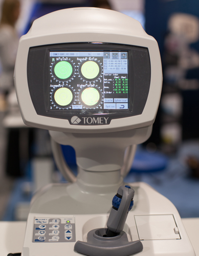
Tomey RT-7000 Autorefractor Keratometer Topographer For Sale

$11,350.00

The Tomey Refractometer Keratometer and Topographer only in 1 unit senior citizens and children can also get the inspection with out any trouble because they dont have to move the seat for three measurements.

Other people want this. 15 people have this in their carts right now.

Description

Tomey RT-7000 Autorefractor Keratometer Topographer

-Three Functions in One Instrument

-Easy & Speedy Touch Screen Alignment

-6.4 inches color TFT LCD

-Dual  CCD technology for Refractometry

-IOL/ Cataract Mode

-New incites for Keratometry KAI, KRI

-Auto Alignment / Auto Shot of Topography

Corneal Topography

– Auto Alignment / Auto Shot

Any one can easily capture measurement with Auto Alignment and Auto Shot. The Measurement variation is significantly reduced no matter what the skill level is of the operator.

CL Fitting Simulation

-A pseudo-fluorescent pattern of the contact lens selected from the built-in data base can be confirmed and the fitting can be stimulated before the patient wears  the trial lens.

Corneal Eccentricity Index

-The index of CEI (Corneal Eccentricity Index) indicates the ratio of flattering in percentage from the central cornea to the periphery.

Three Function in One Instrument

– Refractometer Keratometer and Topographer only in 1 unit senior citizens and children can also get the inspection with out any trouble because they don’t have to move the seat for three measurements. Switch from Ref-Kerato Mode to Corneal Topography Mode with only ONE TOUCH. The light come appears with only one button from the measuring head and the Ref-Keratometer is transformed to the topographer.

Refractometry

– Dual CCD technology for Refractometry

Two CCD cameras are used to capture images for observation and for measurement while providing highly accurate measurement data.

Keratometory

– New Indices for Keratometry

The Corneal Irregular astigmatism display function is installed to expand the possibility of Keratometer. This is the new function to measure the level of corneal irregular astigmatism which was difficult in the past.

SPECIFICATIONS

MeasurmentRanges

Refraction Measuremnt

Sphere  -25.00D to ±22.00DIVD=12.0mm

Cylinder  0D to ± 10.00DIVD=12.0mm

Axis  0 to 180

Corneal Curvature Measurement  5.00mm to 11.00mm

Measurement Zone 3mm R:8.00mm

Corneal Mapping

Display Range  9 to 100 D

-Normal Mode

1.0 to 8.0mm (R:8.00mm)

-Special Mode

0.9 to 7.0 mm (R:8.00mm)

Measurement Time

REF 0.2 Seconds

KRT 0.1 Seconds

Minimum Pupil Diameter  2.2mm

Minimum Measurment Step


Refraction Measurement

Sphere  0.01D

Cylinder  0.01D

Axis  1

Corneal Curvature Measurement  0.01mm

PD (Pupil Distance)  50mm to 86mm

Recording  Built-In Thermal Printer

Vertex Distance  0mm, 12.0mm, 13.5mm, 14.0mm, 15.5mm, 16.0mm

Exterior Output  (AN port / 4USB ports)

Dimensions Tomey RT-7000 Autorefractor Keratometer Topographer

High  466mmm or 18 Inches

Width  307mm or 12 Inches

Depth  490mm or 19 Inches

Weight  Approx 20,000Kg / 44.4 Pounds

Display  6.4 Inches color liquid crystal

Power Supply

Voltage  AC 100 to 240V

Frequency  50/60 Hz

Consumption Power  120VA to 150VA

Temperature  +10C to + 40C

Display  30% to 75%

Reviews

There are no reviews yet.

Be the first to review “Tomey RT-7000 Autorefractor Keratometer Topographer For Sale”

Your email address will not be published. Required fields are marked *

Quick Comparison

SettingsTomey RT-7000 Autorefractor Keratometer Topographer For Sale removeEssilor Mr Blue 2.0 Lens Edgers and Tracer For Sale removeEssilor Neksia Lens Edging System For Sale removeNidek CEM-530 Specular Microscope For Sale removeNidek RS-330 Retina Scan Duo 2 with FAF removeTopcon KR8000PA Autorefractor Keratometer For Sale remove
NameTomey RT-7000 Autorefractor Keratometer Topographer For Sale removeEssilor Mr Blue 2.0 Lens Edgers and Tracer For Sale removeEssilor Neksia Lens Edging System For Sale removeNidek CEM-530 Specular Microscope For Sale removeNidek RS-330 Retina Scan Duo 2 with FAF removeTopcon KR8000PA Autorefractor Keratometer For Sale remove
Image
SKU
Rating
Price $11,350.00 $3,600.00 $4,240.00 $3,250.00 $3,150.00 $4,100.00
Stock
Availability
Add to cart

Add to cart

Add to cart

Add to cart

Add to cart

Add to cart

Add to cart

DescriptionThe Tomey Refractometer Keratometer and Topographer only in 1 unit senior citizens and children can also get the inspection with out any trouble because they dont have to move the seat for three measurements.Mr Blue 2.0 is on the cutting edge of innovation – serving as a genuine ECP partner since its launch. Now featuring a whole range of new functions and even greater flexibility, Mr Blue is the perfect solution for demanding workshop.workflow, because some letters inspire confidence.With Neksia, Essilor combines years of experience in high-performance and precision edging into one of the most user-friendly and productive systems in the world.

Combination of auto and manual analyses

All three manual analysis methods can be performed on the same image and also on auto-analyzed images. The versatility of combining automated and manual analyses allows analysis of the range of pathology in a comprehensive practice.
The CEM-530 with new advanced software and enhanced image capture system allows rapid image acquisition. 
The Nidek RS-330 OCT is a combination of a High-Definition OCT and fundus camera. Optional delivery with FAF (Auto-fluorescence function)
The new tri-functional Autorefractor Keratometer Topcon KR-8000PA Supra combines an autorefractor, an autokeratometer, and a computerized color corneal mapping system all in a single, compact unit! This enables the instrument to provide three times the diagnostic capabilities in one-third the space, at a fraction of the cost of the individual instruments.
Content

Tomey RT-7000 Autorefractor Keratometer Topographer

-Three Functions in One Instrument-Easy & Speedy Touch Screen Alignment-6.4 inches color TFT LCD-Dual  CCD technology for Refractometry-IOL/ Cataract Mode-New incites for Keratometry KAI, KRI-Auto Alignment / Auto Shot of TopographyCorneal Topography– Auto Alignment / Auto ShotAny one can easily capture measurement with Auto Alignment and Auto Shot. The Measurement variation is significantly reduced no matter what the skill level is of the operator.CL Fitting Simulation-A pseudo-fluorescent pattern of the contact lens selected from the built-in data base can be confirmed and the fitting can be stimulated before the patient wears  the trial lens.Corneal Eccentricity Index-The index of CEI (Corneal Eccentricity Index) indicates the ratio of flattering in percentage from the central cornea to the periphery.Three Function in One Instrument– Refractometer Keratometer and Topographer only in 1 unit senior citizens and children can also get the inspection with out any trouble because they don’t have to move the seat for three measurements. Switch from Ref-Kerato Mode to Corneal Topography Mode with only ONE TOUCH. The light come appears with only one button from the measuring head and the Ref-Keratometer is transformed to the topographer.Refractometry– Dual CCD technology for RefractometryTwo CCD cameras are used to capture images for observation and for measurement while providing highly accurate measurement data.Keratometory– New Indices for KeratometryThe Corneal Irregular astigmatism display function is installed to expand the possibility of Keratometer. This is the new function to measure the level of corneal irregular astigmatism which was difficult in the past.SPECIFICATIONSMeasurmentRangesRefraction MeasuremntSphere  -25.00D to ±22.00DIVD=12.0mmCylinder  0D to ± 10.00DIVD=12.0mmAxis  0 to 180Corneal Curvature Measurement  5.00mm to 11.00mmMeasurement Zone 3mm R:8.00mmCorneal MappingDisplay Range  9 to 100 D-Normal Mode1.0 to 8.0mm (R:8.00mm)-Special Mode0.9 to 7.0 mm (R:8.00mm)Measurement TimeREF 0.2 SecondsKRT 0.1 SecondsMinimum Pupil Diameter  2.2mmMinimum Measurment Step Refraction MeasurementSphere  0.01DCylinder  0.01DAxis  1Corneal Curvature Measurement  0.01mmPD (Pupil Distance)  50mm to 86mmRecording  Built-In Thermal PrinterVertex Distance  0mm, 12.0mm, 13.5mm, 14.0mm, 15.5mm, 16.0mmExterior Output  (AN port / 4USB ports)Dimensions Tomey RT-7000 Autorefractor Keratometer TopographerHigh  466mmm or 18 InchesWidth  307mm or 12 InchesDepth  490mm or 19 InchesWeight  Approx 20,000Kg / 44.4 PoundsDisplay  6.4 Inches color liquid crystalPower SupplyVoltage  AC 100 to 240VFrequency  50/60 HzConsumption Power  120VA to 150VATemperature  +10C to + 40CDisplay  30% to 75%
Essilor Mr Blue 2.0 Lens Edgers and TracerComplete Control of Lens Mounting
  • Optimal monitoring of all trajectories, irrespective of finishing materials or shapes
  • Absolute axis control thanks to hybrid edging technology: grinding + milling
  • Ideally suited for quality finishing even on highest base curves
  • Fast and accurate drilling with data acquired in a single operation
  • Automatic centering and blocking for simple operation
Unrivalled Ergonomics
  • Completely integrated functions without external peripheral devices
  • Vertical architecture with <Heads-up displays> for optimum comfort
  • Storage features ensure all accessories are within reach.
  • Intelligent interface adapts to the way you work.
Highest Quality Guaranteed Essilor Mr Blue 2.0 Lens Edgers and TracerPerfect Centering
  • Binocular 3-D tracing of full-rim frames up to base 9*
  • Automatic adjustment of centering data based on the frames’ 30D parameters*
Aesthetic and Robust Mountings
  • FIT-4-Frame technology with dedicated small wheel*
  • Drilling and grooving tools can be tilted up to 30o, making it safe to mount lenses of up to base 10. *Essilor patents and exclusives
Safe Process
  • Roughing cycle automatically adapts to the curves of lenses up to base 10, avoiding all risk of breakage
  • Bevel placement and shelf depth optimized for safe mounting without undue stress of lens
Aesthetically Pleasing and Easy-to-Mount Bevels
  • The bevel heights are adjustable to perfectly adjust to the thinnest grooves
  • The shelf bevel heights are optimized to follow the frame’s shape perfectly
  • The step bevel is particularly suited for sport eyeglasses with deep, rectangular or uneven groove profile
Mr Blue 2.0 is on the cutting edge of innovation – serving as a genuine ECP partner since its launch. Now featuring a whole range of new functions and even greater flexibility, Mr Blue is the perfect solution for demanding workshop.
Essilor Neksia Lens Edging SystemWork More Easily
  • A touch of efficiency – The 8.4-inch color touch-screen display makes navigation a breeze. And you can select the icon-based functions quickly and easily with just the touch of a finger.
  • Fast-access settings – With all job settings located conveniently on the centering part of the system, you can define your finishing settings quickly and launch edging jobs in just a few clicks. If necessary, you can still adjust settings after the feeler measurement.
  • A user-friendly 1,000-position job database – Save precious time by easily accessing or storing complete jobs in the database via the touch screen or optional barcode reader.
Experience Workflow Efficiency
  • Fast, easy and extended frame tracking – Equipped with auto frame-type selection, simple clamping and rapid tracing cycles, Neksia helps you trace any type of frame effortlessly, including children’s frames, small B-size and large A-size frames
  • Automatic drilling-hole recognition – Achieve even greater productivity thanks to optional features dedicated to automatic detection of drilling holes and flat bevel shapes.
  • Convenient centering and blocking – Neksia arms you with valuable features that make centering and blocking straight-forward and precise.
    • Handle lenses easily with the wide-open centering area
    • Increase precision with intelligent targeting and parallax-free working
    • Benefit from the intuitive on-screen interface
Achieve High Performance Essilor Neksia Lens Edging System
  • Improve axis accuracy – By combining new edging cycle algorithms and cutting-force regulation, you are guaranteed optimal results whatever the edging situation.
    • Optimized edging speed based on the lens material, shape and lens thickness guarantees fast cycle times, lens positioning and alignment.
    • The New Edging Assisted System cycle avoids axis deviation on new- generation hydrophobic lenses, rectangular shapes or fragile lenses.
  • Best automatic cycles – For 90% of your jobs, all it takes are a few clicks to achieve a perfectly calculated bevel, groove or drilling cycle. The lens is well positioned for an aesthetic and perfectly fitted result, no matter the material or frame type.
  • Consistent intelligent chamfering – With Neksia’s patented flexible chamfering wheel, the pressure applied to the wheel adapts seamlessly to the lens edge depending on the shape and curve of the lens.

Nidek CEM-530 Specular Microscope

The analysis results with graphic and color-coded cell images helps the clinician to rapidly and effectively evaluate the endothelial cell layer.In addition to conventional central and peripheral specular microscopy, the CEM-530 includes a unique function to capture paracentral images.
  • Paracentral specular microscopy
  • Faster measurements and two-second auto analysis
  • Comprehensive analysis
  • Advanced manual analysis functions
  • Easy operation
  • Additional features with CEM Viewer for NAVIS-EX
Combination of auto and manual analyses
All three manual analysis methods can be performed on the same image and also on auto-analyzed images. The versatility of combining automated and manual analyses allows analysis of the range of pathology in a comprehensive practice.Features
  • Multi area specular microscopy
  • Enhanced usability and quick analysis
  • Advanced manual analysis functions
  • Combination of auto and manual analyses
  • Additional features with CEM Viewer for NAVIS-EX
Multi area specular microscopy
In addition to conventional central and peripheral specular microscopy, the CEM-530 includes a NIDEK original function that captures paracentral images. The combination of central, paracentral, and peripheral imaging provides a broader, overall view that can be used for detailed morphological and quantitative evaluation of the endothelial layer and individual cells.


Enhanced usability and quick analysis

The 3D auto tracking and auto shot functions result in a user friendly and patient friendly experience. Data analysis within 2 seconds allows efficient patient flow.

Detail analysis


Advanced manual analysis functions
Center point

Select the approximate center of a cell. The cells are detected based on the surrounding points. This method is effective for areas where groups of cells are clumped together.


Corner point

Trace the outlines of the cells to be analyzed by selecting the corners of each cell. This method is suitable for detailed identification of the size and dimension of isolated cells.


Pattern select

Select a hexagonal reference pattern that is similar to the cell size and drag it onto the cell to be analyzed. This method is effective for rough identification of the size and dimension of the cells.


 Combination of auto and manual analyses 
All three manual analysis methods can be performed on the same image and on auto-analyzed images. The versatility of combining automated and manual analysis on the same image allows for better clinical interpretation of the diverse range of pathology in a comprehensive practice.


Additional features with CEM Viewer for NAVIS-EX* (optional)
CEM Viewer is software used for viewing and working with CEM-530 data via NAVIS-EX. This function enhances the capability of the CEM-530 with additional features and increases the efficiency of any clinic. *NAVIS-EX is optional software and is required for use of the CEM Viewer.
Data management and endothelial cell count

Unlimited NAVIS-EX database is available for review on the CEM Viewer. The basic functions of the CEM-530 such as endothelial cell count are available on the CEM Viewer.


Progression follow-up and comparison

Multiple examination data sets are displayed in chronological order for follow-up. Additionally, two data sets are displayed for comparison. Endothelial changes can be monitored over time with this function.


Paracentral display with peripheral
The images and analyses of the paracentral and peripheral areas are displayed providing a comprehensive image of endothelial cells.

Nidek RS-330 Retina Scan Duo 2 with FAF

The Retina Scan Duo 2 is a combined OCT and fundus camera system that incorporates new features enhancing screening and clinical efficiency in addition to user-friendly features that were incorporated from the previous model.FEATURES : Nidek RS-330 Retina Scan Duo 2 with FAF
  • Fundus image acquisition with macula and disc capture in one image on OCT
  • Combined diagnosis of macular and disc pathologies
  • Wide area scan (12 x 9 mm) Wide area normative database (macula: 9 x 9 mm, disc: 6 x 6 mm): A 12 x 9 mm wide area image can be acquired. The retina map captures both the macula and disc in a single shot. The normative database provides a wide area color-coded map comparing the patient’s macular thickness to a population of normal eyes.
  • Denoising technique with deep learning: A new image enhancement technique using deep learning automatically displays a denoised image once B-scan acquisition is complete. With deep learning of a large data set of images averaged from 120 images, this denoising technique provides high definition images comparable to a multiple-image-averaging technique. The denoising function generates high definition images from a single frame while decreasing image acquisition time and increasing patient comfort.
  • Quick acquisition of high definition B-scan images from a single-frame image
  • Fundus autofluorescence (FAF): The FAF function is an advanced screening feature that allows non-invasive evaluation of the RPE without contrast dye. *Available for the FAF model
Nidek RS-330 Retina Scan Duo 2– Easy operation with 3-D auto tracking, auto shot, and user friendly interface – High definition images – Wide area scan (12 x 9 mm) / Wide area normative database (9 x 9 mm) – Multiple OCT scan patterns – Value added features – Various reports
Nidek High speed image capture OCT images are captured at scan speeds of 70,000 A-scans/s which is 32% faster than acquisition with the Retina Scan Duo™ using Regular OCT sensitivity*Wide area scan (12 x 9 mm) Wide area normative database (macula: 9 x 9 mm, disc: 6 x 6 mm) A 12 x 9 mm wide area image can be acquired. The retina map captures both the macula and disc in a single shot. The normative database provides a wide area color-coded map comparing the patient’s macular thickness to a population of normal eyes.Denoising using deep learning A new image enhancement technique using deep learning automatically displays a denoised image once B-scan acquisition is complete. With deep learning of a large data set of images averaged from 120 images, this denoising technique provides high definition images comparable to a multiple-image-averaging technique. The denoising function generates high definition images from a single frame while decreasing image acquisition time and increasing patient comfort

Topcon KR8000PA Autorefractor Keratometer

By implementing Topcon’s Autorefractor Keratometer Topcon KR-8000PA Supraexclusive rotary prism technology for the refraction portion of the measurement, the Autorefractor Keratometer Topcon KR-8000PA Supra provides objective data that is unmatched in accuracy and reliability. Auto tracking and auto measurement enhance the ease of operation.

The Autorefractor Keratometer Topcon KR-8000PA Supra unit also incorporates ten placido rings for enhanced testing precision, providing corneal mapping measurements out to 10mm!

Features for Autorefractor Keratometer Topcon KR-8000PA Supra

Topcon Color Mapping 32 Software

This versatile 32-bit color mapping software system enhances clinical evaluations with the output of highly accurate, full color corneal maps. The system offers an enlarged color map, tangential axial maps, sagittal maps, refractive and keratometric data, and peripheral keratometric maps. It provides early phase detection of subclinical keratoconus as well as pre and post-op evaluation. The software also features a new image overlay function, as well as a Multi-Map and Difference Map View, which display multiple image maps for easy comparison.

The color mapping system is an excellent patient education tool and is designed for use with the Topcon KR-7000P or KR-8000PA Auto Refractor/Keratometer. Windows® 95/98/NT Compatible.

The color mapping software also features a contact lens fitting module with adjustable parameters to view the contact lens fit on screen.The Autorefractor Keratometer Topcon KR-8000PA Supra System Benefits Include:Topcon KR8000PA Autorefractor Keratometer
  • Allows Three Functions at One Station: – Refraction – Keratometry – Corneal Mapping
  • Contact Lens Fitting Utility
  • Early Phase Detection of Subclinical Keratoconus
  • Direct Link to CV System
The new tri-functional Autorefractor Keratometer Topcon KR-8000PA Supra combines an autorefractor, an autokeratometer, and a computerized color corneal mapping system all in a single, compact unit! This enables the instrument to provide three times the diagnostic capabilities in one-third the space, at a fraction of the cost of the individual instruments.

WeightN/AN/AN/AN/AN/AN/A
DimensionsN/AN/AN/AN/AN/AN/A
Additional information
You're viewing: Tomey RT-7000 Autorefractor Keratometer Topographer For Sale $11,350.00
Add to cart
Select the fields to be shown. Others will be hidden. Drag and drop to rearrange the order.
  • Image
  • SKU
  • Rating
  • Price
  • Stock
  • Availability
  • Add to cart
  • Description
  • Content
  • Weight
  • Dimensions
  • Additional information
  • Attributes
  • Custom attributes
  • Custom fields
Click outside to hide the comparison bar
Compare
Shopping cart close