| Description | The Tomey Refractometer Keratometer and Topographer only in 1 unit senior citizens and children can also get the inspection with out any trouble because they dont have to move the seat for three measurements. | Combination of auto and manual analysesAll three manual analysis methods can be performed on the same image and also on auto-analyzed images. The versatility of combining automated and manual analyses allows analysis of the range of pathology in a comprehensive practice.
The CEM-530 with new advanced software and enhanced image capture system allows rapid image acquisition. | The Extra – 818 is the outcome of more than a decade of treatments and thousands of devices operated worldwide. It contains It contains unique features:
* Contemporary touch screen control
* Built-in Patch Test procedure for examining the optimal energy level
* A stunning number of 450,000 flashes in both lamps.
* The Large Handpiece has a Push Button in addition to the Foot Switch making
work easier and faster for the therapist. | The Nidek RS-330 OCT is a combination of a High-Definition OCT and fundus camera. Optional delivery with FAF (Auto-fluorescence function) | The Sequoia system’s advanced technologies are designed to make image acquisition easier, reduce exam time and increase overall efficiency. Along with many other included features, the Sequoia system offers Native, a patient specific imaging which adapts both phase and amplitude information of each unique patient’s properties which delivers a consistently better image across a broad range of patients. | CIRRUS 4000 delivers high-speed image capture with HD imaging detail and a wider field of view so you can make more informed decisions and spend more time with the patients who need it. |
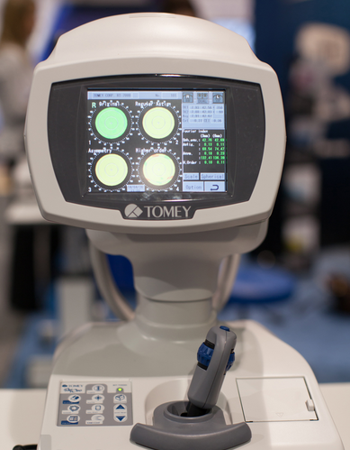
| Content | Tomey RT-7000 Autorefractor Keratometer Topographer
-Three Functions in One Instrument-Easy & Speedy Touch Screen Alignment-6.4 inches color TFT LCD-Dual CCD technology for Refractometry-IOL/ Cataract Mode-New incites for Keratometry KAI, KRI-Auto Alignment / Auto Shot of TopographyCorneal Topography– Auto Alignment / Auto ShotAny one can easily capture measurement with Auto Alignment and Auto Shot. The Measurement variation is significantly reduced no matter what the skill level is of the operator.CL Fitting Simulation-A pseudo-fluorescent pattern of the contact lens selected from the built-in data base can be confirmed and the fitting can be stimulated before the patient wears the trial lens.Corneal Eccentricity Index-The index of CEI (Corneal Eccentricity Index) indicates the ratio of flattering in percentage from the central cornea to the periphery.Three Function in One Instrument– Refractometer Keratometer and Topographer only in 1 unit senior citizens and children can also get the inspection with out any trouble because they don’t have to move the seat for three measurements. Switch from Ref-Kerato Mode to Corneal Topography Mode with only ONE TOUCH. The light come appears with only one button from the measuring head and the Ref-Keratometer is transformed to the topographer.Refractometry– Dual CCD technology for RefractometryTwo CCD cameras are used to capture images for observation and for measurement while providing highly accurate measurement data.Keratometory– New Indices for KeratometryThe Corneal Irregular astigmatism display function is installed to expand the possibility of Keratometer. This is the new function to measure the level of corneal irregular astigmatism which was difficult in the past.SPECIFICATIONSMeasurmentRangesRefraction MeasuremntSphere -25.00D to ±22.00DIVD=12.0mmCylinder 0D to ± 10.00DIVD=12.0mmAxis 0 to 180Corneal Curvature Measurement 5.00mm to 11.00mmMeasurement Zone 3mm R:8.00mmCorneal MappingDisplay Range 9 to 100 D-Normal Mode1.0 to 8.0mm (R:8.00mm)-Special Mode0.9 to 7.0 mm (R:8.00mm)Measurement TimeREF 0.2 SecondsKRT 0.1 SecondsMinimum Pupil Diameter 2.2mmMinimum Measurment Step Refraction MeasurementSphere 0.01DCylinder 0.01DAxis 1Corneal Curvature Measurement 0.01mmPD (Pupil Distance) 50mm to 86mmRecording Built-In Thermal PrinterVertex Distance 0mm, 12.0mm, 13.5mm, 14.0mm, 15.5mm, 16.0mmExterior Output (AN port / 4USB ports)Dimensions Tomey RT-7000 Autorefractor Keratometer TopographerHigh 466mmm or 18 InchesWidth 307mm or 12 InchesDepth 490mm or 19 InchesWeight Approx 20,000Kg / 44.4 PoundsDisplay 6.4 Inches color liquid crystalPower SupplyVoltage AC 100 to 240VFrequency 50/60 HzConsumption Power 120VA to 150VATemperature +10C to + 40CDisplay 30% to 75% | Nidek CEM-530 Specular Microscope
The analysis results with graphic and color-coded cell images helps the clinician to rapidly and effectively evaluate the endothelial cell layer.In addition to conventional central and peripheral specular microscopy, the CEM-530 includes a unique function to capture paracentral images.- Paracentral specular microscopy
- Faster measurements and two-second auto analysis
- Comprehensive analysis
- Advanced manual analysis functions
- Easy operation
- Additional features with CEM Viewer for NAVIS-EX
Combination of auto and manual analysesAll three manual analysis methods can be performed on the same image and also on auto-analyzed images. The versatility of combining automated and manual analyses allows analysis of the range of pathology in a comprehensive practice. Features- Multi area specular microscopy
- Enhanced usability and quick analysis
- Advanced manual analysis functions
- Combination of auto and manual analyses
- Additional features with CEM Viewer for NAVIS-EX
Multi area specular microscopyIn addition to conventional central and peripheral specular microscopy, the CEM-530 includes a NIDEK original function that captures paracentral images. The combination of central, paracentral, and peripheral imaging provides a broader, overall view that can be used for detailed morphological and quantitative evaluation of the endothelial layer and individual cells.
Enhanced usability and quick analysis
The 3D auto tracking and auto shot functions result in a user friendly and patient friendly experience.
Data analysis within 2 seconds allows efficient patient flow.
  Detail analysis
Advanced manual analysis functions
Center point
Select the approximate center of a cell. The cells are detected based on the surrounding points. This method is effective for areas where groups of cells are clumped together.
Corner point
Trace the outlines of the cells to be analyzed by selecting the corners of each cell. This method is suitable for detailed identification of the size and dimension of isolated cells.
Pattern select
Select a hexagonal reference pattern that is similar to the cell size and drag it onto the cell to be analyzed. This method is effective for rough identification of the size and dimension of the cells.
Combination of auto and manual analyses All three manual analysis methods can be performed on the same image and on auto-analyzed images. The versatility of combining automated and manual analysis on the same image allows for better clinical interpretation of the diverse range of pathology in a comprehensive practice.
Additional features with CEM Viewer for NAVIS-EX* (optional)
CEM Viewer is software used for viewing and working with CEM-530 data via NAVIS-EX. This function enhances the capability of the CEM-530 with additional features and increases the efficiency of any clinic.
*NAVIS-EX is optional software and is required for use of the CEM Viewer.
Data management and endothelial cell count
Unlimited NAVIS-EX database is available for review on the CEM Viewer. The basic functions of the CEM-530 such as endothelial cell count are available on the CEM Viewer.
Progression follow-up and comparison
Multiple examination data sets are displayed in chronological order for follow-up. Additionally, two data sets are displayed for comparison. Endothelial changes can be monitored over time with this function.
Paracentral display with peripheralThe images and analyses of the paracentral and peripheral areas are displayed providing a comprehensive image of endothelial cells. | Active Optical Extra 818 IPL Skin Rejuvenation & Hair RemovalHandpieces:Integrated dual handpieces
Chilled Tip:Up to 4c
Pulse Interval:0.5-2 Seconds
Output Power:11-31 joulse/cm2, upto 4,000W
Cooling System:Revolutionary dual cooling
Control:Intuitive touch screen control
Lamp’s Life Span:250,000 Flashes (Universal) 200,000 Flashes (Facial)
Treatment Area:4.5 cm2 (15*50mm) Universal, 4.5 cm2 (15*30mm) Facial
Wavelength:420-100 nm
Weight:42Kg (including Stand)
Dimensions:44cm x 45cm x 100cmExtra-818 Active Optical Extra 818 IPL Skin Rejuvenation & Hair Removal
The Extra – 818 is the outcome of more than a decade of treatments and thousands of devices operated worldwide. It contains It contains unique features:
* Contemporary touch screen control
* Built-in Patch Test procedure for examining the optimal energy level
* A stunning number of 450,000 flashes in both lamps.
* The Large Handpiece has a Push Button in addition to the Foot Switch making
work easier and faster for the therapist.The Extra – 818, based on Active’s GEM-PL technology, sets sets a specific Pulse Pattern according to the selected treatment type, skin color, skin photosensitivity and treatment depth.
The treatment protocols and different pulse patterns take into account the tissue’s TRT (Thermal Relaxation Time) and the combined knowledge accumulated from thousands of treatments done during the years and clinical investigations done to confirm the findings.The combination of tested treatment protocols with high end engineering makes the Extra – 818 the perfect device for leading clinics.Revolutionary Cooling System
The Extra – 818 has a dual cooling system, controlled and monitored in order to ensure long lifetime to the lamp and a comfortable treatment to the patient.Competitive cooling systems “freeze” the skin so that during the treatment the skin temperature will rise to a bearable level. However, this “freezing” temperature might limit the results because the follicles will not reach the coagulation temperature. To ensure optimal results Active installed a special Processor that prevents overcooling the skin and maintains a uniform low temperature of the “glass” throughout the treatment. This revolutionary system enables the Extra – 818 to provide best results with optimal patient comfort.Research Program
For clinical investigations, the self setting program allows researchers to set the treatment’s parameters according to their needs. Allowing advanced research with the device.
The energy levels in the self setting program can reach 31Jcm2 with high intensity of 4,000W. | Nidek RS-330 Retina Scan Duo 2 with FAFThe Retina Scan Duo 2 is a combined OCT and fundus camera system that incorporates new features enhancing screening and clinical efficiency in addition to user-friendly features that were incorporated from the previous model. FEATURES : Nidek RS-330 Retina Scan Duo 2 with FAF- Fundus image acquisition with macula and disc capture in one image on OCT
- Combined diagnosis of macular and disc pathologies
- Wide area scan (12 x 9 mm)
Wide area normative database (macula: 9 x 9 mm, disc: 6 x 6 mm): A 12 x 9 mm wide area image can be acquired. The retina map captures both the macula and disc in a single shot. The normative database provides a wide area color-coded map comparing the patient’s macular thickness to a population of normal eyes.
- Denoising technique with deep learning: A new image enhancement technique using deep learning automatically displays a denoised image once B-scan acquisition is complete. With deep learning of a large data set of images averaged from 120 images, this denoising technique provides high definition images comparable to a multiple-image-averaging technique. The denoising function generates high definition images from a single frame while decreasing image acquisition time and increasing patient comfort.
- Quick acquisition of high definition B-scan images from a single-frame image
- Fundus autofluorescence (FAF): The FAF function is an advanced screening feature that allows non-invasive evaluation of the RPE without contrast dye.
*Available for the FAF model
Nidek RS-330 Retina Scan Duo 2– Easy operation with 3-D auto tracking, auto shot, and user friendly interface
– High definition images
– Wide area scan (12 x 9 mm) / Wide area normative database (9 x 9 mm)
– Multiple OCT scan patterns
– Value added features
– Various reports
OCT images are captured at scan speeds of 70,000
A-scans/s which is 32% faster than acquisition with the
Retina Scan Duo™ using Regular OCT sensitivity* Wide area scan (12 x 9 mm)
Wide area normative database (macula: 9 x 9 mm, disc: 6 x 6 mm)
A 12 x 9 mm wide area image can be acquired. The retina
map captures both the macula and disc in a single shot.
The normative database provides a wide area color-coded
map comparing the patient’s macular thickness to a
population of normal eyes. Denoising using deep learning
A new image enhancement technique using deep learning automatically displays a denoised image once
B-scan acquisition is complete. With deep learning of a large data set of images averaged from 120 images,
this denoising technique provides high definition images comparable to a multiple-image-averaging
technique. The denoising function generates high definition images from a single frame while decreasing
image acquisition time and increasing patient comfort | Siemens Acuson Sequoia 512 LCD Ultrasound Machine18″ Flat panel LCD Monitor–Progressive Media & Display II Package, Cardiac/OB/Vascular Calc. Package, Signature II Option, AUX, CPS Cardiology, CPS General Cadene Trigg Burst, Calc Data to MO, Card Cadence II Contrast Quant, Cornoary Flow reserve, Dicome Store/Print, DTI (Doppler Tissue Imaging), High HR ECG, MIS/MIF, Modality Worklist Native TEQ, Native TEQ, NTHI (Native Tissue Harmonics), Spectral TEQ, TEQ (Tissue Equalization), VVIFeatures- Shared Service
- Native Patient Specific Imaging
- Four Sight TEE view
- Axius quantitative strain rate imaging
- Cadence contrast pulse sequencing technology
- Native Tissue Harmonic Imaging
- Native TEQ ultrasound technology
- Color doppler, Color angio and b-color
- PW/CW Doppler
- M-Mode
- Folding Flat Panel Display
- DICOM capabilities
Applications- Abdominal
- Breast
- OB/GYN
- Vascular
- Pediatrics
- Cardiology
- Musculoskeletal
- Neonatal
Acuson Sequoia 512 Features:- Imaging: Gray-scale 2-D, M-mode, SST Color Doppler, Solo Spectral Doppler, 3-D Surface Rendering, Native Tissue Harmonic Imaging, Auto Doppler, M-Mode, PW Doppler, Color Doppler, Color Power Doppler
- 19" color CRT monitor
- Native tissue harmonic imaging (NTHI)
- DIMAQ: image management
- Advanced vascular analysis
- Advanced spatial compounding
- Native patient specific imaging
- Mitral valve assessment
- CPS contrast pulse sequencing technology
- Clarifying vascular enhancement technology
- High resolution color flow
- Vascular, and General Imaging calculation packages
- Coherent beamformer technology/spatial compounding
- Space/time resolution control
- Cine and zoom/RES
Compatible Ultrasound Probes and Transducers:- Acuson V5Ms TEE Probe
- Acuson 4C1 Curved Array Probe
- Acuson 6C2 Curved Array Probe
- Acuson 8C4 Curved Array Probe
- Acuson 4V1 Vector Array Probe
- Acuson EV-8C4 Endocavitary Probe
- Acuson EC-10C5 Endocavitary Probe
- Acuson 6L3 Linear Array Probe
- Acuson 8L5T Intraoperative Probe
- Acuson 8L5 Linear Array Probe
- Acuson 9L4 Linear Array Probe
- Acuson 15L8 Linear Array Probe
- Acuson 17L5 Linear Array Probe
The Sequoia 512 with LCD ultrasound machine uniquely minimizes automatic image optimization time by up to 80% to make way for an easier and speedier ultrasound test time as well as allow for a more confident diagnosis each time. As a patient-specific imaging ultrasound, it has a unique matched response capacity for accurate image quality, sharp clinical specificity, and optimized diagnostic outcomes that makes infinitesimal pathologies and other anatomical detail in the most difficult-to-image patients becomes more vivid and visible. | Carl Zeiss Cirrus HD-OCT 4000
The Cirrus HD-OCT is a non-contact, high resolution tomographic and biomicroscopic imaging device. It is indicated for in-vivo viewing, axial cross-sectional, and three-dimensional imaging and measurement of anterior and posterior ocular structures, including cornea, retina, retinal nerve fiber layer, ganglion cell plus inner plexiform layer, macula, and optic nerve head. The Cirrus normative databases are quantitative tools for the comparison of retinal nerve fiber layer thickness, macular thickness, ganglion cell plus inner plexiform layer thickness, and optic nerve head measurements to a database of normal subjects. The Cirrus HD-OCT is intended for use as a diagnostic device to aid in the detection and management of ocular diseases including, but not limited to, macular holes, cystoid macular edema, diabetic retinopathy, age-related macular degeneration, and glaucoma.The ZEISS Certified Factory Direct Cirrus 4000 HD-OCT also includes the full Accessory Kit
External Fixation Light
Test Eye
Mini Keyboard
Optical Mouse
Dust Cover
Metric 5A/250v Fuse
14 Ft. CAT 5e Network Cable
USB Cable
IEC 320 to NEMA 12 inch Power Cable
IEC 320 39in. Power Cable
Install Disc including Roxio Basic
Symbol Sheet
OCT Primer Guide
Quick Start GuideHD-OCT ImagingMethodology: Spectral Domain OCT
Optical Source: superluminescent diode (SLD), 840 nm
Optical Power: < 725 μW at the cornea
Scan Speed: 27,000 A-scans per second
A-Scan depth: 2.0 mm (in tissue), 1024 points
Axial resolution: 5 μm (in tissue)
Transverse resolution: 15 μm (in tissue)Zeiss Cirrus HD-OCT 4000 OCT Spectral Domain OCT HD Fundus ImagingMethodology: Line scanning ophthalmoscope
Live Fundus Image: During alignment and during OCT scan
Optical Source: Superluminescent diode (SLD), 750 nm
Optical Power: < 1.5 mW at the cornea
Field of View: 36 degrees W x 30 degrees H
Frame rate: >20 Hz
Transverse resolution: 25 μm (in tissue)Carl Zeiss Cirrus HD-OCT 4000 Iris ImagingMethodology: CCD Camera
Resolution: 1280 x 1024
Live iris image: During alignmentElectrical, Physical and EnvironmentalWeight: 38kg (83 lbs)
Dimensions: 65L x 44W x 53H (cm)
Fixation: Internal, external
Internal fixation focus adjustment: -20D to +20D (diopters)
Input devices: Keyboard, mouse
Electrical rating (115V) Single Phase, 100/120V~ systems:50-60Hz, 5A
Fuse rating (115V) T 5A 250V
Electrical rating (230V) Single Phase, 220/240V~ systems:50-60 Hz, 2.5A
Fuse rating (230V) Fuse rating: T 5A 250V
Convenience Receptacle output ratings 115V~, 0.5 A Max, 50-60 Hz
Temperature (transport and storage) -40º to +70º C
Relative humidity (transport and storage) 10% to 100%, including condensation
Atmospheric pressure (transport and storage): 500 hPa to 1060 hPa
Temperature (operation) +10º to +35º C
Relative humidity (operation) 30% to 75%, excluding condensation
Atmospheric Pressure (operation) 700 hPa to 1060 hPa |




Reviews
There are no reviews yet.