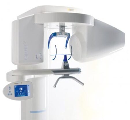
| Description | With the integrated FaceScanner in GALILEOS you can now record the facial surface parallel to the x-ray image. And that, as opposed to traditional face scanners without the use of lasers, which is therefore gentler on the patients. Thanks to the simultaneous x-ray image and surface depiction, the fully automated overlaying of the data is extremely precise. In addition, texture and coloring in the software allow for a particularly realistic depiction of the soft tissue proportions – an ideal tool for patient consultation! | The inEos X5 can be used to scan impressions and models of all or part of the jaw manually or automatically. The new operating concept increases flexibility for the dental technician so that the scanning process can be optimally integrated into the workflow of the laboratory. While the manual scanning mode saves time for simpler operations, the fully automatic scanning mode has its benefits especially with regard to extensive operations | Designed especially for general dentists, the Sirona Orthophos XG3 combines top-tier image quality and exceptional ease-of-use in an affordable, digital panoramic x-ray solution. | Elite in Image Quality
CDR Elite images provide bold bone tribeculation, crisp lamina dura and a clear, clean DEJ to meet the diagnostic needs of every clinician.
Developed with guidance from a panel of leading dental radiologists and validated by an extensive range of dental practitioners from all fields, CDR Elite takes Schick’s commitment to optimum quality for diagnostic imaging to a new level. | Elite in Image Quality
CDR Elite images provide bold bone tribeculation, crisp lamina dura and a clear, clean DEJ to meet the diagnostic needs of every clinician.
Developed with guidance from a panel of leading dental radiologists and validated by an extensive range of dental practitioners from all fields, CDR Elite takes Schick’s commitment to optimum quality for diagnostic imaging to a new level. | NOMAD Pro 2 is designed with safety as the number one priority. The external backscatter shield and internal radiation shielding are specially designed to protect the operator from radiation exposure. In fact, the exposure from using NOMAD Pro 2 is less than one percent of allowed occupational doses. |
| Content | Sirona GALILEOS Comfort/Compact| GALILEOS Comfort | GALILEOS Compact | | Oral and maxillofacial surgeons
Dental clinics
Private clinics
Orthodontists | General practioners
Dental implant practices
Oral surgeons |  |  | | Comparison Table of Galileos Comfort/ Compact & Orthophos XG 3D X-Ray systems | | Technical overview | GALILEOS Comfort | GALILEOS Compact | ORTHOPHOS
XG 3D | | Field of view | (15 x 15 x 15) cm³ | (12 x 15 x 15) cm³ | 8 cm x 8 cm (Ø x Höhe) | | 3D Resolution (isotropic voxel size) | 0.3/0.15 mm | 0.3 mm | 0,2 mm; 0,1 mm (available at the end of 201) | | Scan time / exposure time | 14 s / 2 – 6 s | 14 s / 2 – 6 s | 2–5 s | | Reconstruction time | 2.5 – 4.5 min | 4.5 min | 4,5 min | | Patient position | standing / seated | standing / seated | Standing/seated, chin rest/bite block, occlusal bite block for automatic patient positioning with 2D PAN images | | X-ray generator | | kV | 85 | 85 | 60–90 | | Effective dosage | 29 µSv / 68 µSv **
(21 mAs, 85 kV) | < 29 µSv / 68 µSv **
(21 mAs, 85 kV) | 43–175 μSv (Schulze)
(Standard: 100 μSv) | | Space requirements (minimum) | 1,6 x 1,6 x 2,25 m (d x w x h) | 1,6 x 1,6 x 2,25 m(d x w x h) | 1,5 m x 1,1 m x 2,25 m (Pan ), | | Space requirements (recommended) | >1,8 x 1,8 x 2,5 m (d x w x h) | 1,8 x 1,8 x 2,5 m (d x w x h) | 1,7 m x 1,3 m x 2,5 m (Pan ) | | User Interface | Full color touchscreen | Multipad | EasyPad | | Patient fixation | Occlusal bite block, forehead | Occlusal bite bock, forehead | | | Positioner | support, chin rest, head positioner *** | support, chin rest | | | Upgradable to Comfort version | |  | | | Optional floor stand | | | | | Wheelchair accessible | | | | | Remote control | | | | | Programs | | 150 µm resolution for close-up reconstruction | | | | | 300 µm resolution for standard volumes | | | | | High contrast mode for optimized hard tissue display | | | | | Views | PAN with 3D slice navigation | PAN with 3D slice navigation | Tiltable 2D slices, custom | | TSA, axial, sagittal, coronal, CEPH lat., CEPH pa/ap | TSA, axial, sagittal, coronal | 3D slicing, TSA, LSA, axial, sagittal, coronal | | 3D model, 1-click OP reports | 3D model, 1-click OP reports | 3D model, implant-oriented view, 1-Click OP reports |
With the integrated FaceScanner in GALILEOS you can now record the facial surface parallel to the x-ray image. And that, as opposed to traditional face scanners without the use of lasers, which is therefore gentler on the patients. Thanks to the simultaneous x-ray image and surface depiction, the fully automated overlaying of the data is extremely precise. In addition, texture and coloring in the software allow for a particularly realistic depiction of the soft tissue proportions – an ideal tool for patient consultation! |
| Sirona InLab InEos X5 Scanner
High-tech camera with many advantagesLike its predecessor inEos Blue, the fast scanning speed of the inEos X5 is unsurpassed. The large image field allows the camera to capture four to five teeth per image and an entire jaw with a total of five images. This is how it digitalizes one single crown in less than ten seconds, a three-unit bridge construction in only 30 seconds, as well as an entire jaw in less than one minute. A completely new optical system is used for the scan, which is based on the digital pattern projection: The considerably improved accuracy of less than 12 µm and the camera’s autofocus guarantee high-quality scan data that is also suitable for procedures that require the highest degree of accuracy, such as the most demanding implant operations. Due to the camera’s greater depth of definition, the inEos X5 can also capture the entire jaw including the palate, which makes it possible to create a digital construction of model casting works.Improved handling for new and experienced usersWith an innovative five-axis technology including rotation arm and intelligent scan planning, the inEos X5 positions and captures models with all indications automatically. This standardizes the images, eliminates user error and speeds up the imaging process. That means that new users in particular can digitalize traditional procedures without requiring significant training. Altogether, the inEos X5 stands out due to its easy handling: The large and open operating area allows all standard articulators to be positioned as well as quick access to the model. With the universal model and impression tray holder, all conventional model support and split cast systems, as well as impression trays in all sizes, can also be used. The multi-die scanning offers advantages in cases where the proximal contacts cannot be seen well and in the fabrication of frameworks and copings for single restorations. With this function, up to four prepared stumps can be positioned in a special holder and digitalized at the same time.
The inEos X5 has been available at retailers since May 2013. The list price including PC and inLab software as well as the OPEN inEos interface is EUR 19,990.Overview of new inEos X5 features:Sirona InLab InEos X5 Scanner- A combination of manual and fully automatic operation
- High accuracy
- Time savings and improved workflow due to the five-axis technology and autofocus
- Large scan field and good depth of definition
- Multi-die scanning for up to four individual stumps
- Universal model and articulator holders
- Easy handling
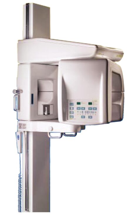
| SIRONA XG3 Digital Panoramic X-Ray
The ORTHOPHOS XG 3 is designed for the general practice dentist who would like to work with a basic X-ray system. The XG 3 offers you an outstanding price-performance ratio and provides the following benefits:- High image quality
- Extremely simple operation
- Solid workmanship
Accurate positioningThe ORTHOPHOS XG 3 uses proven, logical Sirona principles for exceptional imaging:- Exact, immediate positioning of the anterior teeth in the focal layer
- Effective, comfortable patient stabilisation with forehead and temple supports to prevent motion blurring
Proven TechnologyThe advanced technology from the XG product family enables the ORTHOPHOS XG 3 to produce high quality diagnostic images.
The benefits include:- Specific focal layers and orbital paths for anatomically optimised exposures
- Very detailed display of the anterior tooth region through automatic kV adaption as X-ray beam passes through spinal region
- 16 bit image acquisition technology provides more gray scales
- SIDEXIS XG imaging software with useful analysis tools
Features:SIRONA XG3 Digital Panoramic X-Ray- High-quality, digital panoramic and TMJ imaging power with advanced patient communication tools
- Outstanding price-performance ratio for seamless transition from film to direct digital imaging
- Easy operation with the Multipad; effective, three-point patient stabilization for sharp images
- Direct network capability for exceptional connectivity
SIRONA ORTHOPHOS XG 3The horizontally and vertically reduced pediatric panoramic program offers outstanding image quality with the lowest dose. Ideal for practices that treat a lot of children. The unit’s easy operation and seamless integration into your practice’s daily routines will save you valuable time. The high image quality and proven Sidexis software guarantee that you will come to a safe diagnosis quickly and easily.Quality from the very startHigh quality. Durable. Safe: the Orthophos XG 3 X-ray unit is the perfect basic equipment for general dentists. The unit’s easy operation and seamless integration into your practice’s daily routines will save you valuable time. The high image quality and proven Sidexis software guarantees that you will reach a safe diagnosis quickly and easily.The positioning can be very easily reproduced since image parameters such as the bite block height are saved together with the image data. A bitewing program is available, which helps in special cases and patient situations where intraoral images cannot be used and a shorter panoramic program with a reduced dose, which is suitable for treating children, is also available. | Schick CDR Elite Sensor Kit Size 2
Elite in Design
Based on input from Schick’s customer base – digital intraoral radiography’s largest – CDR Elite is designed to focus on ease of use, diagnostic image quality and durability. It provides an intraoral radiography solution for all clinicians that is truly Elite.
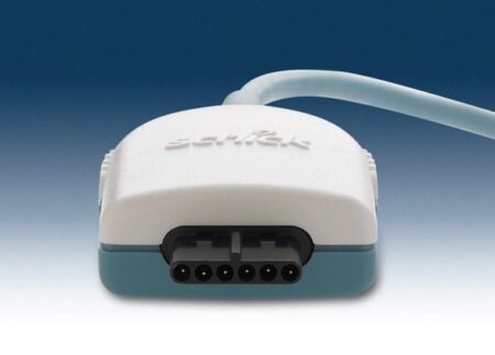
Embracing the success of Schick’s unique removable cable technology (introduced with the CDR PlusWire), CDR Elite incorporates this technology on all three sensor sizes, ensuring that every clinician and every dental practice can enjoy the simplicity and convenience of a l-step cable-replacement process.
Simple and easier sensor placement, even for vertical bitewings, comes from an optimally located sensor-cable interface and a new color scheme that provides high visibility in the oral cavity.
CDR Elite’s Smart Remote System has been developed to allow simple, accurate, in-office diagnosis to ensure peek performance of the entire system.Seamless Integration
Ideal complement to your existing Schick intra-oral and extraoral equipment, and integrates with Schick’s holder system for simple and easy positioning.
Integrates fully with Schick’s intuitive and easy-to-use CDR DICOM imaging software, which features multiple tools for enhanced diagnostic capabilities and patient communication.Specifications
Product CDR Elite Sensor
Size 2
Cable style Field Replaceable
Sensor interface High performance low force gold-plated contact system
Remote interface High speed USB 2.0
Sensor life Greater than 400,000 dosesPackage Inclusion
Schick CDR Elite Sensor Kit – Size 2:CDR Elite Sensor Size 2
CDR Elite USB Remote HS (with clip holder mounting)
CDR DICOM 3.5 Software Multi-user
USB Remote HS Cable 5M with ferrite
Schick Universal Sensor Holster (blue)
Peel’N’Stick Sensor Positioning Starter Kit:Centre Aiming Ring
Posterior Aiming Ring
Anterior Arm
Bitewing Arm
Posterior Arm
Schick Wired Sensor Sheaths Size 1 (300 pieces)
Universal Bite Tab Holder (15 pieces)
Universal Bitewing Holder (15 pieces)
Universal Endodontic Holder (15 pieces)
Universal Anterior Holder (15 pieces)
Universal Periapical Holder (15pieces)Elite in Image Quality
CDR Elite images provide bold bone tribeculation, crisp lamina dura and a clear, clean DEJ to meet the diagnostic needs of every clinician.
Developed with guidance from a panel of leading dental radiologists and validated by an extensive range of dental practitioners from all fields, CDR Elite takes Schick’s commitment to optimum quality for diagnostic imaging to a new level. | Schick CDR Elite Sensor Kit Size 1
Elite in Design
Based on input from Schick’s customer base – digital intraoral radiography’s largest – CDR Elite is designed to focus on ease of use, diagnostic image quality and durability. It provides an intraoral radiography solution for all clinicians that is truly Elite.
Embracing the success of Schick’s unique removable cable technology (introduced with the CDR PlusWire), CDR Elite incorporates this technology on all three sensor sizes, ensuring that every clinician and every dental practice can enjoy the simplicity and convenience of a l-step cable-replacement process.
Simple and easier sensor placement, even for vertical bitewings, comes from an optimally located sensor-cable interface and a new color scheme that provides high visibility in the oral cavity.
CDR Elite’s Smart Remote System has been developed to allow simple, accurate, in-office diagnosis to ensure peek performance of the entire system.Seamless Integration
Ideal complement to your existing Schick intra-oral and extraoral equipment, and integrates with Schick’s holder system for simple and easy positioning.
Integrates fully with Schick’s intuitive and easy-to-use CDR DICOM imaging software, which features multiple tools for enhanced diagnostic capabilities and patient communication.Specifications
Product CDR Elite Sensor
Size 1
Cable style Field Replaceable
Sensor interface High performance low force gold-plated contact system
Remote interface High speed USB 2.0
Sensor life Greater than 400,000 dosesPackage Inclusion
Schick CDR Elite Sensor Kit – Size 1:CDR Elite Sensor Size 1
CDR Elite USB Remote HS (with clip holder mounting)
CDR DICOM 3.5 Software Multi-user
USB Remote HS Cable 5M with ferrite
Schick Universal Sensor Holster (blue)
Peel’N’Stick Sensor Positioning Starter Kit:Centre Aiming Ring
Posterior Aiming Ring
Anterior Arm
Bitewing Arm
Posterior Arm
Schick Wired Sensor Sheaths Size 1 (300 pieces)
Universal Bite Tab Holder (15 pieces)
Universal Bitewing Holder (15 pieces)
Universal Endodontic Holder (15 pieces)
Universal Anterior Holder (15 pieces)
Universal Periapical Holder (15pieces)Elite in Image Quality
CDR Elite images provide bold bone tribeculation, crisp lamina dura and a clear, clean DEJ to meet the diagnostic needs of every clinician.
Developed with guidance from a panel of leading dental radiologists and validated by an extensive range of dental practitioners from all fields, CDR Elite takes Schick’s commitment to optimum quality for diagnostic imaging to a new level. | NOMAD Pro2 Handheld Portable Dental X-Ray AribexNOMAD Pro2 Handheld Portable Dental X-Ray
• Entire unit weighs just over five pounds
• Handheld and portable design moves from room to room, eliminating the need for multiple units
• Perfect for use with children and special needs patients
• Ideal for clinical, hospital, home-health, nursing home, and out-of-office use
• Includes a one-year product warrantyModern Radiation Shielding
The external backscatter shield and internal radiation shielding are specially designed to protect the operator from radiation exposure. In fact, the exposure from using the NOMAD Pro is less than 1% of allowed occupational doses.Time Savings NOMAD Pro2 Handheld Portable Dental X-Ray Aribex
The NOMAD Pro color LCD screen, simple touch controls, and handheld design saves time.• Handheld design allows operator to stay with the patient, thereby reducing time required for X-ray procedures
• Quickly adjust exposure time with simple controls
• Ability to save custom exposure settings for fast recall
• Icons quickly identify tooth, receptor, and patient typeClear Images
The NOMAD Pro produces the highest quality images.• No blur from hand movement
• 0.4 mm focal spot produces sharp, high resolution images
• High-frequency, 60kV DC X-ray generator produces clear images while reducing patient doseWeight: 4.6 lbThe NOMAD Pro 2, now part of the trusted brand of KaVo, provides reliability and efficiency for busy dental offices as well as powerful freedom in workflow that is unattainable with wall-mount units. While the unit allows for the operator to move freely between operatories, it also helps with fearful or challenging patients since the assistant can stay in the room during the radiographic process.The most succinct diagnosis starts with the best image quality. NOMAD Pro 2 utilizes the latest in X-ray technology for precise, repeatable exposures that help to produce sharp, clear radiographic images. And the best part is that all of the benefits of this handheld unit can be attained with an eye on ALARA. With its intuitive user interface, the team member can select the right dose for each patient’s particular need.The unique, lead-infused acrylic shield protects the operator from direct and scatter radiation, and the internal proprietary housing encases the X-ray tube to block radiation leakage. When the KaVo NOMAD Pro 2 is used as directed, these shields create a “safe zone” for the operator throughout the acquisition process. |




Reviews
There are no reviews yet.