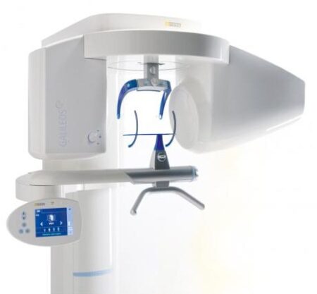
| Description | With the integrated FaceScanner in GALILEOS you can now record the facial surface parallel to the x-ray image. And that, as opposed to traditional face scanners without the use of lasers, which is therefore gentler on the patients. Thanks to the simultaneous x-ray image and surface depiction, the fully automated overlaying of the data is extremely precise. In addition, texture and coloring in the software allow for a particularly realistic depiction of the soft tissue proportions – an ideal tool for patient consultation! | NOMAD Pro 2 is designed with safety as the number one priority. The external backscatter shield and internal radiation shielding are specially designed to protect the operator from radiation exposure. In fact, the exposure from using NOMAD Pro 2 is less than one percent of allowed occupational doses. | At only 4.2 pounds, the portable x-ray, is considered to be the lightest x-ray generator on the market. Many design features are included with an LCD Display, and High Frequency Tube, a perfect balance of technology and simplicity. There is no doubt that the EXARO is the best, handheld and portable x-ray, available. | The APOLLO DI is our very latest camera. It impresses with its simple handling, small and lightweight camera and outstanding value for money. | Benefits:- Faster Scan Speed
- Superb Image Quality
- Light Guidance System
- Portable for Easy Sharing
- Lightweight Hand Piece
| Cost-effective production
The inLab software positions your zirconium oxide restorations optimally in the material block – for efficient use of blocks, attractive unit prices and the best possible machine utilization, e.g. by milling or grinding overnight.
Wet grinding of sintered NPM blocks
“inLab goes metal.” With the inLab MC XL and the new inCoris CC sintered metal blocks you can handle a complete spectrum of metal and ceramic materials. The unique wet grinding function rules out any health risks. |
| Content | Sirona GALILEOS Comfort/Compact| GALILEOS Comfort | GALILEOS Compact | | Oral and maxillofacial surgeons
Dental clinics
Private clinics
Orthodontists | General practioners
Dental implant practices
Oral surgeons |  |  | | Comparison Table of Galileos Comfort/ Compact & Orthophos XG 3D X-Ray systems | | Technical overview | GALILEOS Comfort | GALILEOS Compact | ORTHOPHOS
XG 3D | | Field of view | (15 x 15 x 15) cm³ | (12 x 15 x 15) cm³ | 8 cm x 8 cm (Ø x Höhe) | | 3D Resolution (isotropic voxel size) | 0.3/0.15 mm | 0.3 mm | 0,2 mm; 0,1 mm (available at the end of 201) | | Scan time / exposure time | 14 s / 2 – 6 s | 14 s / 2 – 6 s | 2–5 s | | Reconstruction time | 2.5 – 4.5 min | 4.5 min | 4,5 min | | Patient position | standing / seated | standing / seated | Standing/seated, chin rest/bite block, occlusal bite block for automatic patient positioning with 2D PAN images | | X-ray generator | | kV | 85 | 85 | 60–90 | | Effective dosage | 29 µSv / 68 µSv **
(21 mAs, 85 kV) | < 29 µSv / 68 µSv **
(21 mAs, 85 kV) | 43–175 μSv (Schulze)
(Standard: 100 μSv) | | Space requirements (minimum) | 1,6 x 1,6 x 2,25 m (d x w x h) | 1,6 x 1,6 x 2,25 m(d x w x h) | 1,5 m x 1,1 m x 2,25 m (Pan ), | | Space requirements (recommended) | >1,8 x 1,8 x 2,5 m (d x w x h) | 1,8 x 1,8 x 2,5 m (d x w x h) | 1,7 m x 1,3 m x 2,5 m (Pan ) | | User Interface | Full color touchscreen | Multipad | EasyPad | | Patient fixation | Occlusal bite block, forehead | Occlusal bite bock, forehead | | | Positioner | support, chin rest, head positioner *** | support, chin rest | | | Upgradable to Comfort version | |  | | | Optional floor stand | | | | | Wheelchair accessible | | | | | Remote control | | | | | Programs | | 150 µm resolution for close-up reconstruction | | | | | 300 µm resolution for standard volumes | | | | | High contrast mode for optimized hard tissue display | | | | | Views | PAN with 3D slice navigation | PAN with 3D slice navigation | Tiltable 2D slices, custom | | TSA, axial, sagittal, coronal, CEPH lat., CEPH pa/ap | TSA, axial, sagittal, coronal | 3D slicing, TSA, LSA, axial, sagittal, coronal | | 3D model, 1-click OP reports | 3D model, 1-click OP reports | 3D model, implant-oriented view, 1-Click OP reports |
With the integrated FaceScanner in GALILEOS you can now record the facial surface parallel to the x-ray image. And that, as opposed to traditional face scanners without the use of lasers, which is therefore gentler on the patients. Thanks to the simultaneous x-ray image and surface depiction, the fully automated overlaying of the data is extremely precise. In addition, texture and coloring in the software allow for a particularly realistic depiction of the soft tissue proportions – an ideal tool for patient consultation! |
| NOMAD Pro2 Handheld Portable Dental X-Ray AribexNOMAD Pro2 Handheld Portable Dental X-Ray
• Entire unit weighs just over five pounds
• Handheld and portable design moves from room to room, eliminating the need for multiple units
• Perfect for use with children and special needs patients
• Ideal for clinical, hospital, home-health, nursing home, and out-of-office use
• Includes a one-year product warrantyModern Radiation Shielding
The external backscatter shield and internal radiation shielding are specially designed to protect the operator from radiation exposure. In fact, the exposure from using the NOMAD Pro is less than 1% of allowed occupational doses.Time Savings NOMAD Pro2 Handheld Portable Dental X-Ray Aribex
The NOMAD Pro color LCD screen, simple touch controls, and handheld design saves time.• Handheld design allows operator to stay with the patient, thereby reducing time required for X-ray procedures
• Quickly adjust exposure time with simple controls
• Ability to save custom exposure settings for fast recall
• Icons quickly identify tooth, receptor, and patient typeClear Images
The NOMAD Pro produces the highest quality images.• No blur from hand movement
• 0.4 mm focal spot produces sharp, high resolution images
• High-frequency, 60kV DC X-ray generator produces clear images while reducing patient doseWeight: 4.6 lbThe NOMAD Pro 2, now part of the trusted brand of KaVo, provides reliability and efficiency for busy dental offices as well as powerful freedom in workflow that is unattainable with wall-mount units. While the unit allows for the operator to move freely between operatories, it also helps with fearful or challenging patients since the assistant can stay in the room during the radiographic process.The most succinct diagnosis starts with the best image quality. NOMAD Pro 2 utilizes the latest in X-ray technology for precise, repeatable exposures that help to produce sharp, clear radiographic images. And the best part is that all of the benefits of this handheld unit can be attained with an eye on ALARA. With its intuitive user interface, the team member can select the right dose for each patient’s particular need.The unique, lead-infused acrylic shield protects the operator from direct and scatter radiation, and the internal proprietary housing encases the X-ray tube to block radiation leakage. When the KaVo NOMAD Pro 2 is used as directed, these shields create a “safe zone” for the operator throughout the acquisition process. | Xray2go Hand Held Dental Portable X-Ray System| FDA Registered. Welcome to the new way of taking X-rays in Dental Offices: x-ray2go is the way to go! x-ray2go is a portable hand held camera that uses a high frequency inverter to generate a stable X-ray output with DC high voltage. This effective DC unit enables patient and operator to be exposed to the least amount of radiation. The integrated compact “Camera like” generator increases the user’s confidence and decrease the negative perception patients have about X-rays. It is small: 5.6″ (W) x 3.3″ (D) x 1.2″ (H). Best of all it only weighs 4.18 Lbs (1.9 Kgs), the lightest on the market.• Portability – room to room ease
• No installation necessary versus complex, expensive and time consuming on-wall installation.
• No need for pass troughs
• Large LCD display – easy to read
• Simple touch controls
• Adjustable exposure time
• Hundreds of images from one (1) battery charge
• High resolution diagnostic images
• No blur from hand movement high-frequency generator produces clear images while reducing
patient dose
• Shield protects the operator from scattered radiation – small dose of radiation emitted
• Leading technology provides the highest level of safety, quality and patient care
• Perfect for special needs patients, home health care patients, nursing home patients, children and
veterinary practices
• Perfect for small operatories
• Perfect for mobile dentistry where room is a big factor |
| | | | |
| | | | Specifications Xray2go Hand Held Dental Portable X-Ray System- Portable, cordless x-ray device
· FDA Registered – K082875
· Safe to use-leakage and scatter radiation are less than conventional wall mounted units
· Small & lightweight (4.2 lbs)
· Camera-like ergonomic design
· 100’s of images on single charge!
· Compatible with both digital & film use
· Simple operation
· Highest image quality
· Wide exposure range
· Lowest patient exposure
· Compact outside—smart inside
· Great staff acceptance
· Convenient for children and “x-ray phobic” patients
· Helps eliminate patient fear and reduces x-ray rejection
| X-ray installation | Tube voltage: 60KV
Tube current: 2mA
Heat Capacity: 8.5KHU | | X-ray Generator | Type: High Frequency Inverter
Power Output: 100W
X-ray Tube | | X-ray Tube | Anode Type: Stationary
Anode Angle: 20°
Focal Spot: 0.8mm | | X-ray Control | Exposure time set 0.01 ~ 1.6sec (0.01 sec step) | | Power Requirement | 25.2VDC | | Weight | 4.2 lbs. | | Dimensions | 6.7”(W) x 6.1”(D) x 5.25”(H) |
|
|
| Sirona Apollo DI Intra Oral Scanner
APOLLO DI:
The Sirona Apollo DI Intra Oral Scanner features easy handling, precise imaging, and the proven Sirona Connect workflow. The impression system includes an imaging unit, APOLLO Connect software, and the APOLLO DI intraoral
camera,with which users can make digital impressions of the clinical situation in a seamless workflow. To do this, a moisture-insensitive high-contrast spray is sprayed on the teeth very finely. Fine particles in the spray ensure high contrast and thus very precise images.The most cost-effective start to digital impression-taking.
APOLLO DI
APOLLO DI, the specially developed intraoral scanner for cost-efficient digital impressions.
Easy handling thanks to multitouch control
Small and lightweight camera
Export of scan-data in the laboratory
No follow-up costs
OPEN APOLLO DI*:
Export of digital impression data (captured with the APOLLO DI in the practice and received via the Sirona Connect Portal) in an open ST L format for processing in other CAD/CAM systems.Designed to provide an economical entry point into CAD/CAM dentistry, Sirona’s APOLLO DI digital impression system includes an imaging unit, APOLLO Connect software and the APOLLO DI intraoral camera.The system provides precise imaging with the use of a contrast spray, and digital impression data can be sent to dental labs via the Sirona Connect system where it can be turned into a digital restoration design in Sirona’s inLab software or compatible 3rd party software applications, according to a press release.The new APOLLO DI is a complement to the company’s existing CEREC Bluecam and CEREC Omnicam options which each use a different imaging technology, but send the digital impressions through the same workflow. APOLLO DI is designed for practices looking to take digital impressions but continue to work with a lab, while the Bluecam and Onmicam can each be purchased as stand-alone digital impression systems, or with a mill for complete chairside CAD/CAM dentistry. | Carestream CS 3600 Intraoral ScannerAcquire true color:With the CS 3600 Intraoral Scanner, practitioners can access 2D and 3D images that can be used with CS Restore software to design restorations within their practice. Colored 3D scans allow practitioners to easily define the margin lines and identify the differences between natural tooth structure and existing restorations. This makes restoration planning more accurate for practitioners.No external heater, powder or trolley system needed:The CS 3600 does not require you to spray powder of liquid over the patient’s teeth or gingival tissue before scanning. It features high-angulation scanning of up to 45 degrees and to a depth from -2 to +13m.Features a unique light guidance systemYet another feature of the CS 3600 that assists it in the capture of the data during the image acquisition process. This feature allows the user to place more focus on the patient’s mouth rather than the monitor.This device is easily shared:The CS 3600 can be easily shared between operatories because there is no heavy trolley to push around. Additionally, the intraoral scanner connects via USB 2 cable* to any PC workstation or laptop. * Windows 7, 32-bit minimum; 64-bit recommendedEasy to use and hold:The CS 3600’s sleek, lightweight design (295 grams) makes it comfortable and easy for users to navigate the intraoral scanner around patients’ mouths, especially during full arch acquisitions.Benefits:Carestream CS 3600 Intraoral Scanner- Faster Scan Speed
- Superb Image Quality
- Light Guidance System
- Portable for Easy Sharing
- Lightweight Hand Piece
- Features
- High Speed Continuous Scanning
- Fast, Simple and Smooth user experience
- Freefill Missing Scan Data at your Leisure
- Scan History allows you to remove excess scanned tissue for more refined final digital impression
- Three Workflows
- user friendly interface
- Autoclavable reusable tips in two interchangeable styles
- Accurate full 3D HD Color scanning
| Sirona Cerec InLab MCXL Milling Unit
The inLab MC XL milling and grinding unit opens up the widest possible range of production options for your dental laboratory. You profit from high speed and precision. You can switch from grinding to milling in just a few simple steps. And thanks to the large milling volume and broad spectrum of applications, you will reap substantial economic benefits.
inLab MC XL
Large range of materials:
Processing of zirconium oxide, resin, silicate ceramics and NP-metal
High-speed milling:
For example, a four-unit zirconium oxide bridge can be completed in just 40 minutes
Large milling capacity: For restorations consisting of up to 12 units and ceramic blocks measuring 85 x 40 x 22 mm
Four milling motors: Flexible selection of milling burs for different materials
Milling and grinding: For high precision regardless of the materialand indication
Milling* of zirconium oxide and polymers
In addition to grinding, you can also mill zirconium oxide and polymers on the inLab MC XL. You benefit from an enhanced initial fit and a faster production process.Flexible and efficient Sirona Cerec InLab MCXL Milling UnitinLab MC XL is the fast wet milling and grinding unit with many production options for your dental laboratory. You benefit from high speed and precision and can switch from grinding to milling in just a few steps. The large selection of materials and many uses give you particularly flexible and efficient production options. inLab high-speed grindingGlass- and hybrid ceramic restorations can be produced at a previously unachievable speed with simultaneous double four-axis processing. A fully contoured Celtra Duo crown takes less than 10 minutes. This is a key success factor for potential new business models that can call for providing digital impression orders within an hour. Precision processingThe inLab MC XL is characterized by precise wet machining. Especially when processing glass ceramics, the grinders used can be as small as 0.6 mm — for restorations with maximum detail in the occlusal and interproximal areas and at the preparation margin. Wide selection of materialsAs with all CAD/CAM production units from Dentsply Sirona, inLab MC XL lets you benefit from a the large selection of materials. Dentsply Sirona CAD/CAM materials and those of our material partners are optimally adapted for high-speed processing. |


Reviews
There are no reviews yet.