View cart “Sirona Sirolaser Soft Tissue Laser For Sale” has been added to your cart.
-38%
Availability: In Stock
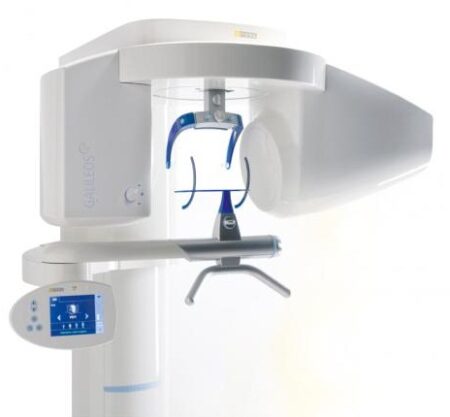
Sirona GALILEOS Comfort/Compact For Sale
$11,000.00$6,800.00
With the integrated FaceScanner in GALILEOS you can now record the facial surface parallel to the x-ray image. And that, as opposed to traditional face scanners without the use of lasers, which is therefore gentler on the patients. Thanks to the simultaneous x-ray image and surface depiction, the fully automated overlaying of the data is extremely precise. In addition, texture and coloring in the software allow for a particularly realistic depiction of the soft tissue proportions – an ideal tool for patient consultation!
Other people want this. 15 people have this in their carts right now.
3D model, implant-oriented view, 1-Click OP reports
With the integrated FaceScanner in GALILEOS you can now record the facial surface parallel to the x-ray image. And that, as opposed to traditional face scanners without the use of lasers, which is therefore gentler on the patients. Thanks to the simultaneous x-ray image and surface depiction, the fully automated overlaying of the data is extremely precise. In addition, texture and coloring in the software allow for a particularly realistic depiction of the soft tissue proportions – an ideal tool for patient consultation!
Reviews (0)
Reviews
There are no reviews yet.
Be the first to review “Sirona GALILEOS Comfort/Compact For Sale” Cancel reply
With the integrated FaceScanner in GALILEOS you can now record the facial surface parallel to the x-ray image. And that, as opposed to traditional face scanners without the use of lasers, which is therefore gentler on the patients. Thanks to the simultaneous x-ray image and surface depiction, the fully automated overlaying of the data is extremely precise. In addition, texture and coloring in the software allow for a particularly realistic depiction of the soft tissue proportions – an ideal tool for patient consultation!
At only 4.2 pounds, the portable x-ray, is considered to be the lightest x-ray generator on the market. Many design features are included with an LCD Display, and High Frequency Tube, a perfect balance of technology and simplicity. There is no doubt that the EXARO is the best, handheld and portable x-ray, available.
WaterLase iPlus includes the all-new SureFire™ delivery system, with redesigned optics that can more efficiently deliver precise amounts of Er,Cr:YSGG laser energy to cut enamel, dentin and bone. The SureFire delivery system is optimized to provide the dentist with more up time with a replaceable, disposable shield that protects the vital optical components in the fiber, resulting in greater dependability.
TRIOS Color automatically measures the teeth shades as you scan. The dentist has the option to evaluate and highlight relevant areas for the lab. The complete shade information sis sent to the lab together with the impression.
Milling Unit:• Choose from CEREC MC, CEREC MC X or CEREC MC XL
• Get access to the entire range of chairside treatment with up to 40 mm block size, including bridges and abutments
• Create fully anatomic, precise treatments quickly
• CEREC Guide 2 surgical guide
• Dry mill ready
• Includes CEREC software
CEREC now provides a completely new process to dental practices: by combining the new CEREC SpeedFire furnace and CEREC Zirconia material, dentists can now deliver full contour crowns and small bridges made of the full-strength high-quality zirconium oxide in their own practice while the patient waits.
3D model, implant-oriented view, 1-Click OP reports
With the integrated FaceScanner in GALILEOS you can now record the facial surface parallel to the x-ray image. And that, as opposed to traditional face scanners without the use of lasers, which is therefore gentler on the patients. Thanks to the simultaneous x-ray image and surface depiction, the fully automated overlaying of the data is extremely precise. In addition, texture and coloring in the software allow for a particularly realistic depiction of the soft tissue proportions – an ideal tool for patient consultation!
FDA Registered. Welcome to the new way of taking X-rays in Dental Offices: x-ray2go is the way to go! x-ray2go is a portable hand held camera that uses a high frequency inverter to generate a stable X-ray output with DC high voltage. This effective DC unit enables patient and operator to be exposed to the least amount of radiation. The integrated compact “Camera like” generator increases the user’s confidence and decrease the negative perception patients have about X-rays. It is small: 5.6″ (W) x 3.3″ (D) x 1.2″ (H). Best of all it only weighs 4.18 Lbs (1.9 Kgs), the lightest on the market.• Portability – room to room ease
• No installation necessary versus complex, expensive and time consuming on-wall installation.
• No need for pass troughs
• Large LCD display – easy to read
• Simple touch controls
• Adjustable exposure time
• Hundreds of images from one (1) battery charge
• High resolution diagnostic images
• No blur from hand movement high-frequency generator produces clear images while reducing
patient dose
• Shield protects the operator from scattered radiation – small dose of radiation emitted
• Leading technology provides the highest level of safety, quality and patient care
• Perfect for special needs patients, home health care patients, nursing home patients, children and
veterinary practices
• Perfect for small operatories
• Perfect for mobile dentistry where room is a big factor
Specifications Xray2go Hand Held Dental Portable X-Ray System
Portable, cordless x-ray device
· FDA Registered – K082875
· Safe to use-leakage and scatter radiation are less than conventional wall mounted units
· Small & lightweight (4.2 lbs)
· Camera-like ergonomic design
· 100’s of images on single charge!
· Compatible with both digital & film use
· Simple operation
· Highest image quality
· Wide exposure range
· Lowest patient exposure
· Compact outside—smart inside
· Great staff acceptance
· Convenient for children and “x-ray phobic” patients
· Helps eliminate patient fear and reduces x-ray rejection
The WaterLase iPlus all-tissue dental laser from the global leader in dental lasers is designed to deliver practice growth to dentists by offering an enhanced experience for patients, while raising the standard of overall reliability and quality for the #1-preferred all-tissue laser.Enhancing the patient experience
WaterLase iPlus is the latest addition to the WaterLase family, which has been striving to enhance the dental experience since 2001. With an estimated 27 million patients treated, more than 97% would recommend their WaterLase dentist to a friend – and WaterLase iPlus 2.0 offers a host of benefits that deliver an even better patient experience. Improvements to key components boost the availability of the laser when it’s needed most; and a new, minimally invasive laser therapy for periodontal disease offers dentists a clinical protocol that can generate excellent clinical outcomes and a vastly different type of patient experience versus traditional methods.SureFire Delivery System Biolase Waterlase iplus LaserWaterLase iPlus includes the all-new SureFire delivery system, with redesigned optics that can more efficiently deliver precise amounts of Er,Cr:YSGG laser energy to cut enamel, dentin and bone. The SureFire delivery system is optimized to provide the dentist with more up time with a replaceable, disposable shield that protects the vital optical components in the fiber, resulting in greater dependability.REPaiR for Reliably Treating PeriodontitisWaterLase iPlus offers new benefits to dentists and patients with the addition of REPaiR, the first Er,Cr:YSGG-based protocol for safe, effective treatment of moderate to severe periodontitis. It is an achievable, repeatable, minimally-invasive procedure that is less traumatic than traditional surgical techniques and delivers better and gentler experiences for patients. Clinically proven treatment for moderate to severe periodontitis Exclusive to WaterLase iPlus 2.0Includes:
Trunk Fiber
2 Hand Pieces (Turbo & Gold)
Manual (DVD)
Pair of Laser Goggles
Air Line
Power Cord
Tip Cleaning Kit
3Shape Trios 3 Pod ColorNew ! 3Shape TRIOS Color POD Digital impressions3Shape TRIOS Pod is a new configuration solution and an alternative to the TRIOS cart. It enables scanning with the TRIOS handheld scanner and software using selected laptop PCs. The solution offers a high degree of mobility and flexibility for dentists working in multiple locations or for clinics with limited space. The TRIOS Pod lets users control scanning from an iPad or mirror the 3D view on other displays in the clinic such as monitors integrated in the chair.Connect to your laptop or PC
Use the Pod with your laptop or the PC in your treatment room. Simply connect to the USB port and start scanning.FEATURES : 3Shape Trios 3 Pod Color
– RealColor scans with many clinical advantages: With TRIOS color you can scan in natural colors to clearly distinguish between teeth, gingiva, and restorative materials. Easily identify true preparation margins and improve scanning experience.
– Powder-free for optimal accuracy and comfort: Applying powder is technique demanding, can ruin scan accuracy, is uncomfortable for patients, and prolongs chair time.
– Autoclaveable tip with anti-mist heater: Achieve optimal hygiene and meet clinical requirements. The integrated antimist heater automatically ensures an optimal temperature for undistorted and crystal clear scanning.
– Extreme Mobility: The light and handy Pod solution is easy to share among treatment rooms or different locations
– Use your iPad with scanning: Control TRIOS from your iPad or mirror the live 3D view directly on other displays in the clinic such as monitors integrated in the chair.
– Connect to multiple laptops or PCs: Use with laptops, PCs in your treatment rooms, or with the PCs integrated in your chair units. Simply connect to the USB port and start scanning. Note: Use with approved PCs.
– Small footprint: The compact TRIOS Pod can be placed anywhere in the treatment room or even in rooms where space is very limited.Included : Mac laptop
CEREC Primescan AC Intraoral scanner
Cerec Primescan from Dentsply Sirona is the most advanced digital scanner on the market. It’s providing a level of accuracy never before seen in dentistry. At the peak of innovation, it is widening the gap between those with and without digital dentistry. The Prime Scan is the fastest most accurate intraoral scanner to date. With new scanning technology, it is the ultimate in scanning. Prime Scan and CEREC 5.0 combinations are greatly reduced because more data allows many applications to become automated.For more than 30 years, CEREC® has provided technological precision, superior design and excellent performance to thousands of practices. Now, experience a brand-new fully integrated solution with CEREC Primescan AC. Acquire precise digital impressions with Primescan, design your proposal on the Primescan AC using the intelligent software, then produce precise, smooth restorations with your choice of milling unit. Find the next level of productivity and patient care – it’s all here.How Does CEREC Primescan Work?
CEREC Primescan is an alternative to traditional intraoral dental impressions, which use a putty-like substance to take physical impressions of your teeth. This method does not allow for digital assessment of your mouth, and some patients find it the process of physical impressions to be uncomfortable.CEREC Primescan uses a wand-like device, loaded with powerful imaging sensors which can capture more than 50,000 images of your mouth per second. These images are quickly stitched and compiled into a 3D image of your teeth, jaw, gums, and mouth, and displayed on a high-definition screen for immediate assessment by our doctors.Using this sensor, our doctors can image your entire mouth up to 20mm in depth, including gums, blood, and bone structures, quickly and accurately. The Primescan software (as compared to the Omnicam) creates high-resolution 3D models to assess your oral health and create the very best crowns, implants, removable prosthesis, restorations, and even 3D prints. With just a few quick sweeps of the CEREC Primescan device, we’ll be able to generate an in-depth, modifiable 3D image of your mouth in minutes.The Benefits of CEREC Primescan:
An excellent choice for outstanding results: Primescan is your perfect starting point in digital dentistry. No matter how you would like to design your workflows, Primescan is the enabler for efficient digital workflows – both chairside in your practice and with your preferred partners.Well designed and perfectly compatible: Along with the high-performance intraoral scanner comes the new Acquisition Center. Together, they form a highly effective and most comfortable system – meeting your needs with two individual software configurations:So, why is CEREC Primescan such a big deal? Here are just a few improvements to this sophisticated system:Shorter wait times – We can quickly image your mouth without waiting for dental impressions to set, allowing us to treat each patient more quickly and effectively. A full jaw impression only takes 2 minutes. This adds up to less time spent in the dental chair.Real-time image processing – Our dentists can quickly adjust and modify scanned images using filters and other tools, leading to more accurate diagnostics and better overall patient outcomes.Extremely versatile – CEREC Primescan can be used for almost any dental procedure. From implant dentistry to the creation of CEREC same-day crowns, the straightening of teeth, whitening, development of dentures and partial dentures.CEREC Primescan AC with CEREC Software Sirona Advantages:
Enables all kinds of treatment, from a single tooth to full arch
With Primescan intraoral scanning is now more accurate, faster and easier
Allows an efficient workflow, both chairside and with preferred partners
Along with the high-performance intraoral scanner comes the new Acquisition Center, together forming an effective and most comfortable system, meeting your needs with two individual software configurations: Primescan AC with Connect Software (supports the connection to established partners workflows) and CEREC Primescan AC with CEREC Software (ideal for cabinets)
Smart Pixel Sensor technology – allows superior precision and excellent detail reproduction, processes more than 1 million 3D points per second, producing photorealistic and highly accurate data
Due to its ability to scan at an incredible data density, it delivers complete 3D structures of everything in its field of view
Its dynamic depth scan technology enables perfect sharpness and outstanding precision, even at a measuring depth up to 20 mm
Easy to use
Easy scanning of all dental materials and hard to access areas
Allows you to start scanning right away
Self-heating for fog-free scanning
The increased field of view visualizes larger areas with fewer sweeps and immediate precision
Faster scanning with a smooth scan flow
Consolidating more than 50.000 images per second and fast processing of exactly the data the software needs Easy capturing and quicker processing of more data in higher resolution
Complete 3D-scan models are displayed immediately, no matter how fast you scan
Accelerate your workflow: Due to seamless, validated and open data transfer options, labs and other third parties are supplied with high-resolution models at an instant
3 different sleeves (disposable and stainless steel, with sapphire glass or autoclavable)
Acquisition Center – Smart features and greater comfort: intuitive use via movable 16:9 touchscreen and touchpad for perfect ergonomics
Kinematics for perfect ergonomic positioning
Smart Hygiene concept for fast and easy disinfection
Full mobility with more than 60-min. battery buffer
CEREC Primescan AC with CEREC Software – complete chairside system, which offers flexible data export options, with automatized workflow thanks to Artificial Intelligence and touch-enabled and intuitive user interface
Milling Unit:• Choose from CEREC MC, CEREC MC X or CEREC MC XL
• Get access to the entire range of chairside treatment with up to 40 mm block size, including bridges and abutments
• Create fully anatomic, precise treatments quickly
• CEREC Guide 2 surgical guide
• Dry mill ready
• Includes CEREC softwareStriking in every detail: The new Acquisition CenterThe new Acquisition Center is a work station designed for modern dentistry and with the dentist in mind. It comes with a touchpad and a 16:9 wide-format movable touchscreen, offering you a highly intuitive and ergonomic work platform.Your benefits at a glance:
Touchscreen & Touchpad for most comfortable, intuitive use
Kinematics for perfect ergonomic positioning
Smart Hygiene concept for fast and easy disinfection
Mobility concept for full mobility with more than 60-min. battery buffer
The best choice for your practice: Take digital impressions and continue working with your trusted partners or enjoy the benefits of complete chairside workflows.Primescan meets your needs with two individual software configurations:Primescan AC with Connect Software:
Supports data transfer options to your preferred partners
Secure and encrypted data transfer through Connect Case Center Inbox
Easy upgradability to full chairside workflow
Touch-enabled and intuitive user interface
CEREC Primescan AC with CEREC Software:
Supports full chairside workflows for single-visit dentistry
Flexible data export options
Automatized workflow thanks to Artifical Intelligence
Touch-enabled and intuitive user interface
Technical Data:CEREC Primescan AC Intraoral scanner
CAD system for high-precision intraoral optical impressions
High-resolution, heated intraoral 3D scanner
Integrated image processing
High processing power due to state-of-the-art processor
Trackball or touchpad
Hand and foot controlled enter keys
Ethernet port and WLAN
USB interfaces
The high-resolution 3D intraoral scanner allows image acquisition with the image control inside the scanner, the image data transfer is made through USB 2.0 interface
21.5″ inch TFT LED flat-screen display, HD resolution: 1920 x 1080 pixels
Mobile housing, with no water or air connection required
Voltage: 100 – 240 VAC/50-60Hz
Nominal current: 5.0 – 2.1 A
Type of protection against electric shock: Class I device
High strength, short manufacturing process
The greatest benefit of CEREC Zirconia is the high flexural strength of the material. It is suitable for individual crowns as well as small bridges and can be processed in thin wall thicknesses. Since these restorations are manufactured in monolithic form, there is no risk of chipping. Another benefit for dentists is that zirconium oxide can be cemented conventionally.CEREC Zirconia is a pre-shaded translucent zirconium oxide available in 10 shades on the basis of the VITA Classic Shade Guide®. The material is milled in an enlarged form and then densely sintered to its final size in the new sintering furnace CEREC Speed-Fire. The oversized milling facilitates a new level of milling accuracy leading to superb, precisely fitting restorations. The sintering process takes just 10-15 minutes for crowns and 25 minutes for bridges. The subsequent glaze firing gives the restoration a high gloss finish.The short process to produce CEREC Zirconia restorations is both convenient and economical. With this market launch, all CEREC milling/grinding units now provide wet and dry milling. Dry milling reduces the overall processing time for zirconia and, combined with the world’s fastest sintering cycles, enables the chairside procedure.The workflow is easy to learn since the CEREC Software 4.4.1 guides the dentist through the entire process, and even sends the sintering and glazing information to the furnace. No programming of the furnace is required – it is all handled automatically by the software. A high-performance material and a specially tailored workflow ensure a simple process and high-quality treatment.
CEREC meets patients’ needsSirona CEREC Zirconia SpeedFire Milling UnitEighty three percent of patients in a recent survey said they prefer single visit dentistry to traditional treatment. The majority said they would even be willing to pay more for single visit dentistry. Two thirds indicated they’d be happy to travel further than they currently do, in order to receive treatment in one session. Another two thirds said they would even be prepared to change their dentist. The advantages of single visit dentistry are obvious: Patients avoid impression material, receive fewer anesthetics and don’t need a temporary. Reducing the number of visits, injections and making the overall prosthetic procedure more comfortable influences patients to opt for treatment with CEREC.In addition to CEREC Zirconia, many other high-performance materials can be processed with CEREC. The range of materials with CEREC also expands clinical indications since the dentist can easily select the material suited for each indication.
Investment for a successful futureThere have always been good reasons to start using digital dentistry with CEREC. CEREC Zirconia completes the range of indications for chairside application for nearly
every situation, thus giving dentists greater choice and allowing them to increase the value of their practice’s offering.It is obvious that advanced technologies in automobiles, computers and smartphones make our daily lives easier. CEREC is also a technology that further develops a dental practice and can make it well positioned for the future. Especially now, as the system is highly flexible, it enables dentists to expand their offering in implant dentistry and orthodontics.


 An excellent choice for outstanding results: Primescan is your perfect starting point in digital dentistry. No matter how you would like to design your workflows, Primescan is the enabler for efficient digital workflows – both chairside in your practice and with your preferred partners.Well designed and perfectly compatible: Along with the high-performance intraoral scanner comes the new Acquisition Center. Together, they form a highly effective and most comfortable system – meeting your needs with two individual software configurations:So, why is CEREC Primescan such a big deal? Here are just a few improvements to this sophisticated system:Shorter wait times – We can quickly image your mouth without waiting for dental impressions to set, allowing us to treat each patient more quickly and effectively. A full jaw impression only takes 2 minutes. This adds up to less time spent in the dental chair.Real-time image processing – Our dentists can quickly adjust and modify scanned images using filters and other tools, leading to more accurate diagnostics and better overall patient outcomes.Extremely versatile – CEREC Primescan can be used for almost any dental procedure. From implant dentistry to the creation of CEREC same-day crowns, the straightening of teeth, whitening, development of dentures and partial dentures.CEREC Primescan AC with CEREC Software Sirona Advantages:
 The new Acquisition Center is a work station designed for modern dentistry and with the dentist in mind. It comes with a touchpad and a 16:9 wide-format movable touchscreen, offering you a highly intuitive and ergonomic work platform.Your benefits at a glance:


Reviews
There are no reviews yet.