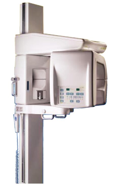
| Content | Sirona GALILEOS Comfort/Compact| GALILEOS Comfort | GALILEOS Compact | | Oral and maxillofacial surgeons
Dental clinics
Private clinics
Orthodontists | General practioners
Dental implant practices
Oral surgeons |  |  | | Comparison Table of Galileos Comfort/ Compact & Orthophos XG 3D X-Ray systems | | Technical overview | GALILEOS Comfort | GALILEOS Compact | ORTHOPHOS
XG 3D | | Field of view | (15 x 15 x 15) cm³ | (12 x 15 x 15) cm³ | 8 cm x 8 cm (Ø x Höhe) | | 3D Resolution (isotropic voxel size) | 0.3/0.15 mm | 0.3 mm | 0,2 mm; 0,1 mm (available at the end of 201) | | Scan time / exposure time | 14 s / 2 – 6 s | 14 s / 2 – 6 s | 2–5 s | | Reconstruction time | 2.5 – 4.5 min | 4.5 min | 4,5 min | | Patient position | standing / seated | standing / seated | Standing/seated, chin rest/bite block, occlusal bite block for automatic patient positioning with 2D PAN images | | X-ray generator | | kV | 85 | 85 | 60–90 | | Effective dosage | 29 µSv / 68 µSv **
(21 mAs, 85 kV) | < 29 µSv / 68 µSv **
(21 mAs, 85 kV) | 43–175 μSv (Schulze)
(Standard: 100 μSv) | | Space requirements (minimum) | 1,6 x 1,6 x 2,25 m (d x w x h) | 1,6 x 1,6 x 2,25 m(d x w x h) | 1,5 m x 1,1 m x 2,25 m (Pan ), | | Space requirements (recommended) | >1,8 x 1,8 x 2,5 m (d x w x h) | 1,8 x 1,8 x 2,5 m (d x w x h) | 1,7 m x 1,3 m x 2,5 m (Pan ) | | User Interface | Full color touchscreen | Multipad | EasyPad | | Patient fixation | Occlusal bite block, forehead | Occlusal bite bock, forehead | | | Positioner | support, chin rest, head positioner *** | support, chin rest | | | Upgradable to Comfort version | |  | | | Optional floor stand | | | | | Wheelchair accessible | | | | | Remote control | | | | | Programs | | 150 µm resolution for close-up reconstruction | | | | | 300 µm resolution for standard volumes | | | | | High contrast mode for optimized hard tissue display | | | | | Views | PAN with 3D slice navigation | PAN with 3D slice navigation | Tiltable 2D slices, custom | | TSA, axial, sagittal, coronal, CEPH lat., CEPH pa/ap | TSA, axial, sagittal, coronal | 3D slicing, TSA, LSA, axial, sagittal, coronal | | 3D model, 1-click OP reports | 3D model, 1-click OP reports | 3D model, implant-oriented view, 1-Click OP reports |
With the integrated FaceScanner in GALILEOS you can now record the facial surface parallel to the x-ray image. And that, as opposed to traditional face scanners without the use of lasers, which is therefore gentler on the patients. Thanks to the simultaneous x-ray image and surface depiction, the fully automated overlaying of the data is extremely precise. In addition, texture and coloring in the software allow for a particularly realistic depiction of the soft tissue proportions – an ideal tool for patient consultation! |
| Kodak 9000 3D Extraoral Imaging System
Add cephalometric imaging abilities to your 9000 3D System for even more abilities. Through our revolutionary one-shot technology, cephalometric image acquisition takes less than a second—thereby reducing exposure time and the risk of retakes due to patient movement. With the 9000C 3D System, you can perform a wider range of diagnoses and treatments right in your office, rather than referring patients to imaging centers. Reduce multiple visits, prevent rescheduling issues, and enhance patient care with three versatile imaging abilities right at your fingertips: panoramic, cephalometric, and 3D imaging.Superb image quality, all three modalitiesVariable focal trough adapts to patient’s jaw morphology – generating high-quality, true panoramic images Best-in-class cephalometric image quality with reduced risk of motion blur Ultra-high resolution, low-dose, focused-field CBCT images provide superior visualization of fine bony anatomy, such as radicular structures
Optimized for radiology workflowsDR technology reduces exam time and eliminates steps, such as film processing and cassette handling One-shot cephalometric technology captures images in less than a second, reducing exam time and risk of retakes Dedicated sensors for each modality save technologist time, reduce risk of breakage DICOM conformance ensures easy integration with PACS, RIS, and printing systems
Simple by design Kodak 9000 3D Extraoral Imaging SystemStreamlined user interface, preset programs and face-to-face positioning makes exams quick and simple Accommodates patients of all shapes and sizes and wheelchair accessible Intuitive, easy-to-use imaging software with improved features for reviewing, reporting and sharing images Includes a comprehensive implant planning model to help referring dentists identify exact measurements and create simulations of proposed treatments
A trusted company, proven productsProven product reliability – even in demanding, high-volume environments Broad range of Carestream equipment and professional services such equipment service agreements, site planning and DICOM integration services. - One-stop shopping for digital imaging and IT solutions from a company with over a century of medical and dental imaging expertise
| Panoramic Modality | Cephalometric Modality | Cone Beam CT Modality | Technology CCD – Optical | CCD – Optical Fiber Sensor | One-Shot with CCD Sensor with Optical Fiber | Cone Beam with CMOS Sensor with Optical Fiber | Gray Scale | 14 bit | 14 bit | 15 bit | 3D Field-of-View | n/a | n/a | • Single Volume: 5 cm x 3.75 cm • Stitched Volumes (up to 3): 9 cm x 7 cm x 3.75 cm | Exam Options | • Panoramic • Child Mode • Segmented Panoramic • Maxillary Sinus • LA TMJ x 2 • LA TMJ x 4 | • Cephalometric • Lateral • Oblique • Frontal (AP/PA) • Submento-Vertex • Carpus • Multiple Cephalometric Field Choices: 30 x 30, 24 x 30, 24 x 24, 18 x 24, 18 x 18 | • CBCT • Resolution Options: 76, 100, or 200 micrometers • Location of Exam: User-defined (mandible, maxilla, TMJ) | Input Voltage | 230/240 V – 60 Hz | 230/240 V – 60 Hz | 230/240 V – 60 Hz | Tube Voltage | 60-90 kV (max) | 60-90 kV (max) | 60-90 kV (max) | Tube Current | 2-15 mA (max | 2-15 mA (max | 2-15 mA (max | Frequency | 140 kHZ (max) | 140 kHZ (max) | 140 kHZ (max) | Total Filtration | > 3.5 mm eq. AL | > 3.5 mm eq. AL | > 3.5 mm eq. AL | Weight | 353 lb (160 kg) | 438 lb (199 kg) | 353 lb (160 kg) |
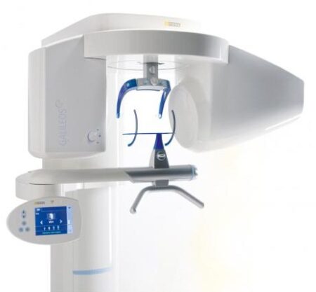
| SIRONA GALILEOS COMFORT PLUS
GALILEOS Comfort Plus is an advanced CBCT that provides seamless work flow integration. With an optional HD mode and a 14 second scan, the GALIELOS offers clinicians and specialists numerous options for diagnosis, treatment and patient consultation with superior image quailtiy and low radiation dose. It includes Integrated Implantology and FaceScan, which helps patients better understand and accept treatment recommendations. SICAT Function enables diagnosis and treatment of TMD, and SICAT Air (coming soon) software allows you to anyalze airways.Features:High-Quality Images:
This dental imaging system is able to position itsef and capture a superior quality of images with each scan, ultimately ensuring that you are receiving the detailed images you need, every time.Panoramic:
This imaging system provides crisp panoramic images to your practice with sensors built for high-quality 2D images. With multiple modes, it’s meant to give you the images you need.3D Conebeam:
Equipped with a high-quality, 3D sensor, this machine is capable of producing crisp and detailed representations of what you need to see. It’s cross-sectional images provide you with the high precision you need.Medium Field of View:
The 15×12 cm field of view option on this machine function allows for the versatility of a standard field of view.Sirona Galileos Dental Cone Beam Imaging System /Full Complete System Software and Hardware :
– Phantom
– Workstation
– Display Monitor
– Software
– ManualsAccessories
– 3D
– Galileos Comfort includes the following 3D views: pan with 3D slice navigation, TSA, axial, sagittal, coronal, 3D model, implant alignment and one-click op reports.
– MEDIUM FOV
– The system’s ellipsoid field of of view (15 x 12 cm) captures the complete dentition, as well as the ascending rami and sinus regions.
– ADJUSTABLE FOV
– Adjustable collimation can save time and reduce patient exposure by allowing you to scan only the regions of interest.
– STANDING DESIGN
– The unit features standing, face-to-face image programming as well as a patented head positioner, ensuring patient comfort and proper positioning.Sirona Galileos Comfort PlusCompact Model D3437Approximately 1100 CT images takenOperating PC includedFOV of the Gallileos Compact is 12×12 cmCrystal Clear 3D/CT imagesSeamless Cerec Integration | Sirona Apollo DI Intra Oral Scanner
APOLLO DI:
The Sirona Apollo DI Intra Oral Scanner features easy handling, precise imaging, and the proven Sirona Connect workflow. The impression system includes an imaging unit, APOLLO Connect software, and the APOLLO DI intraoral
camera,with which users can make digital impressions of the clinical situation in a seamless workflow. To do this, a moisture-insensitive high-contrast spray is sprayed on the teeth very finely. Fine particles in the spray ensure high contrast and thus very precise images.The most cost-effective start to digital impression-taking.
APOLLO DI
APOLLO DI, the specially developed intraoral scanner for cost-efficient digital impressions.
Easy handling thanks to multitouch control
Small and lightweight camera
Export of scan-data in the laboratory
No follow-up costs
OPEN APOLLO DI*:
Export of digital impression data (captured with the APOLLO DI in the practice and received via the Sirona Connect Portal) in an open ST L format for processing in other CAD/CAM systems.Designed to provide an economical entry point into CAD/CAM dentistry, Sirona’s APOLLO DI digital impression system includes an imaging unit, APOLLO Connect software and the APOLLO DI intraoral camera.The system provides precise imaging with the use of a contrast spray, and digital impression data can be sent to dental labs via the Sirona Connect system where it can be turned into a digital restoration design in Sirona’s inLab software or compatible 3rd party software applications, according to a press release.The new APOLLO DI is a complement to the company’s existing CEREC Bluecam and CEREC Omnicam options which each use a different imaging technology, but send the digital impressions through the same workflow. APOLLO DI is designed for practices looking to take digital impressions but continue to work with a lab, while the Bluecam and Onmicam can each be purchased as stand-alone digital impression systems, or with a mill for complete chairside CAD/CAM dentistry. | 3Shape Trios 3 Pod ColorNew ! 3Shape TRIOS Color POD Digital impressions3Shape TRIOS Pod is a new configuration solution and an alternative to the TRIOS cart. It enables scanning with the TRIOS handheld scanner and software using selected laptop PCs. The solution offers a high degree of mobility and flexibility for dentists working in multiple locations or for clinics with limited space. The TRIOS Pod lets users control scanning from an iPad or mirror the 3D view on other displays in the clinic such as monitors integrated in the chair.Connect to your laptop or PC
Use the Pod with your laptop or the PC in your treatment room. Simply connect to the USB port and start scanning.FEATURES : 3Shape Trios 3 Pod Color
– RealColor scans with many clinical advantages: With TRIOS color you can scan in natural colors to clearly distinguish between teeth, gingiva, and restorative materials. Easily identify true preparation margins and improve scanning experience.
– Powder-free for optimal accuracy and comfort: Applying powder is technique demanding, can ruin scan accuracy, is uncomfortable for patients, and prolongs chair time.
– Autoclaveable tip with anti-mist heater: Achieve optimal hygiene and meet clinical requirements. The integrated antimist heater automatically ensures an optimal temperature for undistorted and crystal clear scanning.
– Extreme Mobility: The light and handy Pod solution is easy to share among treatment rooms or different locations
– Use your iPad with scanning: Control TRIOS from your iPad or mirror the live 3D view directly on other displays in the clinic such as monitors integrated in the chair.
– Connect to multiple laptops or PCs: Use with laptops, PCs in your treatment rooms, or with the PCs integrated in your chair units. Simply connect to the USB port and start scanning. Note: Use with approved PCs.
– Small footprint: The compact TRIOS Pod can be placed anywhere in the treatment room or even in rooms where space is very limited.Included : Mac laptop | Schick CDR PanX Laser
FeaturesCDRPanX features proven Schick CDR digital imaging technology and renders crisp, clinically accurate images
Only slightly more expensive than most film-based systems, CDRPanX eliminates costly panoramic film and processing – increasing your economic efficiency
Eight different imaging modes and three laser alignment beams for optimal exposures
Superior vertical travel allows it to be used with patients of any height, including those in wheelchairs
Patient Positioning: 3 laser alignment beams (mid-sagittal, Frankfort, focal trough) bite guide, chin rest, optional temple support
SpecificationsGenerator: Multipulse DC
X-Ray Tube: OCX105
Focal spot size: 0.5 IEC 336
Minimum total filtration: 2.5 mm Al
Anode Voltage: 60-86kV (2kV increments)
Anode Current: 4, 5, 6.3, 8, and 10mA
Exposure time: 19s – adult panoramic
Power Supply: 115V, 50/60Hz
Fusing: T8A
Weight: 474lbs
Electrical Safety Classification: Class 1, type B
Imaging modes (Exposure times): adult (19s), child (15s), left half (10s), right half (10s), anterior (8s), TMJ opened and closed (4x4s), frontal sinuses (8s)
Patient Positioning: 3 laser alignment beams (mid-sagittal, Frankfort, focal trough) bite guide, chin rest, optional temple supportFeatures- CDRPanX features proven Schick CDR digital imaging technology and renders crisp, clinically accurate images
- Only slightly more expensive than most film-based systems, CDRPanX eliminates costly panoramic film and processing – increasing your economic efficiency
- Eight different imaging modes and three laser alignment beams for optimal exposures
- Superior vertical travel allows it to be used with patients of any height, including those in wheelchairs
- Patient Positioning: 3 laser alignment beams (mid-sagittal, Frankfort, focal trough) bite guide, chin rest, optional temple support
Specifications Schick CDR PanX Laser| Generator: | Multipulse DC | | X-Ray Tube: | OCX105 | | Focal spot size: | 0.5 IEC 336 | | Minimum total filtration: | 2.5 mm Al | | Anode Voltage: | 60-86kV (2kV increments) | | Anode Current: | 4, 5, 6.3, 8, and 10mA | | Exposure time: | 19s – adult panoramic | | Power Supply: | 115V, 50/60Hz | | Fusing: | T8A | | Weight: | 474lbs | | Electrical Safety Classification: | Class 1, type B | | Imaging modes (Exposure times): | adult (19s), child (15s), left half (10s), right half (10s), anterior (8s), TMJ opened and closed (4x4s), frontal sinuses (8s) | | Patient Positioning: | 3 laser alignment beams (mid-sagittal, Frankfort, focal trough) bite guide, chin rest, optional temple support |
|


Reviews
There are no reviews yet.