| Description | Designed especially for general dentists, the Sirona Orthophos XG3 combines top-tier image quality and exceptional ease-of-use in an affordable, digital panoramic x-ray solution. | The Identica T300 dramatically improves the fundamental aspects of scanner performance and the latest software technology provides the clearest, sharpest data. Identica T300 is customized to deliver the finest data quality using its dual 2.0 MP cameras. Identica T300 will save valuable time and money by minimizing trial & error when adjusting restoration fits. | The 3Shape D710 is optimized for scanning both the impression and gypsum models and enables high throughput for both advanced and standard indications. These innovative scanners are so easy to use that staff training is barely required. Item fixation is fast, and the scan starts with a click on the program. | Perform a 3D body scan in seconds using just a simple turntable and 3D camera. In only 35 seconds, we extract millions of measurements with 2mm accuracy and reconstruct your body in 3D. | Sirona Galileos Dental Cone Beam Imaging System The system captures images using the lowest possible radiation dose and is designed to provide high-quality images for implant planning, treatment guide design, jaw function analysis and more. | Caries diagnostic aid for surface caries.
TRIOS 4 has built-in fluorescent technology that aids in the identification of possible caries. Using TRIOS 4, dental professionals can now be aided in the early-detection of surface caries, without the need for an additional scanning device.
Caries diagnostic aid for interproximal caries.
TRIOS 4 will also feature a dedicated transillumination smart tip later this year. The smart tip will aid in the identification of possible interproximal caries undetectable to the eye, without emitting radiation. |
| Content | SIRONA XG3 Digital Panoramic X-Ray
The ORTHOPHOS XG 3 is designed for the general practice dentist who would like to work with a basic X-ray system. The XG 3 offers you an outstanding price-performance ratio and provides the following benefits:- High image quality
- Extremely simple operation
- Solid workmanship
Accurate positioningThe ORTHOPHOS XG 3 uses proven, logical Sirona principles for exceptional imaging:- Exact, immediate positioning of the anterior teeth in the focal layer
- Effective, comfortable patient stabilisation with forehead and temple supports to prevent motion blurring
Proven TechnologyThe advanced technology from the XG product family enables the ORTHOPHOS XG 3 to produce high quality diagnostic images.
The benefits include:- Specific focal layers and orbital paths for anatomically optimised exposures
- Very detailed display of the anterior tooth region through automatic kV adaption as X-ray beam passes through spinal region
- 16 bit image acquisition technology provides more gray scales
- SIDEXIS XG imaging software with useful analysis tools
Features:SIRONA XG3 Digital Panoramic X-Ray- High-quality, digital panoramic and TMJ imaging power with advanced patient communication tools
- Outstanding price-performance ratio for seamless transition from film to direct digital imaging
- Easy operation with the Multipad; effective, three-point patient stabilization for sharp images
- Direct network capability for exceptional connectivity
SIRONA ORTHOPHOS XG 3The horizontally and vertically reduced pediatric panoramic program offers outstanding image quality with the lowest dose. Ideal for practices that treat a lot of children. The unit’s easy operation and seamless integration into your practice’s daily routines will save you valuable time. The high image quality and proven Sidexis software guarantee that you will come to a safe diagnosis quickly and easily.Quality from the very startHigh quality. Durable. Safe: the Orthophos XG 3 X-ray unit is the perfect basic equipment for general dentists. The unit’s easy operation and seamless integration into your practice’s daily routines will save you valuable time. The high image quality and proven Sidexis software guarantees that you will reach a safe diagnosis quickly and easily.The positioning can be very easily reproduced since image parameters such as the bite block height are saved together with the image data. A bitewing program is available, which helps in special cases and patient situations where intraoral images cannot be used and a shorter panoramic program with a reduced dose, which is suitable for treating children, is also available. | Medit Identica T300 Dental 3D Scanner
Identica T300 to meet all of your needs. With powerful scan engine and affordable pricing, the new Identica T300 is a perfect fit for both performance enthusiasts and entry level users. Its evolutionary design and high tech features will help your lab rediscover productivity. Bundled colLab 2017 scan software makes scan data processing easy and efficient.Quality & affordability- 2.0 MP cameras, Blue LED
- Accuracy : 7 μm (ISO)
- Full arch scan time 24 sec
- 2-Axis Manual
Identica T300 dramatically improves the most important aspects of scanner performance. The latest software technology provides you with the cleanest and sharpest data. T300 is specially designed to bring out the finest data quality using its 2.0 MP cameras. T300 will save your precious time and money by minimizing trial & error to adjust restoration fits.Identica T300 is an upgrade version of Identica Blue, provided at lowest pricing. Giving better value for the affordable scanner users. Identica T300 has enhanced data quality compared to Identica Blue.Introducing the Next Generation of 3D Dental Scanners: Our T-Series Medit represents the apex in 3D dental scanning. With unsurpassed speed, accuracy, and reliability, Medit helps you rediscover your productivity. The T-Series’ new powerful scan engine and affordable pricing are the perfect fit for performance enthusiasts and entry-level users, helping your lab rediscovering productivity thanks to its convenient design and high-tech features. Medit T300 is the latest Medit scanner with an enhanced SW scan. It uses Blue Light technology, which enables quick scanning and provides excellent precision (7μm).The program is user-friendly, because it takes you through simple and fast individual steps, from inserting the model into the scanner to exporting the file to the CAD design program. FEATURES:Medit Identica T300 Dental 3D Scanner- Scanned printing: 2 manual axes.
- Touch sensor: no.
- Full arcade scan speed: 24 ´´
- Resolution of the camera: 2 x 2 MP camera.
- Scan volume: 90 x 72 x 60 mm.
- Scanning principle: Optical change phase.
- Dimensions: 290 x 290 x 340 mm.
- Weight: 12Kg.
- Light source: Blue LED.
- Connectivity: Type USB 3.0 B.
- Power: 100- 240 vac, 50-60 HZ.
- Warranty: 2 years.
ADVANCED DATA PROCESSING AND HIGHER RESOLUTION DUAL CAMERAS The Identica T300 dramatically improves the fundamental aspects of scanner performance and the latest software technology provides the clearest, sharpest data. Identica T300 is customized to deliver the finest data quality using its dual 2.0 MP cameras. Identica T300 will save valuable time and money by minimizing trial & error when adjusting restoration fits. | 3shape D710 Dental Scanner3Shape D710 Scanner series can scan gypsum and impression versions. The series scanners can integrate into the clinic with minimum staff training. The scanners provide greater practice productivity, a comprehensive list of signals, and the most extensive 3D scans.Where flexibility and productivity are the key standards 3Shape chain scanners are for medium to large labs.The 3Shape D710 is optimized for scanning both the impression and gypsum models and enables high throughput for both advanced and standard indications. These innovative scanners are so easy to use that staff training is barely required. Item fixation is fast, and the scan starts with a click on the program.3SHAPE D710 For Sale3Shape D700 Scanner is both a scanner and impression scanner in 1 unit. It utilizes three measurement movement and two cameras a laser and creates another pass to especially hone. Just one device can be scanned by the apparatus in under 30 seconds and a whole model in about a minute with a degree of accuracy of 20 microns.Features:
-3-axis motion system
-Red-laser Tech
-Gypsum Version scanning
-Impression scanning Alternative
-Multi-die scanningBenefits:
-Easy operation, extreme Precision, and Rapid Scan
-Enables impressions, deep inlays, and Complete undercuts to scan Efficiently
-Supply labs Using ultra-efficient batches and multi-case processingHigh-speed scanning
The D710 is optimized for scanning gypsum and impression versions and enables high throughput for both advanced and standard indications3Shape – leading impression scanning technologies
3Shape’s patented adaptive scanning technology, combined with the scanner’s 3-axis motion and 2 cameras with reduced angle, facilitate the unique impression scanning capability.Adaptive scanning intelligently detects incomplete areas and automatically builds
adaptive scan sequences for capturing full object geometry. | Styku 3D Body scanner
Styku 3D is a professional, full-body 3D scanner. It is a high-precision measurement tool able to scan people in just 30 seconds. It uses Infrared Depth Sensor technology for high-resolution scanning results. It is composed of an automatic turntable and a sensor tower. Its small footprint and aluminum structure make it easy to move and reposition.This powerful scanner is simple and convenient to use. It can capture the full body of a person in three dimensions. The person only has to stand on the rotating plate for 30 seconds holding the pose. The scanner is able to extract millions of data points in no time thanks to the efficient Microsoft Kinect v2 3D camera. It lets you perform complete body scans, view 3D models, extract exact body measurements, track posture changes, and more. After generating the 3D avatar, the advanced scanning and feature recognition technology automatically detects specific landmarks, which are used to extract accurate data related to the volume, circumference, and surface of each body area.Styku is mainly used for fitness and professional health purposes. It consents to view and analyze the body shape, measurements, and other health indicators. It allows tracking the evolution of the body shape or posture of a person through several measurements. Find out more reading our complete review.Styku is often used to track body changes. For example, it is generally adopted by people who want to capture and compare before and after images of themselves in order to analyze their physical evolution.Specifications:- Scan accuracy
- Scan volume
- Scanner controls
- What’s in the box
- Look and feel
- Price
Scan accuracyStyku 3D body scanner is a professional, full-body 3D scanner. It features a depth data resolution of ~2mm (.08in). It grants a torso measurement precision of +- 2.5-5mm (.1-.2in) and can perform a complete scan in 30/40 seconds. It is engineered with advanced feature recognition and a 3D visualization system for automatic detection of landmark points and measurement extraction. It is capable of stitching more than 600 surfaces to create a three-dimensional avatar in seconds. The Kinect v2 3D camera has a depth image resolution of 512 x 424 pixels.Scan volume It features a minimum scanning area of 8.4 x 5.5 ft ( 251 x 168 cm). It works at a recommended distance of the sensor tower from the turntable of 46 inches (117 cm). It requires a minimum clearance of 21 inches (53 cm) around the turntable. In case of white walls or glossy surfaces, it might require more clearance.Scanner controlsThe scanner is controlled via an external computer.The proprietary Styku intuitive, multi-platform software allows performing a complete scan and all the measurements with ease. The generated avatar can be rotated, panned, and zoomed to evaluate each body part in detail. The collected data are strictly private. You can decide who can view them by setting appropriate export and sharing options. The software is compatible with Windows 10 and is optimized for touchscreens. It features a minimum scanning area of 8.4 x 5.5 ft ( 251 x 168 cm). It works at a recommended distance of the sensor tower from the turntable of 46 inches (117 cm). It requires a minimum clearance of 21 inches (53 cm) around the turntable. In case of white walls or glossy surfaces, it might require more clearance.Scanner controlsThe scanner is controlled via an external computer.The proprietary Styku intuitive, multi-platform software allows performing a complete scan and all the measurements with ease. The generated avatar can be rotated, panned, and zoomed to evaluate each body part in detail. The collected data are strictly private. You can decide who can view them by setting appropriate export and sharing options. The software is compatible with Windows 10 and is optimized for touchscreens.What’s in the box
- Essential parts required to operate the Styku 3D body scanner
- Rotating platform (max. weight 250kg)
- Sensor tower
- Microsoft Kinect v2 3D camera
- Aluminum enclosure and stand to protect the 3D Camera
- All the necessary cables
For some items, package content may change. In case of any questions, please get in touch.Look and feel
Styku 3D body scanner is available in white with red trims. Its stylish look makes it ideal for fitness clubs, gyms, healthcare facilities, and fashion companies.The scanner has an overall footprint of 101 x 66 inches ( 257cm x 168cm). The rotating platform measures 24 x 24 x 5 inches (66cm x 66cm x 12.5 cm) and weighs 23 lb (10.5 kg). It withstands a max load of 550 lb (250 kg). The sensor tower measures 10 x 10 x 46 inches ( 25.4 x 25.4 x 117 cm). It weighs 13 lb (6 kg). | SIRONA GALILEOS COMFORT PLUS
GALILEOS Comfort Plus is an advanced CBCT that provides seamless work flow integration. With an optional HD mode and a 14 second scan, the GALIELOS offers clinicians and specialists numerous options for diagnosis, treatment and patient consultation with superior image quailtiy and low radiation dose. It includes Integrated Implantology and FaceScan, which helps patients better understand and accept treatment recommendations. SICAT Function enables diagnosis and treatment of TMD, and SICAT Air (coming soon) software allows you to anyalze airways.Features:High-Quality Images:
This dental imaging system is able to position itsef and capture a superior quality of images with each scan, ultimately ensuring that you are receiving the detailed images you need, every time.Panoramic:
This imaging system provides crisp panoramic images to your practice with sensors built for high-quality 2D images. With multiple modes, it’s meant to give you the images you need.3D Conebeam:
Equipped with a high-quality, 3D sensor, this machine is capable of producing crisp and detailed representations of what you need to see. It’s cross-sectional images provide you with the high precision you need.Medium Field of View:
The 15×12 cm field of view option on this machine function allows for the versatility of a standard field of view.Sirona Galileos Dental Cone Beam Imaging System /Full Complete System Software and Hardware :
– Phantom
– Workstation
– Display Monitor
– Software
– ManualsAccessories
– 3D
– Galileos Comfort includes the following 3D views: pan with 3D slice navigation, TSA, axial, sagittal, coronal, 3D model, implant alignment and one-click op reports.
– MEDIUM FOV
– The system’s ellipsoid field of of view (15 x 12 cm) captures the complete dentition, as well as the ascending rami and sinus regions.
– ADJUSTABLE FOV
– Adjustable collimation can save time and reduce patient exposure by allowing you to scan only the regions of interest.
– STANDING DESIGN
– The unit features standing, face-to-face image programming as well as a patented head positioner, ensuring patient comfort and proper positioning.Sirona Galileos Comfort PlusCompact Model D3437Approximately 1100 CT images takenOperating PC includedFOV of the Gallileos Compact is 12×12 cmCrystal Clear 3D/CT imagesSeamless Cerec Integration | 3Shape TRIOS 4 Intraoral ScannerOur most powerful intraoral scanner to date Take your dentistry beyond treatment
Our intraoral scanning solutions allow you to take your dentistry beyond. The new 3Shape TRIOS 4 combines our established superior scanning technology with groundbreaking caries diagnostic aid technology* for both surface and interproximal caries. No additional scanning device needed. Smart tips 3Shape TRIOS 4 Intraoral ScannerThe new generation of TRIOS smart tips feature instant-heat technology for optimized scanning.Be scan-ready in seconds with instant-heat technology.Scan 2-3 times as many patients with instant-heat in combination with 30% more battery life.Optimal scanning due to automatic use counter and tip-change alert.The TRIOS system is a complete in-house ecosystem including a scanner and extensive design software. This means there are plenty of options depending on your needs. Those who are just looking for a scanner and want to utilize labs may look into the basic, cheaper options. Those who want to design crowns, bridges and other prosthetics can opt for design modules. The only missing component is a milling machine, which if you want to mill restorations in-house, will have to be from a third party. This proves to be a weak link in the digital chain.At iDD, we have been using the TRIOS 3 for over four years in our clinic, spending literally thousands of hours learning the workflow. We have carried out all aspects of dentistry using the TRIOS system including same-day crowns, implant surgical guides, implant restorations and digital smile design.Disclaimer: This is an impartial review of the scanner and we do not have any ties to the company. The team at iDD remain unwaveringly committed to providing you with objective and trustworthy information and therefore do not seek any sponsorship. 3Shape kindly loaned us a TRIOS 4 scanner, due to a personal interest in upgrading our older TRIOS 3 model. Our team at iDD are constantly approached by scanner companies to review their scanners and it is always made clear that they will have no part in writing reviews or restricting any conclusions iDD makes in our thorough analysis and clinical use of these products. |


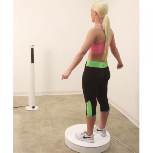 It features a minimum scanning area of 8.4 x 5.5 ft ( 251 x 168 cm). It works at a recommended distance of the sensor tower from the turntable of 46 inches (117 cm). It requires a minimum clearance of 21 inches (53 cm) around the turntable. In case of white walls or glossy surfaces, it might require more clearance.Scanner controlsThe scanner is controlled via an external computer.
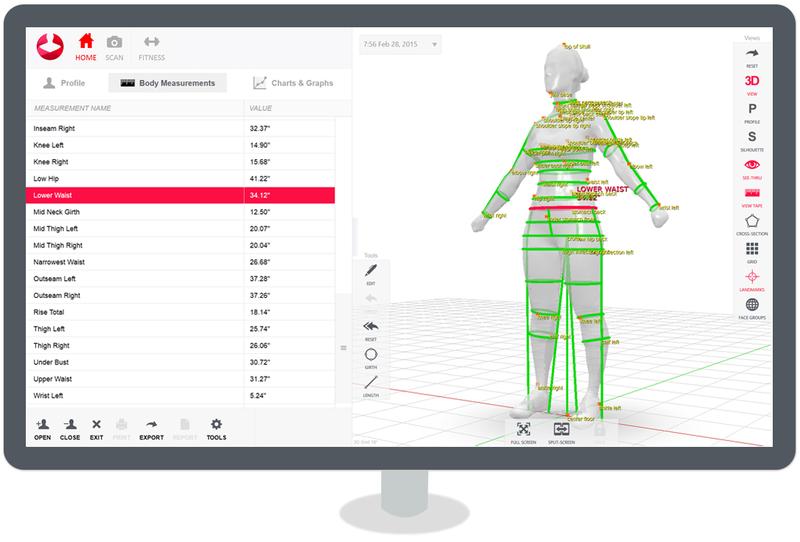 The proprietary Styku intuitive, multi-platform software allows performing a complete scan and all the measurements with ease. The generated avatar can be rotated, panned, and zoomed to evaluate each body part in detail. The collected data are strictly private. You can decide who can view them by setting appropriate export and sharing options. The software is compatible with Windows 10 and is optimized for touchscreens.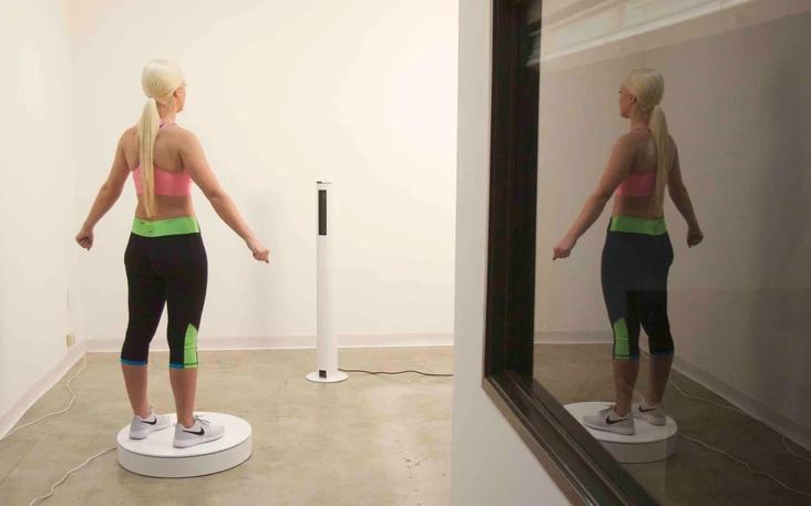
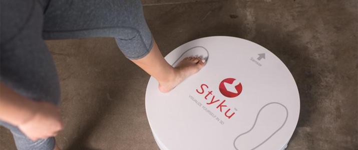 Styku 3D body scanner is available in white with red trims. Its stylish look makes it ideal for fitness clubs, gyms, healthcare facilities, and fashion companies.The scanner has an overall footprint of 101 x 66 inches ( 257cm x 168cm). The rotating platform measures 24 x 24 x 5 inches (66cm x 66cm x 12.5 cm) and weighs 23 lb (10.5 kg). It withstands a max load of 550 lb (250 kg). The sensor tower measures 10 x 10 x 46 inches ( 25.4 x 25.4 x 117 cm). It weighs 13 lb (6 kg).


Reviews
There are no reviews yet.