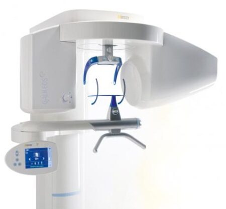
| Description | With the integrated FaceScanner in GALILEOS you can now record the facial surface parallel to the x-ray image. And that, as opposed to traditional face scanners without the use of lasers, which is therefore gentler on the patients. Thanks to the simultaneous x-ray image and surface depiction, the fully automated overlaying of the data is extremely precise. In addition, texture and coloring in the software allow for a particularly realistic depiction of the soft tissue proportions – an ideal tool for patient consultation! | The iTero Element Scanner is a digital impression system that eliminates the need for messy putty in your mouth.Improving iTero Intraoral ScannersWhile it’s already a leader in dental technology, iTero is constantly refining its intraoral scanning options. Its Element Itraoral Scanner, introduced in March 2015, captures 6,000 frames per second, up to 20 times faster than its predecessor. Its wand is also smaller and lighter, with in-built controls for more intuitive operation. Intraoral scanners from iTero are renowned for their accuracy, but the increased capture speed improves the company’s already impressive statistics. | Sirona offers your lab greater freedom in terms of indications, materials and for processing third-party CAD/CAM data. The open 5-axis milling and grinding machine inLab MC X5 is designed specifically to meet dental lab requirements for cost-efficient production today and in the future. | Sirona Galileos Dental Cone Beam Imaging System The system captures images using the lowest possible radiation dose and is designed to provide high-quality images for implant planning, treatment guide design, jaw function analysis and more. | The E4 boasts double the speed, double the accuracy, and double as many cameras as its predecessor the E3, delivering improved efficiency and consistency with every scan. From small labs seeking the best-of-the-best, to high-volume, full-service labs striving for a leaner workflow, our E4 lab scanner is the right choice for you. | CEREC now provides a completely new process to dental practices: by combining the new CEREC SpeedFire furnace and CEREC Zirconia material, dentists can now deliver full contour crowns and small bridges made of the full-strength high-quality zirconium oxide in their own practice while the patient waits. |
| Content | Sirona GALILEOS Comfort/Compact| GALILEOS Comfort | GALILEOS Compact | | Oral and maxillofacial surgeons
Dental clinics
Private clinics
Orthodontists | General practioners
Dental implant practices
Oral surgeons |  |  | | Comparison Table of Galileos Comfort/ Compact & Orthophos XG 3D X-Ray systems | | Technical overview | GALILEOS Comfort | GALILEOS Compact | ORTHOPHOS
XG 3D | | Field of view | (15 x 15 x 15) cm³ | (12 x 15 x 15) cm³ | 8 cm x 8 cm (Ø x Höhe) | | 3D Resolution (isotropic voxel size) | 0.3/0.15 mm | 0.3 mm | 0,2 mm; 0,1 mm (available at the end of 201) | | Scan time / exposure time | 14 s / 2 – 6 s | 14 s / 2 – 6 s | 2–5 s | | Reconstruction time | 2.5 – 4.5 min | 4.5 min | 4,5 min | | Patient position | standing / seated | standing / seated | Standing/seated, chin rest/bite block, occlusal bite block for automatic patient positioning with 2D PAN images | | X-ray generator | | kV | 85 | 85 | 60–90 | | Effective dosage | 29 µSv / 68 µSv **
(21 mAs, 85 kV) | < 29 µSv / 68 µSv **
(21 mAs, 85 kV) | 43–175 μSv (Schulze)
(Standard: 100 μSv) | | Space requirements (minimum) | 1,6 x 1,6 x 2,25 m (d x w x h) | 1,6 x 1,6 x 2,25 m(d x w x h) | 1,5 m x 1,1 m x 2,25 m (Pan ), | | Space requirements (recommended) | >1,8 x 1,8 x 2,5 m (d x w x h) | 1,8 x 1,8 x 2,5 m (d x w x h) | 1,7 m x 1,3 m x 2,5 m (Pan ) | | User Interface | Full color touchscreen | Multipad | EasyPad | | Patient fixation | Occlusal bite block, forehead | Occlusal bite bock, forehead | | | Positioner | support, chin rest, head positioner *** | support, chin rest | | | Upgradable to Comfort version | |  | | | Optional floor stand | | | | | Wheelchair accessible | | | | | Remote control | | | | | Programs | | 150 µm resolution for close-up reconstruction | | | | | 300 µm resolution for standard volumes | | | | | High contrast mode for optimized hard tissue display | | | | | Views | PAN with 3D slice navigation | PAN with 3D slice navigation | Tiltable 2D slices, custom | | TSA, axial, sagittal, coronal, CEPH lat., CEPH pa/ap | TSA, axial, sagittal, coronal | 3D slicing, TSA, LSA, axial, sagittal, coronal | | 3D model, 1-click OP reports | 3D model, 1-click OP reports | 3D model, implant-oriented view, 1-Click OP reports |
With the integrated FaceScanner in GALILEOS you can now record the facial surface parallel to the x-ray image. And that, as opposed to traditional face scanners without the use of lasers, which is therefore gentler on the patients. Thanks to the simultaneous x-ray image and surface depiction, the fully automated overlaying of the data is extremely precise. In addition, texture and coloring in the software allow for a particularly realistic depiction of the soft tissue proportions – an ideal tool for patient consultation! |
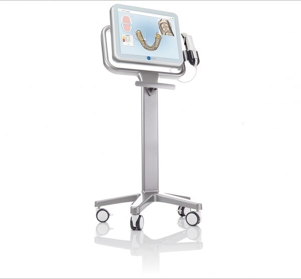
| iTero Element Intraoral Scanner
What Are iTero Intraoral Scanners?Intraoral scanners from iTero scan the mouths of patients, capturing images to create three-dimensional dental images in minutes. Intraoral scanners are simple to use and can be operated by one person. Their user-friendly nature helps dental professionals get the best results. The scans they produce are also more detailed than the traditional two-dimensional images they replace.Intraoral digital scans help dental professionals create accurate physical dental models for restorative work, including crowns, veneers, and implants. They also help orthodontists diagnose orthodontic problems and develop the best treatment plans.The company’s digital ecosystem software works seamlessly with its intraoral scanners, improving workflow for dental professionals working on orthodontic and restorative cases. However, unlike many early intraoral scanners, they are open systems. This feature gives dental professionals more flexibility about how they use their digital scan files. Intraoral digital scans from iTero scanners can be easily shared with other dental professionals and third-party providers such as Invisalign. When all relevant parties have the scans, they can communicate better to improve patient outcomes.What iTero Intraoral Scanners DoIntraoral scanners feature a wand, which the dental professional moves around a patient’s mouth. In the latest versions, the wand captures thousands of frames per second which are pieced together to create a three-dimensional visualisation of the patient’s mouth. The wands on iTero intraoral scanners are smaller than early intraoral scanners, allowing them to scan molars in the back of the mouth which were traditionally difficult to reach. Dental professionals using small wands also aren’t limited by how wide their patients can open their mouths. The small wands are also less likely to make patients gag than older forms of scanning technology.Intraoral scanners also have screens which display the digital dental images as they’re captured in real time. The screens show whether the scan is good or not before it’s saved and submitted to the lab. This feature can be a real time-saver for dental professionals who, in the past, could receive word from the lab two or three weeks later that their scans were inadequate. By providing immediate feedback, intraoral scanners can save dental professionals and patients time and frustration.Unlike many intraoral scanners, patients don’t need to cover their teeth in titanium dioxide powder before an iTero intraoral scan. This benefit further improves the scanning process for dental patients.How Intraoral Scanners Make Invisalign Orthodontics Easier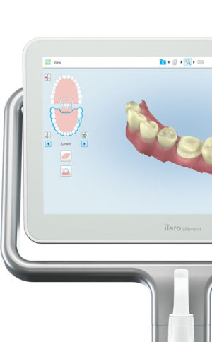 Invisalign clear aligners are one of the most popular teeth-straightening aids used today as they’re effective, removable, and virtually undetectable. Unlike many intraoral scanners, iTero intraoral scanners have open architecture which makes them compatible with the Invisalign system, including its Invisalign Outcome Simulator. Orthodontists can scan their patients’ mouths with an iTero intraoral scanner, then show them how their Invisalign treatment will look. This technology improves the patient experience because patients can know what to expect and feel more confident in their diagnosis and treatment plan. It also makes the ClinCheck setup three times faster.After setup, speed is still on the iTero intraoral scanners’ side. iTero states ClinCheck treatment plans submitted with its scans are usually posted to the Invisalign Doctor Site three times faster than traditional polyvinyl siloxane scans. As a result, your Invisalign aligners are created and posted back to your orthodontist sooner so that you can start treatment faster. Since the iTero intraoral scanning system is open, orthodontists can also send the scan files to any laboratory of their choosing for the creation of a retainer, which reinforces your treatment after the Invisalign process, and other dental tools.Orthodontists can also create better Invisalign treatment plans for their patients using iTero intraoral scans. Align Technology research shows orthodontists who use the scans have 10 times fewer rejections and seven times fewer issues with the fit of the Invisalign aligners. These results may be because iTero intraoral scanners can help orthodontists track their patients’ progress. Regular scans throughout Invisalign treatment can help orthodontists compare expected outcomes with results. If results aren’t as expected, orthodontists can use the scans to educate their patients about their treatment and the importance of complying with their recommendations.Improving iTero Intraoral Scanners iTero Element Intraoral ScannerWhile it’s already a leader in dental technology, iTero is constantly refining its intraoral scanning options. Its Element Itraoral Scanner, introduced in March 2015, captures 6,000 frames per second, up to 20 times faster than its predecessor. Its wand is also smaller and lighter, with in-built controls for more intuitive operation. Intraoral scanners from iTero are renowned for their accuracy, but the increased capture speed improves the company’s already impressive statistics. Invisalign clear aligners are one of the most popular teeth-straightening aids used today as they’re effective, removable, and virtually undetectable. Unlike many intraoral scanners, iTero intraoral scanners have open architecture which makes them compatible with the Invisalign system, including its Invisalign Outcome Simulator. Orthodontists can scan their patients’ mouths with an iTero intraoral scanner, then show them how their Invisalign treatment will look. This technology improves the patient experience because patients can know what to expect and feel more confident in their diagnosis and treatment plan. It also makes the ClinCheck setup three times faster.After setup, speed is still on the iTero intraoral scanners’ side. iTero states ClinCheck treatment plans submitted with its scans are usually posted to the Invisalign Doctor Site three times faster than traditional polyvinyl siloxane scans. As a result, your Invisalign aligners are created and posted back to your orthodontist sooner so that you can start treatment faster. Since the iTero intraoral scanning system is open, orthodontists can also send the scan files to any laboratory of their choosing for the creation of a retainer, which reinforces your treatment after the Invisalign process, and other dental tools.Orthodontists can also create better Invisalign treatment plans for their patients using iTero intraoral scans. Align Technology research shows orthodontists who use the scans have 10 times fewer rejections and seven times fewer issues with the fit of the Invisalign aligners. These results may be because iTero intraoral scanners can help orthodontists track their patients’ progress. Regular scans throughout Invisalign treatment can help orthodontists compare expected outcomes with results. If results aren’t as expected, orthodontists can use the scans to educate their patients about their treatment and the importance of complying with their recommendations.Improving iTero Intraoral Scanners iTero Element Intraoral ScannerWhile it’s already a leader in dental technology, iTero is constantly refining its intraoral scanning options. Its Element Itraoral Scanner, introduced in March 2015, captures 6,000 frames per second, up to 20 times faster than its predecessor. Its wand is also smaller and lighter, with in-built controls for more intuitive operation. Intraoral scanners from iTero are renowned for their accuracy, but the increased capture speed improves the company’s already impressive statistics. |
The new freedom in detailEvery aspect of inLab MC X5 has been designed to address the CAD/CAM needs encountered in dental labs. Intensive technical and technological exchange with many dental technicians has led to the creation of a production machine fulfilling practically all requirements. For the reliable, independent production of dental prostheses.OpeninLab MC X5 is an open production unit. It is the perfect complement to the inLab components inEos X5 and inLab software, but it can also be used to process other STL restoration data. 1 With its own CAM module, the machine can be connected to a wide range of other CAD systems at no additional cost. DesignThe new inLab MC X5 has more than just outstanding functionality and simple operation: its extremely stable construction and modern design are also highly attractive. Its compact, elegant shape will be a real eye catcher in your lab and complements perfectly the award-winning inEos X5 extraoral scanner.Great variety of materialsinLab MC X5 is designed as a universal production unit for processing zirconium oxide, polymers, composites, wax, titanium and metals 2 as well as glass and hybrid ceramics. inLab MC X5 can be used for wet, dry, or dry/wet combination production during the same machining operation depending on material and indication.Sirona has drawn upon its 30 years of experience in the wet processing of glass ceramics to create the ultimate machine for the wet grinding of fully anatomical restorations from high-strength monolithic materials.Switching between wet and dry production e.g. from glass ceramics to zirconium oxide is fast and uncomplicated.inLab MC X5 – efficient and flexible inLab MC X5 is a universal 5-axis production unit for wet and dry production of blocks and disks. It was designed to meet dental requirements in labs and focus on economic and effective production processes.You benefit from a wide range of materials and can select from the high-quality materials of Dentsply Sirona and its CAD/CAM material partners for which the production processes of inLab MC X5 are optimized. | SIRONA GALILEOS COMFORT PLUS
GALILEOS Comfort Plus is an advanced CBCT that provides seamless work flow integration. With an optional HD mode and a 14 second scan, the GALIELOS offers clinicians and specialists numerous options for diagnosis, treatment and patient consultation with superior image quailtiy and low radiation dose. It includes Integrated Implantology and FaceScan, which helps patients better understand and accept treatment recommendations. SICAT Function enables diagnosis and treatment of TMD, and SICAT Air (coming soon) software allows you to anyalze airways.Features:High-Quality Images:
This dental imaging system is able to position itsef and capture a superior quality of images with each scan, ultimately ensuring that you are receiving the detailed images you need, every time.Panoramic:
This imaging system provides crisp panoramic images to your practice with sensors built for high-quality 2D images. With multiple modes, it’s meant to give you the images you need.3D Conebeam:
Equipped with a high-quality, 3D sensor, this machine is capable of producing crisp and detailed representations of what you need to see. It’s cross-sectional images provide you with the high precision you need.Medium Field of View:
The 15×12 cm field of view option on this machine function allows for the versatility of a standard field of view.Sirona Galileos Dental Cone Beam Imaging System /Full Complete System Software and Hardware :
– Phantom
– Workstation
– Display Monitor
– Software
– ManualsAccessories
– 3D
– Galileos Comfort includes the following 3D views: pan with 3D slice navigation, TSA, axial, sagittal, coronal, 3D model, implant alignment and one-click op reports.
– MEDIUM FOV
– The system’s ellipsoid field of of view (15 x 12 cm) captures the complete dentition, as well as the ascending rami and sinus regions.
– ADJUSTABLE FOV
– Adjustable collimation can save time and reduce patient exposure by allowing you to scan only the regions of interest.
– STANDING DESIGN
– The unit features standing, face-to-face image programming as well as a patented head positioner, ensuring patient comfort and proper positioning.Sirona Galileos Comfort PlusCompact Model D3437Approximately 1100 CT images takenOperating PC includedFOV of the Gallileos Compact is 12×12 cmCrystal Clear 3D/CT imagesSeamless Cerec Integration | 3Shape E4 Dental Lab ScannerThe E4 is a dental 3D scanner produced by 3Shape. 3Shape is a 3D scanner manufacturer based in Denmark.E4 dental scanner main features- Full arch scan in 9 seconds
- 4-micron accuracy
- Four 5-MP cameras
- Color acquisition
3SHAPE E4
Ultimate productivity with our fastest scanner ever
For those seeking the speed and accuracy to drive lab productivity and growth, look no further. The E4 boasts double the speed, double the accuracy, and double as many cameras as its predecessor the E3, delivering improved efficiency and consistency with every scan. From small labs seeking the best-of-the-best, to high-volume, full-service labs striving for a leaner workflow, our E4 lab scanner is the right choice for you.
4 micron
Scan with an accuracy of 4 microns (ISO 12836) for great case results. Combine this precision with increased speed and you can take on more implant cases, for example.
4 cameras
Four cameras save you time by scanning dies in the model instead of individually. You can even scan impressions instead of producing a gypsum model or combine impression and die at the same time.
5MP cameras
5MP cameras are super high definition cameras. They record 5 megapixel images with enhanced camera sensors, meaning you get superior image clarity and can do high-precision scanning.
HARDWARE SPECIFICS
The E4 is a marriage of iconic 3Shape design and superior technical specifications.
Cameras
Four 5 MP cameras enables efficient die scanning and impression scanning, improving your workflows by saving time
Accuracy
4 microns (ISO 12836), giving you the accuracy and precision you need for implant cases
Full arch scan speed 9 seconds (versus 22 seconds for the 3Shape E3)
Full arch impressions scan speed 45 seconds
Texture Color
Scanning strategy Die-in-model | Sirona CEREC Zirconia SpeedFire Milling Unit
High strength, short manufacturing process
The greatest benefit of CEREC Zirconia is the high flexural strength of the material. It is suitable for individual crowns as well as small bridges and can be processed in thin wall thicknesses. Since these restorations are manufactured in monolithic form, there is no risk of chipping. Another benefit for dentists is that zirconium oxide can be cemented conventionally.CEREC Zirconia is a pre-shaded translucent zirconium oxide available in 10 shades on the basis of the VITA Classic Shade Guide®. The material is milled in an enlarged form and then densely sintered to its final size in the new sintering furnace CEREC Speed-Fire. The oversized milling facilitates a new level of milling accuracy leading to superb, precisely fitting restorations. The sintering process takes just 10-15 minutes for crowns and 25 minutes for bridges. The subsequent glaze firing gives the restoration a high gloss finish.The short process to produce CEREC Zirconia restorations is both convenient and economical. With this market launch, all CEREC milling/grinding units now provide wet and dry milling. Dry milling reduces the overall processing time for zirconia and, combined with the world’s fastest sintering cycles, enables the chairside procedure.The workflow is easy to learn since the CEREC Software 4.4.1 guides the dentist through the entire process, and even sends the sintering and glazing information to the furnace. No programming of the furnace is required – it is all handled automatically by the software. A high-performance material and a specially tailored workflow ensure a simple process and high-quality treatment.
CEREC meets patients’ needsSirona CEREC Zirconia SpeedFire Milling UnitEighty three percent of patients in a recent survey said they prefer single visit dentistry to traditional treatment. The majority said they would even be willing to pay more for single visit dentistry. Two thirds indicated they’d be happy to travel further than they currently do, in order to receive treatment in one session. Another two thirds said they would even be prepared to change their dentist. The advantages of single visit dentistry are obvious: Patients avoid impression material, receive fewer anesthetics and don’t need a temporary. Reducing the number of visits, injections and making the overall prosthetic procedure more comfortable influences patients to opt for treatment with CEREC.In addition to CEREC Zirconia, many other high-performance materials can be processed with CEREC. The range of materials with CEREC also expands clinical indications since the dentist can easily select the material suited for each indication.
Investment for a successful futureThere have always been good reasons to start using digital dentistry with CEREC. CEREC Zirconia completes the range of indications for chairside application for nearly
every situation, thus giving dentists greater choice and allowing them to increase the value of their practice’s offering.It is obvious that advanced technologies in automobiles, computers and smartphones make our daily lives easier. CEREC is also a technology that further develops a dental practice and can make it well positioned for the future. Especially now, as the system is highly flexible, it enables dentists to expand their offering in implant dentistry and orthodontics. |


Reviews
There are no reviews yet.