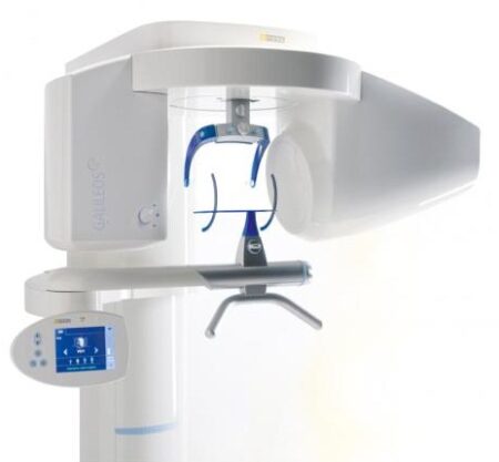
| Description | With the integrated FaceScanner in GALILEOS you can now record the facial surface parallel to the x-ray image. And that, as opposed to traditional face scanners without the use of lasers, which is therefore gentler on the patients. Thanks to the simultaneous x-ray image and surface depiction, the fully automated overlaying of the data is extremely precise. In addition, texture and coloring in the software allow for a particularly realistic depiction of the soft tissue proportions – an ideal tool for patient consultation! | TRIOS Color automatically measures the teeth shades as you scan. The dentist has the option to evaluate and highlight relevant areas for the lab. The complete shade information sis sent to the lab together with the impression. | WaterLase iPlus includes the all-new SureFire™ delivery system, with redesigned optics that can more efficiently deliver precise amounts of Er,Cr:YSGG laser energy to cut enamel, dentin and bone. The SureFire delivery system is optimized to provide the dentist with more up time with a replaceable, disposable shield that protects the vital optical components in the fiber, resulting in greater dependability. | At only 4.2 pounds, the portable x-ray, is considered to be the lightest x-ray generator on the market. Many design features are included with an LCD Display, and High Frequency Tube, a perfect balance of technology and simplicity. There is no doubt that the EXARO is the best, handheld and portable x-ray, available. | The Waterlase MD features Hydro- Photonics?a combination of YSGG laser energy and water?which allows the clinician to perform a wide range of conventional procedures with less anesthetic. Known for its precision and accuracy, the MD enables treatment of targeted areas of both tooth structure and soft tissue while maintaining the structural integrity of the tooth and leaving surrounding areas unaffected. | Cost-effective production
The inLab software positions your zirconium oxide restorations optimally in the material block – for efficient use of blocks, attractive unit prices and the best possible machine utilization, e.g. by milling or grinding overnight.
Wet grinding of sintered NPM blocks
“inLab goes metal.” With the inLab MC XL and the new inCoris CC sintered metal blocks you can handle a complete spectrum of metal and ceramic materials. The unique wet grinding function rules out any health risks. |
| Content | Sirona GALILEOS Comfort/Compact| GALILEOS Comfort | GALILEOS Compact | | Oral and maxillofacial surgeons
Dental clinics
Private clinics
Orthodontists | General practioners
Dental implant practices
Oral surgeons |  |  | | Comparison Table of Galileos Comfort/ Compact & Orthophos XG 3D X-Ray systems | | Technical overview | GALILEOS Comfort | GALILEOS Compact | ORTHOPHOS
XG 3D | | Field of view | (15 x 15 x 15) cm³ | (12 x 15 x 15) cm³ | 8 cm x 8 cm (Ø x Höhe) | | 3D Resolution (isotropic voxel size) | 0.3/0.15 mm | 0.3 mm | 0,2 mm; 0,1 mm (available at the end of 201) | | Scan time / exposure time | 14 s / 2 – 6 s | 14 s / 2 – 6 s | 2–5 s | | Reconstruction time | 2.5 – 4.5 min | 4.5 min | 4,5 min | | Patient position | standing / seated | standing / seated | Standing/seated, chin rest/bite block, occlusal bite block for automatic patient positioning with 2D PAN images | | X-ray generator | | kV | 85 | 85 | 60–90 | | Effective dosage | 29 µSv / 68 µSv **
(21 mAs, 85 kV) | < 29 µSv / 68 µSv **
(21 mAs, 85 kV) | 43–175 μSv (Schulze)
(Standard: 100 μSv) | | Space requirements (minimum) | 1,6 x 1,6 x 2,25 m (d x w x h) | 1,6 x 1,6 x 2,25 m(d x w x h) | 1,5 m x 1,1 m x 2,25 m (Pan ), | | Space requirements (recommended) | >1,8 x 1,8 x 2,5 m (d x w x h) | 1,8 x 1,8 x 2,5 m (d x w x h) | 1,7 m x 1,3 m x 2,5 m (Pan ) | | User Interface | Full color touchscreen | Multipad | EasyPad | | Patient fixation | Occlusal bite block, forehead | Occlusal bite bock, forehead | | | Positioner | support, chin rest, head positioner *** | support, chin rest | | | Upgradable to Comfort version | |  | | | Optional floor stand | | | | | Wheelchair accessible | | | | | Remote control | | | | | Programs | | 150 µm resolution for close-up reconstruction | | | | | 300 µm resolution for standard volumes | | | | | High contrast mode for optimized hard tissue display | | | | | Views | PAN with 3D slice navigation | PAN with 3D slice navigation | Tiltable 2D slices, custom | | TSA, axial, sagittal, coronal, CEPH lat., CEPH pa/ap | TSA, axial, sagittal, coronal | 3D slicing, TSA, LSA, axial, sagittal, coronal | | 3D model, 1-click OP reports | 3D model, 1-click OP reports | 3D model, implant-oriented view, 1-Click OP reports |
With the integrated FaceScanner in GALILEOS you can now record the facial surface parallel to the x-ray image. And that, as opposed to traditional face scanners without the use of lasers, which is therefore gentler on the patients. Thanks to the simultaneous x-ray image and surface depiction, the fully automated overlaying of the data is extremely precise. In addition, texture and coloring in the software allow for a particularly realistic depiction of the soft tissue proportions – an ideal tool for patient consultation! |
| 3Shape Trios 3 Pod ColorNew ! 3Shape TRIOS Color POD Digital impressions3Shape TRIOS Pod is a new configuration solution and an alternative to the TRIOS cart. It enables scanning with the TRIOS handheld scanner and software using selected laptop PCs. The solution offers a high degree of mobility and flexibility for dentists working in multiple locations or for clinics with limited space. The TRIOS Pod lets users control scanning from an iPad or mirror the 3D view on other displays in the clinic such as monitors integrated in the chair.Connect to your laptop or PC
Use the Pod with your laptop or the PC in your treatment room. Simply connect to the USB port and start scanning.FEATURES : 3Shape Trios 3 Pod Color
– RealColor scans with many clinical advantages: With TRIOS color you can scan in natural colors to clearly distinguish between teeth, gingiva, and restorative materials. Easily identify true preparation margins and improve scanning experience.
– Powder-free for optimal accuracy and comfort: Applying powder is technique demanding, can ruin scan accuracy, is uncomfortable for patients, and prolongs chair time.
– Autoclaveable tip with anti-mist heater: Achieve optimal hygiene and meet clinical requirements. The integrated antimist heater automatically ensures an optimal temperature for undistorted and crystal clear scanning.
– Extreme Mobility: The light and handy Pod solution is easy to share among treatment rooms or different locations
– Use your iPad with scanning: Control TRIOS from your iPad or mirror the live 3D view directly on other displays in the clinic such as monitors integrated in the chair.
– Connect to multiple laptops or PCs: Use with laptops, PCs in your treatment rooms, or with the PCs integrated in your chair units. Simply connect to the USB port and start scanning. Note: Use with approved PCs.
– Small footprint: The compact TRIOS Pod can be placed anywhere in the treatment room or even in rooms where space is very limited.Included : Mac laptop | Biolase Waterlase iplus LaserThe WaterLase iPlus all-tissue dental laser from the global leader in dental lasers is designed to deliver practice growth to dentists by offering an enhanced experience for patients, while raising the standard of overall reliability and quality for the #1-preferred all-tissue laser. Enhancing the patient experienceWaterLase iPlus is the latest addition to the WaterLase family, which has been striving to enhance the dental experience since 2001. With an estimated 27 million patients treated, more than 97% would recommend their WaterLase dentist to a friend – and WaterLase iPlus 2.0 offers a host of benefits that deliver an even better patient experience. Improvements to key components boost the availability of the laser when it’s needed most; and a new, minimally invasive laser therapy for periodontal disease offers dentists a clinical protocol that can generate excellent clinical outcomes and a vastly different type of patient experience versus traditional methods. SureFire Delivery System Biolase Waterlase iplus LaserWaterLase iPlus includes the all-new SureFire delivery system, with redesigned optics that can more efficiently deliver precise amounts of Er,Cr:YSGG laser energy to cut enamel, dentin and bone. The SureFire delivery system is optimized to provide the dentist with more up time with a replaceable, disposable shield that protects the vital optical components in the fiber, resulting in greater dependability. REPaiR for Reliably Treating PeriodontitisWaterLase iPlus offers new benefits to dentists and patients with the addition of REPaiR, the first Er,Cr:YSGG-based protocol for safe, effective treatment of moderate to severe periodontitis. It is an achievable, repeatable, minimally-invasive procedure that is less traumatic than traditional surgical techniques and delivers better and gentler experiences for patients. Clinically proven treatment for moderate to severe periodontitis Exclusive to WaterLase iPlus 2.0 Includes:Trunk Fiber 2 Hand Pieces (Turbo & Gold) Manual (DVD) Pair of Laser Goggles Air Line Power Cord Tip Cleaning Kit | Xray2go Hand Held Dental Portable X-Ray System| FDA Registered. Welcome to the new way of taking X-rays in Dental Offices: x-ray2go is the way to go! x-ray2go is a portable hand held camera that uses a high frequency inverter to generate a stable X-ray output with DC high voltage. This effective DC unit enables patient and operator to be exposed to the least amount of radiation. The integrated compact “Camera like” generator increases the user’s confidence and decrease the negative perception patients have about X-rays. It is small: 5.6″ (W) x 3.3″ (D) x 1.2″ (H). Best of all it only weighs 4.18 Lbs (1.9 Kgs), the lightest on the market.• Portability – room to room ease
• No installation necessary versus complex, expensive and time consuming on-wall installation.
• No need for pass troughs
• Large LCD display – easy to read
• Simple touch controls
• Adjustable exposure time
• Hundreds of images from one (1) battery charge
• High resolution diagnostic images
• No blur from hand movement high-frequency generator produces clear images while reducing
patient dose
• Shield protects the operator from scattered radiation – small dose of radiation emitted
• Leading technology provides the highest level of safety, quality and patient care
• Perfect for special needs patients, home health care patients, nursing home patients, children and
veterinary practices
• Perfect for small operatories
• Perfect for mobile dentistry where room is a big factor |
| | | | |
| | | | Specifications Xray2go Hand Held Dental Portable X-Ray System- Portable, cordless x-ray device
· FDA Registered – K082875
· Safe to use-leakage and scatter radiation are less than conventional wall mounted units
· Small & lightweight (4.2 lbs)
· Camera-like ergonomic design
· 100’s of images on single charge!
· Compatible with both digital & film use
· Simple operation
· Highest image quality
· Wide exposure range
· Lowest patient exposure
· Compact outside—smart inside
· Great staff acceptance
· Convenient for children and “x-ray phobic” patients
· Helps eliminate patient fear and reduces x-ray rejection
| X-ray installation | Tube voltage: 60KV
Tube current: 2mA
Heat Capacity: 8.5KHU | | X-ray Generator | Type: High Frequency Inverter
Power Output: 100W
X-ray Tube | | X-ray Tube | Anode Type: Stationary
Anode Angle: 20°
Focal Spot: 0.8mm | | X-ray Control | Exposure time set 0.01 ~ 1.6sec (0.01 sec step) | | Power Requirement | 25.2VDC | | Weight | 4.2 lbs. | | Dimensions | 6.7”(W) x 6.1”(D) x 5.25”(H) |
|
|
| Biolase Waterlase MDX All Tissue LaserThe MDX delivers 300 mJ of energy at 8 W, and it can easily be upgraded to a 450 mJ (9 W) model for 50% faster cutting as clinical skills improve and applications expand (see ?MDX Specs?). This next-generation of the best-selling WaterLase MD features improvements to the laser engine and delivery system ergonomics, and it enables rapid hardtissue cutting speed with unprecedented affordability.Time-Tested Technology Biolase Waterlase MDX All Tissue LaserThe MDX is built on the technologies and reputations of its predecessors, the BIOLASE WaterLase MD and WaterLase iPlus all-tissue lasers.The Waterlase MD features Hydro- Photonics?a combination of YSGG laser energy and water?which allows the clinician to perform a wide range of conventional procedures with less anesthetic. Known for its precision and accuracy, the MD enables treatment of targeted areas of both tooth structure and soft tissue while maintaining the structural integrity of the tooth and leaving surrounding areas unaffected.The Waterlase MD became the company?s flagship product, breaking new ground in the areas of cutting speed, user interface, and ergonomic design. In 2011 that ?flagship? designation was overtaken by the company?s new WaterLase iPlus, an even faster, more powerful machine. It delivers up to 10 W of power at 600 mJ, which makes it the most powerful laser on the global market at the short pulse length of 50 ?s, all through a highly intuitive preprogrammed user interface for everything from routine restorative cases to specialty periodontal, cosmetic, and endodontic procedures.The iPlus features Biolase?s 2780 nm YSGG technology with the versatility of a dual-wavelength configuration; namely, an integrated iLase 940 nm diode laser to handle unexpected soft-tissue cases, better bleeding control, and the potential for temporary pain relief and teeth whitening. As anticipated, the iPlus proved beneficial for experienced laser dentists who had been anxiously awaiting a machine that would deliver more power, control, and versatility. However, the new laser also provided many of the ?uninitiated? with an easier-than-expected, highly intuitive introduction to all-tissue laser dentistry. The MDX takes this one step further, with a price point that puts the technology within reach of nearly any dentist.
No More Excuses: Affordable Pricing, Substantial ROIThe perception of all-tissue laser technology as inaccessible to most dentists is beginning to dissipate. Even so, some dentists remain unconvinced.The reality, however, is that BIOLASE?s positioning of the MDX as an entry-level product, starting under $40,000, has made the technology much more affordable. Combining better and more efficient manufacturing with strategic sourcing of components results in pricing that most clinicians will view as extremely reasonable. In addition, the initial investment will be recouped with a substantial ROI, which can be realized via 3 main areas of application.1. For hard-tissue restorative dentistry? the ?bread and butter? of general practices?the MDX will enable faster, more efficient and, in most cases, virtually painless restorations for all cavity classes in both adults and children.2. The MDX allows dentists to retain patients who may have traditionally been referred out for osseous crown lengthening. Using the MDX, patients reap the benefits of considerably more comfortable soft-tissue procedures without the inconvenience of a referral, while the practice reaps the benefits of a happier and more loyal patient base.3. MDX provides an effective and efficient gateway into periodontics. Specifically, the MDX promotes deep pocket therapy with new attachment and is especially effective for minimally invasive removal of both subgingival inflamed tissue and calculus in patients with moderate to severe periodontal disease.Another DimensionBIOLASE?s comprehensive in-office and online training and support promise to render the learning curve extremely manageable. And, as intimidation is replaced by both comfort and confidence, clinicians will find that all-tissue laser technology will add a new and profitable dimension to their practices. | Sirona Cerec InLab MCXL Milling Unit
The inLab MC XL milling and grinding unit opens up the widest possible range of production options for your dental laboratory. You profit from high speed and precision. You can switch from grinding to milling in just a few simple steps. And thanks to the large milling volume and broad spectrum of applications, you will reap substantial economic benefits.
inLab MC XL
Large range of materials:
Processing of zirconium oxide, resin, silicate ceramics and NP-metal
High-speed milling:
For example, a four-unit zirconium oxide bridge can be completed in just 40 minutes
Large milling capacity: For restorations consisting of up to 12 units and ceramic blocks measuring 85 x 40 x 22 mm
Four milling motors: Flexible selection of milling burs for different materials
Milling and grinding: For high precision regardless of the materialand indication
Milling* of zirconium oxide and polymers
In addition to grinding, you can also mill zirconium oxide and polymers on the inLab MC XL. You benefit from an enhanced initial fit and a faster production process.Flexible and efficient Sirona Cerec InLab MCXL Milling UnitinLab MC XL is the fast wet milling and grinding unit with many production options for your dental laboratory. You benefit from high speed and precision and can switch from grinding to milling in just a few steps. The large selection of materials and many uses give you particularly flexible and efficient production options. inLab high-speed grindingGlass- and hybrid ceramic restorations can be produced at a previously unachievable speed with simultaneous double four-axis processing. A fully contoured Celtra Duo crown takes less than 10 minutes. This is a key success factor for potential new business models that can call for providing digital impression orders within an hour. Precision processingThe inLab MC XL is characterized by precise wet machining. Especially when processing glass ceramics, the grinders used can be as small as 0.6 mm — for restorations with maximum detail in the occlusal and interproximal areas and at the preparation margin. Wide selection of materialsAs with all CAD/CAM production units from Dentsply Sirona, inLab MC XL lets you benefit from a the large selection of materials. Dentsply Sirona CAD/CAM materials and those of our material partners are optimally adapted for high-speed processing. |


Reviews
There are no reviews yet.