View cart “Sirona Apollo DI Intra Oral Scanner For Sale” has been added to your cart.
-38%
Availability: In Stock
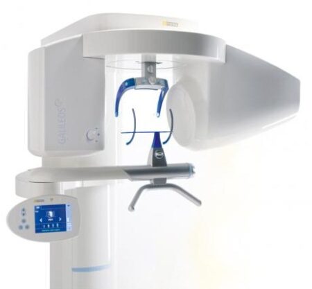
Sirona GALILEOS Comfort/Compact For Sale
$11,000.00$6,800.00
With the integrated FaceScanner in GALILEOS you can now record the facial surface parallel to the x-ray image. And that, as opposed to traditional face scanners without the use of lasers, which is therefore gentler on the patients. Thanks to the simultaneous x-ray image and surface depiction, the fully automated overlaying of the data is extremely precise. In addition, texture and coloring in the software allow for a particularly realistic depiction of the soft tissue proportions – an ideal tool for patient consultation!
Other people want this. 15 people have this in their carts right now.
3D model, implant-oriented view, 1-Click OP reports
With the integrated FaceScanner in GALILEOS you can now record the facial surface parallel to the x-ray image. And that, as opposed to traditional face scanners without the use of lasers, which is therefore gentler on the patients. Thanks to the simultaneous x-ray image and surface depiction, the fully automated overlaying of the data is extremely precise. In addition, texture and coloring in the software allow for a particularly realistic depiction of the soft tissue proportions – an ideal tool for patient consultation!
Reviews (0)
Reviews
There are no reviews yet.
Be the first to review “Sirona GALILEOS Comfort/Compact For Sale” Cancel reply
With the integrated FaceScanner in GALILEOS you can now record the facial surface parallel to the x-ray image. And that, as opposed to traditional face scanners without the use of lasers, which is therefore gentler on the patients. Thanks to the simultaneous x-ray image and surface depiction, the fully automated overlaying of the data is extremely precise. In addition, texture and coloring in the software allow for a particularly realistic depiction of the soft tissue proportions – an ideal tool for patient consultation!
The Waterlase MD features Hydro- Photonics?a combination of YSGG laser energy and water?which allows the clinician to perform a wide range of conventional procedures with less anesthetic. Known for its precision and accuracy, the MD enables treatment of targeted areas of both tooth structure and soft tissue while maintaining the structural integrity of the tooth and leaving surrounding areas unaffected.
The new highly accurate 0.5 mm milling tool reliably delivers first class clinical results. Switch to Extra Fine milling mode for a higher level of detail with occlusal fissures and interdental areas on bridges.
SuniRay 2 achieves maximal image quality with superior diagnostic capabilities while maintaining the lowest radiation levels among all digital sensors. The comprehensive and feature-rich Prof. Suni Advanced Imaging Software supplements the SuniRay 2’s high-quality imaging with a wide array of user-friendly customization and image-enhancement tools. With the SuniRay 2, Suni has created a complete digital radiography system that seamlessly integrates with your entire practice.
Milling Unit:• Choose from CEREC MC, CEREC MC X or CEREC MC XL
• Get access to the entire range of chairside treatment with up to 40 mm block size, including bridges and abutments
• Create fully anatomic, precise treatments quickly
• CEREC Guide 2 surgical guide
• Dry mill ready
• Includes CEREC software
Caries diagnostic aid for surface caries.
TRIOS 4 has built-in fluorescent technology that aids in the identification of possible caries. Using TRIOS 4, dental professionals can now be aided in the early-detection of surface caries, without the need for an additional scanning device.Caries diagnostic aid for interproximal caries.
TRIOS 4 will also feature a dedicated transillumination smart tip later this year. The smart tip will aid in the identification of possible interproximal caries undetectable to the eye, without emitting radiation.
3D model, implant-oriented view, 1-Click OP reports
With the integrated FaceScanner in GALILEOS you can now record the facial surface parallel to the x-ray image. And that, as opposed to traditional face scanners without the use of lasers, which is therefore gentler on the patients. Thanks to the simultaneous x-ray image and surface depiction, the fully automated overlaying of the data is extremely precise. In addition, texture and coloring in the software allow for a particularly realistic depiction of the soft tissue proportions – an ideal tool for patient consultation!
Biolase Waterlase MDX All Tissue LaserThe MDX delivers 300 mJ of energy at 8 W, and it can easily be upgraded to a 450 mJ (9 W) model for 50% faster cutting as clinical skills improve and applications expand (see ?MDX Specs?). This next-generation of the best-selling WaterLase MD features improvements to the laser engine and delivery system ergonomics, and it enables rapid hardtissue cutting speed with unprecedented affordability.Time-Tested Technology Biolase Waterlase MDX All Tissue LaserThe MDX is built on the technologies and reputations of its predecessors, the BIOLASE WaterLase MD and WaterLase iPlus all-tissue lasers.The Waterlase MD features Hydro- Photonics?a combination of YSGG laser energy and water?which allows the clinician to perform a wide range of conventional procedures with less anesthetic. Known for its precision and accuracy, the MD enables treatment of targeted areas of both tooth structure and soft tissue while maintaining the structural integrity of the tooth and leaving surrounding areas unaffected.The Waterlase MD became the company?s flagship product, breaking new ground in the areas of cutting speed, user interface, and ergonomic design. In 2011 that ?flagship? designation was overtaken by the company?s new WaterLase iPlus, an even faster, more powerful machine. It delivers up to 10 W of power at 600 mJ, which makes it the most powerful laser on the global market at the short pulse length of 50 ?s, all through a highly intuitive preprogrammed user interface for everything from routine restorative cases to specialty periodontal, cosmetic, and endodontic procedures.The iPlus features Biolase?s 2780 nm YSGG technology with the versatility of a dual-wavelength configuration; namely, an integrated iLase 940 nm diode laser to handle unexpected soft-tissue cases, better bleeding control, and the potential for temporary pain relief and teeth whitening.
As anticipated, the iPlus proved beneficial for experienced laser dentists who had been anxiously awaiting a machine that would deliver more power, control, and versatility. However, the new laser also provided many of the ?uninitiated? with an easier-than-expected, highly intuitive introduction to all-tissue laser dentistry. The MDX takes this one step further, with a price point that puts the technology within reach of nearly any dentist.
No More Excuses: Affordable Pricing, Substantial ROIThe perception of all-tissue laser technology as inaccessible to most dentists is beginning to dissipate. Even so, some dentists remain unconvinced.The reality, however, is that BIOLASE?s positioning of the MDX as an entry-level product, starting under $40,000, has made the technology much more affordable. Combining better and more efficient manufacturing with strategic sourcing of components results in pricing that most clinicians will view as extremely reasonable. In addition, the initial investment will be recouped with a substantial ROI, which can be realized via 3 main areas of application.1. For hard-tissue restorative dentistry? the ?bread and butter? of general practices?the MDX will enable faster, more efficient and, in most cases, virtually painless restorations for all cavity classes in both adults and children.2. The MDX allows dentists to retain patients who may have traditionally been referred out for osseous crown lengthening. Using the MDX, patients reap the benefits of considerably more comfortable soft-tissue procedures without the inconvenience of a referral, while the practice reaps the benefits of a happier and more loyal patient base.3. MDX provides an effective and efficient gateway into periodontics. Specifically, the MDX promotes deep pocket therapy with new attachment and is especially effective for minimally invasive removal of both subgingival inflamed tissue and calculus in patients with moderate to severe periodontal disease.Another DimensionBIOLASE?s comprehensive in-office and online training and support promise to render the learning curve extremely manageable. And, as intimidation is replaced by both comfort and confidence, clinicians will find that all-tissue laser technology will add a new and profitable dimension to their practices.
DENTSPLY SIRONA CEREC PRIMEMILLSpeedEngineered for speed and precision, the new CEREC Primemill inherits the proven strength of its predecessor and raises its powerful performance to entirely new heights.You can now mill zirconia restorations in Super Fast milling mode in around 5 minutes, cutting processing times by more than half. The improved grinding mode for high quality glass ceramics also grinds the material faster than ever before. The result? Satisfied patients and high productivity.QualityDENTSPLY SIRONA CEREC PRIMEMILL A combination of new electronics, software, motors and mechanical components contributes to finer resolution and enhanced dynamics. This results in outstanding restoration margins and surface details – for extremely precise results.The new highly accurate 0.5 mm milling tool reliably delivers first class clinical results. Switch to Extra Fine milling mode for a higher level of detail with occlusal fissures and interdental areas on bridges.VersatilityDENTSPLY SIRONA CEREC PRIMEMILL Welcome to a world where you‘re spoiled for choice! The new CEREC Primemill combines wet and dry milling and wet grinding. No matter the indication, with CEREC Primemill you can select from a broad range of material options from validated partners providing the flexibility and predictability you need to offer patients the best possible treatment.Choose from the full spectrum of machining options, including dry and wet milled zirconia, or wet grinding of glass and hybrid ceramics.ConvenienceDENTSPLY SIRONA CEREC PRIMEMILL The new 7” touch interface with user guidance guides operators step-by-step through the workflow, key maintenance procedures and other routine tasks, so you can delegate milling with complete peace-of-mind. The integrated block scanner is a time-saving new feature that automatically scans material blocks with compatible data matrix codes and records the information, including type, size, color and zirconia enlargement factor. All CEREC Primemill tools are fitted with a color-coded RFID chip that is read by the RFID tool reader, feeding into the tool status display, alerting the user if a tool needs replacing. This ensures all tools are performing at optimal levels. A LED light strip indicates unit status and milling and grinding progress.The all-new CERECThe latest generation of our CEREC system sets new standards so you can offer your patients an unmatched combination of single-visit dentistry and excellent quality.A unique solutionThe CEREC system comprises world-class components that interact perfectly ensuring seamless workflows. With CEREC Primescan, CEREC Software 5 and CEREC Primemill, digital chairside dentistry is faster, easier and more reliable than ever before.Outstanding resultsAfter 35 years of continuous optimization, CEREC gives you the options you need to treat multiple indications with the confidence that comes from outstanding results.
Designed to stand out – and fit inThe new CEREC Primemill does not just communicate seamlessly with other CEREC devices, but its sleek design is also the perfect match – and the ideal look for a progressive dental practice.
SuniRay 2 Digital Intraoral Sensor Size 1 and Size 2
X-Ray Imaging Properties
Resolution: The pixel resolution of the high resolution (HiRes) sensors exceeds 15 lp/mm. The standard resolution of an X-Ray image, as measured by the modulation transfer function (MTF) exceeds 12 lp/mm. This is measured by using a standard 60 kV intraoral X-ray source.
Does Efficiency: The sensor will produce a high quality image with an X-Ray dose that is only a fraction of the standard dose required by Dental X-Ray film.
Diagnostic Efficacy: The system will produce a superior image quality of images obtained when using standard dental X-Ray film. This allows the dentist to diagnose standard intraoral pathologies encountered during screening procedures.
Wide dynamic range: The sensor pixel well capacity is very high, allowing a higher accommodation of greyscale (bone density) dynamic range.
Sensor Material Biocompatibility SpecificationsThe sensor’s body material and cable material are biocompatible. The sensors and all component materials have been tested to comply with ISO standards, and specifically, the sensor complies with EN IS0-10993 (Biological Evaluation of Medical Devices).
Technical data
Size 1
Size 2
Sensor Dimensions (mm)
39.5 x 26
43.5 x 31.5
Active Area (mm)
31.1 x 20.2
35.2 x 26.2
Sensor Technology
CMOS APS Fiber Plate
CMOS APS Fiber Plate
Maximum Gray Levels
4096
4096
Sensor Cable
3 feet (1m)
3 feet (1m)
Cable Attachment
Reinforced Strain Relief
Reinforced Strain Relief
USB Module
Integrated USB 2.0 Module
Integrated USB 2.0 Module
Software
Prof. Suni
Prof. Suni
X-Ray Imaging Properties SuniRay 2 Digital Intraoral Sensor Size 1 and Size 2
Resolution: The pixel resolution of the high resolution (HiRes) sensors exceeds 15 lp/mm. The standard resolution of an X-Ray image, as measured by the modulation transfer function (MTF) exceeds 12 lp/mm. This is measured by using a standard 60 kV intraoral X-ray source.
Does Efficiency: The sensor will produce a high quality image with an X-Ray dose that is only a fraction of the standard dose required by Dental X-Ray film.
Diagnostic Efficacy: The system will produce a superior image quality of images obtained when using standard dental X-Ray film. This allows the dentist to diagnose standard intraoral pathologies encountered during screening procedures.
Wide dynamic range: The sensor pixel well capacity is very high, allowing a higher accommodation of greyscale (bone density) dynamic range.
CEREC Primescan AC Intraoral scanner
Cerec Primescan from Dentsply Sirona is the most advanced digital scanner on the market. It’s providing a level of accuracy never before seen in dentistry. At the peak of innovation, it is widening the gap between those with and without digital dentistry. The Prime Scan is the fastest most accurate intraoral scanner to date. With new scanning technology, it is the ultimate in scanning. Prime Scan and CEREC 5.0 combinations are greatly reduced because more data allows many applications to become automated.For more than 30 years, CEREC® has provided technological precision, superior design and excellent performance to thousands of practices. Now, experience a brand-new fully integrated solution with CEREC Primescan AC. Acquire precise digital impressions with Primescan, design your proposal on the Primescan AC using the intelligent software, then produce precise, smooth restorations with your choice of milling unit. Find the next level of productivity and patient care – it’s all here.How Does CEREC Primescan Work?
CEREC Primescan is an alternative to traditional intraoral dental impressions, which use a putty-like substance to take physical impressions of your teeth. This method does not allow for digital assessment of your mouth, and some patients find it the process of physical impressions to be uncomfortable.CEREC Primescan uses a wand-like device, loaded with powerful imaging sensors which can capture more than 50,000 images of your mouth per second. These images are quickly stitched and compiled into a 3D image of your teeth, jaw, gums, and mouth, and displayed on a high-definition screen for immediate assessment by our doctors.Using this sensor, our doctors can image your entire mouth up to 20mm in depth, including gums, blood, and bone structures, quickly and accurately. The Primescan software (as compared to the Omnicam) creates high-resolution 3D models to assess your oral health and create the very best crowns, implants, removable prosthesis, restorations, and even 3D prints. With just a few quick sweeps of the CEREC Primescan device, we’ll be able to generate an in-depth, modifiable 3D image of your mouth in minutes.The Benefits of CEREC Primescan:
An excellent choice for outstanding results: Primescan is your perfect starting point in digital dentistry. No matter how you would like to design your workflows, Primescan is the enabler for efficient digital workflows – both chairside in your practice and with your preferred partners.Well designed and perfectly compatible: Along with the high-performance intraoral scanner comes the new Acquisition Center. Together, they form a highly effective and most comfortable system – meeting your needs with two individual software configurations:So, why is CEREC Primescan such a big deal? Here are just a few improvements to this sophisticated system:Shorter wait times – We can quickly image your mouth without waiting for dental impressions to set, allowing us to treat each patient more quickly and effectively. A full jaw impression only takes 2 minutes. This adds up to less time spent in the dental chair.Real-time image processing – Our dentists can quickly adjust and modify scanned images using filters and other tools, leading to more accurate diagnostics and better overall patient outcomes.Extremely versatile – CEREC Primescan can be used for almost any dental procedure. From implant dentistry to the creation of CEREC same-day crowns, the straightening of teeth, whitening, development of dentures and partial dentures.CEREC Primescan AC with CEREC Software Sirona Advantages:
Enables all kinds of treatment, from a single tooth to full arch
With Primescan intraoral scanning is now more accurate, faster and easier
Allows an efficient workflow, both chairside and with preferred partners
Along with the high-performance intraoral scanner comes the new Acquisition Center, together forming an effective and most comfortable system, meeting your needs with two individual software configurations: Primescan AC with Connect Software (supports the connection to established partners workflows) and CEREC Primescan AC with CEREC Software (ideal for cabinets)
Smart Pixel Sensor technology – allows superior precision and excellent detail reproduction, processes more than 1 million 3D points per second, producing photorealistic and highly accurate data
Due to its ability to scan at an incredible data density, it delivers complete 3D structures of everything in its field of view
Its dynamic depth scan technology enables perfect sharpness and outstanding precision, even at a measuring depth up to 20 mm
Easy to use
Easy scanning of all dental materials and hard to access areas
Allows you to start scanning right away
Self-heating for fog-free scanning
The increased field of view visualizes larger areas with fewer sweeps and immediate precision
Faster scanning with a smooth scan flow
Consolidating more than 50.000 images per second and fast processing of exactly the data the software needs Easy capturing and quicker processing of more data in higher resolution
Complete 3D-scan models are displayed immediately, no matter how fast you scan
Accelerate your workflow: Due to seamless, validated and open data transfer options, labs and other third parties are supplied with high-resolution models at an instant
3 different sleeves (disposable and stainless steel, with sapphire glass or autoclavable)
Acquisition Center – Smart features and greater comfort: intuitive use via movable 16:9 touchscreen and touchpad for perfect ergonomics
Kinematics for perfect ergonomic positioning
Smart Hygiene concept for fast and easy disinfection
Full mobility with more than 60-min. battery buffer
CEREC Primescan AC with CEREC Software – complete chairside system, which offers flexible data export options, with automatized workflow thanks to Artificial Intelligence and touch-enabled and intuitive user interface
Milling Unit:• Choose from CEREC MC, CEREC MC X or CEREC MC XL
• Get access to the entire range of chairside treatment with up to 40 mm block size, including bridges and abutments
• Create fully anatomic, precise treatments quickly
• CEREC Guide 2 surgical guide
• Dry mill ready
• Includes CEREC softwareStriking in every detail: The new Acquisition CenterThe new Acquisition Center is a work station designed for modern dentistry and with the dentist in mind. It comes with a touchpad and a 16:9 wide-format movable touchscreen, offering you a highly intuitive and ergonomic work platform.Your benefits at a glance:
Touchscreen & Touchpad for most comfortable, intuitive use
Kinematics for perfect ergonomic positioning
Smart Hygiene concept for fast and easy disinfection
Mobility concept for full mobility with more than 60-min. battery buffer
The best choice for your practice: Take digital impressions and continue working with your trusted partners or enjoy the benefits of complete chairside workflows.Primescan meets your needs with two individual software configurations:Primescan AC with Connect Software:
Supports data transfer options to your preferred partners
Secure and encrypted data transfer through Connect Case Center Inbox
Easy upgradability to full chairside workflow
Touch-enabled and intuitive user interface
CEREC Primescan AC with CEREC Software:
Supports full chairside workflows for single-visit dentistry
Flexible data export options
Automatized workflow thanks to Artifical Intelligence
Touch-enabled and intuitive user interface
Technical Data:CEREC Primescan AC Intraoral scanner
CAD system for high-precision intraoral optical impressions
High-resolution, heated intraoral 3D scanner
Integrated image processing
High processing power due to state-of-the-art processor
Trackball or touchpad
Hand and foot controlled enter keys
Ethernet port and WLAN
USB interfaces
The high-resolution 3D intraoral scanner allows image acquisition with the image control inside the scanner, the image data transfer is made through USB 2.0 interface
21.5″ inch TFT LED flat-screen display, HD resolution: 1920 x 1080 pixels
Mobile housing, with no water or air connection required
Voltage: 100 – 240 VAC/50-60Hz
Nominal current: 5.0 – 2.1 A
Type of protection against electric shock: Class I device
Take your dentistry beyond treatment
Our intraoral scanning solutions allow you to take your dentistry beyond. The new 3Shape TRIOS 4 combines our established superior scanning technology with groundbreaking caries diagnostic aid technology* for both surface and interproximal caries. No additional scanning device needed.Smart tips 3Shape TRIOS 4 Intraoral ScannerThe new generation of TRIOS smart tips feature instant-heat technology for optimized scanning.Be scan-ready in seconds with instant-heat technology.Scan 2-3 times as many patients with instant-heat in combination with 30% more battery life.Optimal scanning due to automatic use counter and tip-change alert.The TRIOS system is a complete in-house ecosystem including a scanner and extensive design software. This means there are plenty of options depending on your needs. Those who are just looking for a scanner and want to utilize labs may look into the basic, cheaper options. Those who want to design crowns, bridges and other prosthetics can opt for design modules. The only missing component is a milling machine, which if you want to mill restorations in-house, will have to be from a third party. This proves to be a weak link in the digital chain.At iDD, we have been using the TRIOS 3 for over four years in our clinic, spending literally thousands of hours learning the workflow. We have carried out all aspects of dentistry using the TRIOS system including same-day crowns, implant surgical guides, implant restorations and digital smile design.Disclaimer: This is an impartial review of the scanner and we do not have any ties to the company. The team at iDD remain unwaveringly committed to providing you with objective and trustworthy information and therefore do not seek any sponsorship. 3Shape kindly loaned us a TRIOS 4 scanner, due to a personal interest in upgrading our older TRIOS 3 model. Our team at iDD are constantly approached by scanner companies to review their scanners and it is always made clear that they will have no part in writing reviews or restricting any conclusions iDD makes in our thorough analysis and clinical use of these products.


 An excellent choice for outstanding results: Primescan is your perfect starting point in digital dentistry. No matter how you would like to design your workflows, Primescan is the enabler for efficient digital workflows – both chairside in your practice and with your preferred partners.Well designed and perfectly compatible: Along with the high-performance intraoral scanner comes the new Acquisition Center. Together, they form a highly effective and most comfortable system – meeting your needs with two individual software configurations:So, why is CEREC Primescan such a big deal? Here are just a few improvements to this sophisticated system:Shorter wait times – We can quickly image your mouth without waiting for dental impressions to set, allowing us to treat each patient more quickly and effectively. A full jaw impression only takes 2 minutes. This adds up to less time spent in the dental chair.Real-time image processing – Our dentists can quickly adjust and modify scanned images using filters and other tools, leading to more accurate diagnostics and better overall patient outcomes.Extremely versatile – CEREC Primescan can be used for almost any dental procedure. From implant dentistry to the creation of CEREC same-day crowns, the straightening of teeth, whitening, development of dentures and partial dentures.CEREC Primescan AC with CEREC Software Sirona Advantages:
 The new Acquisition Center is a work station designed for modern dentistry and with the dentist in mind. It comes with a touchpad and a 16:9 wide-format movable touchscreen, offering you a highly intuitive and ergonomic work platform.Your benefits at a glance:


Reviews
There are no reviews yet.