| Description | The Tomey Refractometer Keratometer and Topographer only in 1 unit senior citizens and children can also get the inspection with out any trouble because they dont have to move the seat for three measurements. | CIRRUS HD-OCT 5000 is configured with the highest resolution visualization capabilities and sophisticated clinical applications such as Advanced RPE analysis to track retinal pigment epithelial integrity and Ganglion Cell Analysis to assess glaucomatous loss in the macula that may not be evident in the peripapillary region. The new FastTrac retinal tracking reduces eye motion artifacts without sacrificing patient throughput with a proprietary scan acquisition strategy, high speed 20 Hz LSO camera, and single-pass alignment scanning. This feature enables the highest resolution B-Scans to be captured in identical locations from visit to visit providing precise assessment of change in targeted pathologies. | The Auto “Refkeratometer” RC-800 is the latest in economical, user-friendly, highly accurate auto “refkeratometry”. The RC-800 provides simple, easy measurement by the operator at an economical cost, without compromising accuracy or reliability. | workflow, because some letters inspire confidence.With Neksia, Essilor combines years of experience in high-performance and precision edging into one of the most user-friendly and productive systems in the world. | Combination of auto and manual analysesAll three manual analysis methods can be performed on the same image and also on auto-analyzed images. The versatility of combining automated and manual analyses allows analysis of the range of pathology in a comprehensive practice.
The CEM-530 with new advanced software and enhanced image capture system allows rapid image acquisition. | The Sequoia system’s advanced technologies are designed to make image acquisition easier, reduce exam time and increase overall efficiency. Along with many other included features, the Sequoia system offers Native, a patient specific imaging which adapts both phase and amplitude information of each unique patient’s properties which delivers a consistently better image across a broad range of patients. |
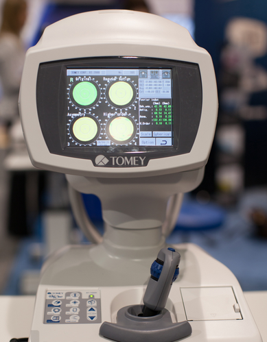
| Content | Tomey RT-7000 Autorefractor Keratometer Topographer
-Three Functions in One Instrument-Easy & Speedy Touch Screen Alignment-6.4 inches color TFT LCD-Dual CCD technology for Refractometry-IOL/ Cataract Mode-New incites for Keratometry KAI, KRI-Auto Alignment / Auto Shot of TopographyCorneal Topography– Auto Alignment / Auto ShotAny one can easily capture measurement with Auto Alignment and Auto Shot. The Measurement variation is significantly reduced no matter what the skill level is of the operator.CL Fitting Simulation-A pseudo-fluorescent pattern of the contact lens selected from the built-in data base can be confirmed and the fitting can be stimulated before the patient wears the trial lens.Corneal Eccentricity Index-The index of CEI (Corneal Eccentricity Index) indicates the ratio of flattering in percentage from the central cornea to the periphery.Three Function in One Instrument– Refractometer Keratometer and Topographer only in 1 unit senior citizens and children can also get the inspection with out any trouble because they don’t have to move the seat for three measurements. Switch from Ref-Kerato Mode to Corneal Topography Mode with only ONE TOUCH. The light come appears with only one button from the measuring head and the Ref-Keratometer is transformed to the topographer.Refractometry– Dual CCD technology for RefractometryTwo CCD cameras are used to capture images for observation and for measurement while providing highly accurate measurement data.Keratometory– New Indices for KeratometryThe Corneal Irregular astigmatism display function is installed to expand the possibility of Keratometer. This is the new function to measure the level of corneal irregular astigmatism which was difficult in the past.SPECIFICATIONSMeasurmentRangesRefraction MeasuremntSphere -25.00D to ±22.00DIVD=12.0mmCylinder 0D to ± 10.00DIVD=12.0mmAxis 0 to 180Corneal Curvature Measurement 5.00mm to 11.00mmMeasurement Zone 3mm R:8.00mmCorneal MappingDisplay Range 9 to 100 D-Normal Mode1.0 to 8.0mm (R:8.00mm)-Special Mode0.9 to 7.0 mm (R:8.00mm)Measurement TimeREF 0.2 SecondsKRT 0.1 SecondsMinimum Pupil Diameter 2.2mmMinimum Measurment Step Refraction MeasurementSphere 0.01DCylinder 0.01DAxis 1Corneal Curvature Measurement 0.01mmPD (Pupil Distance) 50mm to 86mmRecording Built-In Thermal PrinterVertex Distance 0mm, 12.0mm, 13.5mm, 14.0mm, 15.5mm, 16.0mmExterior Output (AN port / 4USB ports)Dimensions Tomey RT-7000 Autorefractor Keratometer TopographerHigh 466mmm or 18 InchesWidth 307mm or 12 InchesDepth 490mm or 19 InchesWeight Approx 20,000Kg / 44.4 PoundsDisplay 6.4 Inches color liquid crystalPower SupplyVoltage AC 100 to 240VFrequency 50/60 HzConsumption Power 120VA to 150VATemperature +10C to + 40CDisplay 30% to 75% | Carl Zeiss Cirrus HD-OCT 5000 Smart Advanced Topography HDAdvancing Smart OCTfor Anterior Segment, Glaucoma and Retina- Anterior Segment Premier Module
- PanoMap Wide-Field Display
- Smart HD Scans
- En face report
Visualization at the speed of CIRRUSAnalyzing a single pathology from multiple views provides comprehensive insight and analysis of the clinical situation. How this helps you: - Spot small areas of pathology. Tightly spaced B-scans, (either 30 or 47 μm apart), in the cube ensure that small areas of pathology are imaged. For reference, a human hair is about 40-120 μm in diameter.
- Visualize the fovea. Scans that are spaced further apart than in the CIRRUS cube may miss the central fovea.
- Fuel for analysis. Millions of data points from the cube are fed into the Zeiss proprietary algorithms for accurate segmentation, reproducible measurements and registration for change analysis.
- Take the pressure off the operator. As long as the scan is placed in the vicinity of the fovea or optic nerve, the software automatically centers the measurements after the capture.
- See the tissue from different perspectives. View the cube data from all angles, with 3D rendering, OCT fundus images and Advanced Visualization™.
- Future ready. Previously captured CIRRUS cubes can be analyzed using new analyses.
Tracking at the speed of CIRRUSFastTrac™ reduces eye motion artifacts without sacrificing patient throughput with a proprietary scan acquisition strategy, high speed 20 Hz LSO camera, and single-pass alignment scanning
With FastTrac, scan at the highest resolution at the same location at each visit. Assessment at the speed of CIRRUSMeasurement centering with FoveaFinder and AutoCenterOn the macula, the unique FoveaFinder technology ensures the ETDRS and ganglion cell plus inner plexiform layer measurement frameworks are centered on the fovea.AutoCenter™ function automatically centers the 3.4 mm diameter peripapillary RNFL calculation circle around the disc for precise placement and repeatable registration. The placement of the circle is not operator dependent. Accuracy, registration and reproducibility are assured. Manage at the speed of CIRRUS Carl Zeiss Cirrus HD-OCT 5000 Smart Advanced Topography HDGuided Progression Analysis (GPA™) for Assessing RNFL and Optic Disc ChangeGPA™ compares RNFL thickness and optic disc measurements over time and determines if statistically significant change has occurred.Focal Progression Analysis – Up to 6 progression maps are compared to two baselines. Areas of statistically significant change are color-coded yellow when first noted and then red when the change is sustained over consecutive visits.
Trend Analysis with Rate of Change – RNFL thickness values are plotted over time. Trends are calculated and confidence intervals are shaded when rate of change is statistically significant. | Tomey RC-800 Auto Refractor Keratometer
Refractometry: Simple handling of the fine adjustment and measurement by yourself. Fogging is applied automatically for each measurement for more accurate results.Keratometry: Short consecutive measurements and time provide reliable data. Measurements at φ3.0mm (R = 8.0mm) give you enough information for contact lens fitting.Diameter Measurement of Cornea and Pupil: Easy measurement by moving the two cursors on the display to the boundaries of Cornea or Pupil.Language Selection: RC-800 has a Multi-language function. You can select your required language from “English”, “Japanese”, “German”, “Chinese”, “Spanish”, “Spanish Latin America”, “Italian”, “Portuguese”, “French” and “Russian”.Features:• Highly Accurate
• Simple to Use
• Diameter Measurement of Cornea and Pupil
• High Speed Printer
• Auto Shot
• Color Touch Screen
• USB connection
• Large Patient Database
• Easy Database Operation
• Multi-Language Operation
• Fourier Refractive Analysis
• Quick Data Reference
• Built-in LCD Alignment
• Keratoconus Screening & Other ApplicationsMeasurement Ranges:Refraction:• Sphere: -25.00D to +22.00D (VD=12.0mm)
• Cylinder: 0D to ±10.00D (VD=12.0mm)
• Axis: 0° to 180°
• Minimum Pupil Dia: Ø2.0mm
• Measurement time: 0.2 second/single eye (data taking time)Keratometry:• Range: 5.00mm to 11.00mm
• Corneal Refraction: 30.68D to 67.50D (n=1.3375)
• Corneal Astigmatism: 0D to 10D (n=1.3375)
• Axis: 0° to 180°
• Measurement area: Ø3mm (at 8.00mm corneal curvature)
• Measurement time: 0.1 second/single eye (data taking time)Other features:Tomey RC-800 Auto Refractor Keratometer• PD Measurement range 50 to 86mm
• Corneal diameter measurement
• Measurement range: 10.0 to 14.0mm
• Display unit: 0.1mm
• Pupil diameter measurement
• Measurement range: 1.0 to 10.0 mm
• Display unit: 0.1mm
• Built-in Printer Thermal printer
• Data output type RS232C / USB-D
• Display 5.7 inch color liquid crystal display
• Dimensions and weight 297(W)x 500(D) x 448(H)mm
• Approx. 17kg
• Power Requirements AC 100V to 240V, 50 / 60 Hz, 80VA to 100VAThe Auto “Refkeratometer” RC-800 is the latest in economical, user-friendly, highly accurate auto “refkeratometry”. The RC-800 provides simple, easy measurement by the operator at an economical cost, without compromising accuracy or reliability. | Essilor Neksia Lens Edging SystemWork More Easily- A touch of efficiency – The 8.4-inch color touch-screen display makes navigation a breeze. And you can select the icon-based functions quickly and easily with just the touch of a finger.
- Fast-access settings – With all job settings located conveniently on the centering part of the system, you can define your finishing settings quickly and launch edging jobs in just a few clicks. If necessary, you can still adjust settings after the feeler measurement.
- A user-friendly 1,000-position job database – Save precious time by easily accessing or storing complete jobs in the database via the touch screen or optional barcode reader.
Experience Workflow Efficiency- Fast, easy and extended frame tracking – Equipped with auto frame-type selection, simple clamping and rapid tracing cycles, Neksia helps you trace any type of frame effortlessly, including children’s frames, small B-size and large A-size frames
- Automatic drilling-hole recognition – Achieve even greater productivity thanks to optional features dedicated to automatic detection of drilling holes and flat bevel shapes.
- Convenient centering and blocking – Neksia arms you with valuable features that make centering and blocking straight-forward and precise.
- Handle lenses easily with the wide-open centering area
- Increase precision with intelligent targeting and parallax-free working
- Benefit from the intuitive on-screen interface
Achieve High Performance Essilor Neksia Lens Edging System- Improve axis accuracy – By combining new edging cycle algorithms and cutting-force regulation, you are guaranteed optimal results whatever the edging situation.
- Optimized edging speed based on the lens material, shape and lens thickness guarantees fast cycle times, lens positioning and alignment.
- The New Edging Assisted System cycle avoids axis deviation on new- generation hydrophobic lenses, rectangular shapes or fragile lenses.
- Best automatic cycles – For 90% of your jobs, all it takes are a few clicks to achieve a perfectly calculated bevel, groove or drilling cycle. The lens is well positioned for an aesthetic and perfectly fitted result, no matter the material or frame type.
- Consistent intelligent chamfering – With Neksia’s patented flexible chamfering wheel, the pressure applied to the wheel adapts seamlessly to the lens edge depending on the shape and curve of the lens.
| Nidek CEM-530 Specular Microscope
The analysis results with graphic and color-coded cell images helps the clinician to rapidly and effectively evaluate the endothelial cell layer.In addition to conventional central and peripheral specular microscopy, the CEM-530 includes a unique function to capture paracentral images.- Paracentral specular microscopy
- Faster measurements and two-second auto analysis
- Comprehensive analysis
- Advanced manual analysis functions
- Easy operation
- Additional features with CEM Viewer for NAVIS-EX
Combination of auto and manual analysesAll three manual analysis methods can be performed on the same image and also on auto-analyzed images. The versatility of combining automated and manual analyses allows analysis of the range of pathology in a comprehensive practice. Features- Multi area specular microscopy
- Enhanced usability and quick analysis
- Advanced manual analysis functions
- Combination of auto and manual analyses
- Additional features with CEM Viewer for NAVIS-EX
Multi area specular microscopyIn addition to conventional central and peripheral specular microscopy, the CEM-530 includes a NIDEK original function that captures paracentral images. The combination of central, paracentral, and peripheral imaging provides a broader, overall view that can be used for detailed morphological and quantitative evaluation of the endothelial layer and individual cells.
Enhanced usability and quick analysis
The 3D auto tracking and auto shot functions result in a user friendly and patient friendly experience.
Data analysis within 2 seconds allows efficient patient flow.
  Detail analysis
Advanced manual analysis functions
Center point
Select the approximate center of a cell. The cells are detected based on the surrounding points. This method is effective for areas where groups of cells are clumped together.
Corner point
Trace the outlines of the cells to be analyzed by selecting the corners of each cell. This method is suitable for detailed identification of the size and dimension of isolated cells.
Pattern select
Select a hexagonal reference pattern that is similar to the cell size and drag it onto the cell to be analyzed. This method is effective for rough identification of the size and dimension of the cells.
Combination of auto and manual analyses All three manual analysis methods can be performed on the same image and on auto-analyzed images. The versatility of combining automated and manual analysis on the same image allows for better clinical interpretation of the diverse range of pathology in a comprehensive practice.
Additional features with CEM Viewer for NAVIS-EX* (optional)
CEM Viewer is software used for viewing and working with CEM-530 data via NAVIS-EX. This function enhances the capability of the CEM-530 with additional features and increases the efficiency of any clinic.
*NAVIS-EX is optional software and is required for use of the CEM Viewer.
Data management and endothelial cell count
Unlimited NAVIS-EX database is available for review on the CEM Viewer. The basic functions of the CEM-530 such as endothelial cell count are available on the CEM Viewer.
Progression follow-up and comparison
Multiple examination data sets are displayed in chronological order for follow-up. Additionally, two data sets are displayed for comparison. Endothelial changes can be monitored over time with this function.
Paracentral display with peripheralThe images and analyses of the paracentral and peripheral areas are displayed providing a comprehensive image of endothelial cells. | Siemens Acuson Sequoia 512 LCD Ultrasound Machine18″ Flat panel LCD Monitor–Progressive Media & Display II Package, Cardiac/OB/Vascular Calc. Package, Signature II Option, AUX, CPS Cardiology, CPS General Cadene Trigg Burst, Calc Data to MO, Card Cadence II Contrast Quant, Cornoary Flow reserve, Dicome Store/Print, DTI (Doppler Tissue Imaging), High HR ECG, MIS/MIF, Modality Worklist Native TEQ, Native TEQ, NTHI (Native Tissue Harmonics), Spectral TEQ, TEQ (Tissue Equalization), VVIFeatures- Shared Service
- Native Patient Specific Imaging
- Four Sight TEE view
- Axius quantitative strain rate imaging
- Cadence contrast pulse sequencing technology
- Native Tissue Harmonic Imaging
- Native TEQ ultrasound technology
- Color doppler, Color angio and b-color
- PW/CW Doppler
- M-Mode
- Folding Flat Panel Display
- DICOM capabilities
Applications- Abdominal
- Breast
- OB/GYN
- Vascular
- Pediatrics
- Cardiology
- Musculoskeletal
- Neonatal
Acuson Sequoia 512 Features:- Imaging: Gray-scale 2-D, M-mode, SST Color Doppler, Solo Spectral Doppler, 3-D Surface Rendering, Native Tissue Harmonic Imaging, Auto Doppler, M-Mode, PW Doppler, Color Doppler, Color Power Doppler
- 19" color CRT monitor
- Native tissue harmonic imaging (NTHI)
- DIMAQ: image management
- Advanced vascular analysis
- Advanced spatial compounding
- Native patient specific imaging
- Mitral valve assessment
- CPS contrast pulse sequencing technology
- Clarifying vascular enhancement technology
- High resolution color flow
- Vascular, and General Imaging calculation packages
- Coherent beamformer technology/spatial compounding
- Space/time resolution control
- Cine and zoom/RES
Compatible Ultrasound Probes and Transducers:- Acuson V5Ms TEE Probe
- Acuson 4C1 Curved Array Probe
- Acuson 6C2 Curved Array Probe
- Acuson 8C4 Curved Array Probe
- Acuson 4V1 Vector Array Probe
- Acuson EV-8C4 Endocavitary Probe
- Acuson EC-10C5 Endocavitary Probe
- Acuson 6L3 Linear Array Probe
- Acuson 8L5T Intraoperative Probe
- Acuson 8L5 Linear Array Probe
- Acuson 9L4 Linear Array Probe
- Acuson 15L8 Linear Array Probe
- Acuson 17L5 Linear Array Probe
The Sequoia 512 with LCD ultrasound machine uniquely minimizes automatic image optimization time by up to 80% to make way for an easier and speedier ultrasound test time as well as allow for a more confident diagnosis each time. As a patient-specific imaging ultrasound, it has a unique matched response capacity for accurate image quality, sharp clinical specificity, and optimized diagnostic outcomes that makes infinitesimal pathologies and other anatomical detail in the most difficult-to-image patients becomes more vivid and visible. |




Reviews
There are no reviews yet.