| Description | Clinical Applications
• Abdominal
• Gynecology
• Small-Parts
• Vascular
• Pediatrics
• Cardiology
• Urology
• Musculoskeletal (MSK)
• Breast | The GE Vivid q portable ultrasound machine is a more powerful version of the Vivid i that adds further quantitative features including ICE, Auto EF, AFI, TVI,TT, TSI, Blood Flow and Blood Flow imaging. The Vivid q is the pinnacle of GE’s portable cardiovascular ultrasounds. | 15″ LCD monitor, 2 Hot-swappable Probe ports, Keyboard with programmable keys, HPRF (High Pulse Repetition Frequency), MBP (Multi-beam Processing), Trapezoidal Imaging, Panoramic Imaging, Advanced THI (Tissue Harmonic Imaging), TDI (Tissue Doppler Imaging) ,MicroScan (Noise & Artefact Reduction), Multi Parameter Compound Imaging, M Tuning (One Touch Image Optimization), Steerable M mode, uScan, Bi-Plane Transrectal Imaging, Multi-Plane Adult and Paediatric TEE/TOE, Endocavitary Imaging with Extended Field-of-View, Stress Echo, anatomic M mode, PDF report, Automatic flow volume analysis, IMT, HQNF (High Q Noise Filter), 2 USB ports, Video in/out ports, Li-ion battery. | Acuson sequoia 512 ultrasound click images to enlarge item description acuson sequoia 512 ultrasoundsold as picturedincludes: 2 probes, 4c1 convex & 6l4 linearsony up 895 md printerunit will be cleaned, function checked and certified for electrical safety. Checked, cleaned, certified for electrical safetyguaranteed working when arrives on the guarantee working we need to be notified in 48 hrs. And the seals must be intact. | The Samsung Accuvix XG was the top of the line system from Samsung prior to the release of the newer. The XG was meant as a direct replacement to the popular Accuvix XQ as a shared service system capable of both 4D OBGYN images as well as full cardiac scans. | M-Turbo is a compact ultrasound imaging system from SonoSite, featuring the image quality and functionality needed to meet your diverse needs. The standard M-Turbo is also still available exclusively from BCF. |
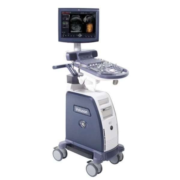
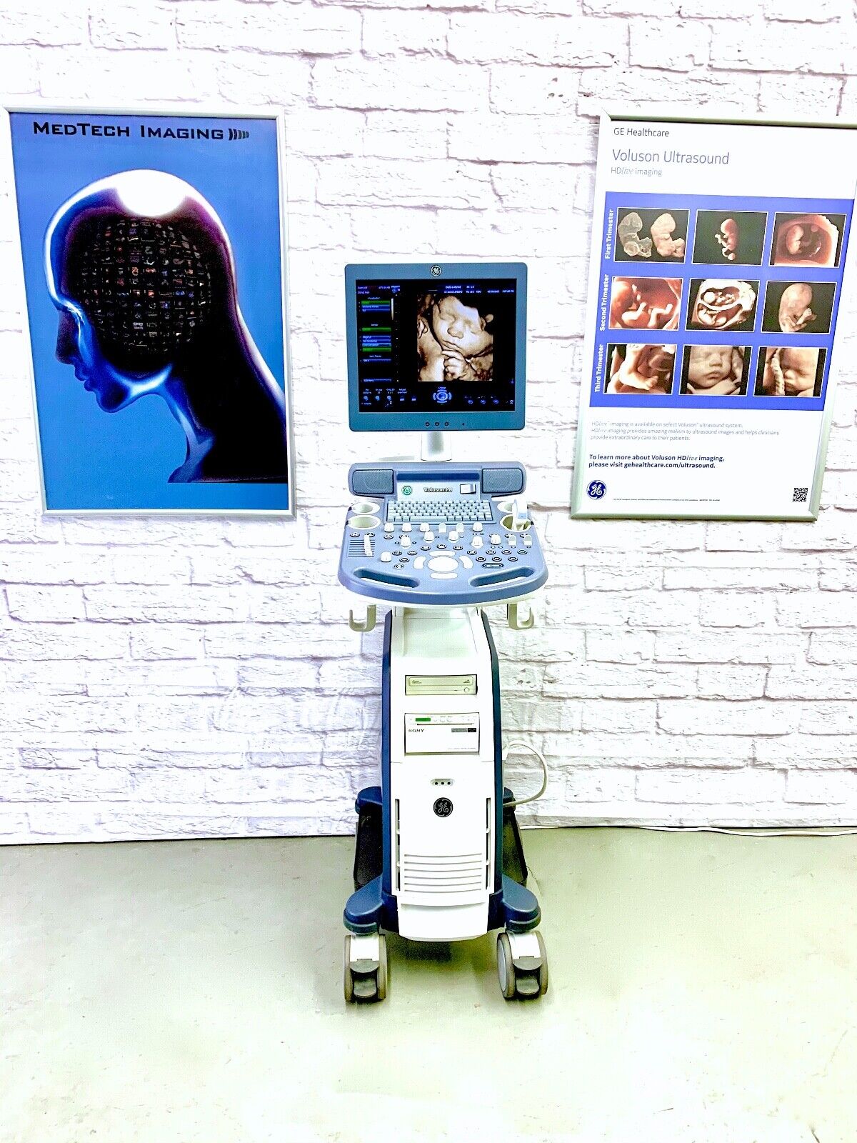
| Content | GE VOLUSON P8 ULTRASOUND
The GE Voluson P8 Ultrasound is an entry-level ultrasound system for women’s healthcare applications. Its automated functions allow physicians in busy practices to maintain high-quality imaging while reducing the time needed to conduct, analyze, and report on exams. The consensus of the GE Voluson P8 is an affordable ultrasound for any medical professional who wants to experience the power of a GE Voluson system for clinical applications. Even though the GE Volsuon P8 system is at a lower price point, it supports in-depth imaging features, such as 3D/4D and HDlive.Dimensions & Weight of the GE Voluson P8Width: 16.5 in (420 mm)
Depth: 626.4 in (70 mm)
Height: 59 in (1495 mm)
Weight: 143 lbs (65 kg)GE Voluson P8 SpecificationsSystem processing channel: Counts up to 161,616 by configuration
Minimum depth of field: 0 – 1 cm (Zoom, probe dependent)
Maximum depth of field: 0 – 30 cm (probe dependent)
Continuous dynamic receive focus/continuous dynamic receive aperture
Adjustable dynamic range: Up to 255 dBGE Voluson P8 Electrical PowerNominal input voltage: 100-240 VAC, frequency 50/60 HzProbes
GE Voluson P8 Probes and Transducers
Convex Probes:
4C-RSLinear Probes:
12L-RSEndocavitary Probes:
E8C-RSPhased Array Probes:
3Sc-RS3D/4D Convex Probes:
RAB2-6-RS3D/4D Endocavitary Probes:
RIC5-9W-RSGE Voluson P8 Features• 17” High Resolution LCD LED Flat Panel Display
• 3 Active Probe Ports
• Integrated HD (500 GB)
• 3 USB Ports for External Peripherals
• 2 USB Ports for On-board Peripherals
• AO(Automatic Optimization)
• CrossXBeam(Spatial Compounding Imaging)
• SRI(Speckle Reduction Imaging)
• B-Steer
• Coded Phased Inversion Harmonic Imaging
• HD-flow
• 3/4D(Real time)
• Virtual Convex
• Patient Information Database
• SonoBiometry
• SonoRender Start
• Raw Data file Standard Voluson P8 Supplies• Aquasonic Ultrasound Gel
• Sono Ultrasound Wipes
• Sony UPP-110HG Thermal Printing Paper
• Sony UPC-21L Color Thermal Printing Package
• Mitsubishi KP95HG Thermal Roll Paper
• Mitsubishi KP65HM-CE High-Density Thermal Paper GE Voluson P8 Ports• 3 USB Ports for External Peripherals
• 2 USB Ports for On-board Peripherals
• RJ45 LAN Port
• 1 HDMI Out Port
• 1 AUDIO Out Port GE Voluson P8 Image Storage Formats & Devices• Storage Formats:– DICOM files (Single- or multi-frame) DCM and DICOM Files with DICOMDIR – Raw Data File (Proprietary format) – Export Data as BMP, TIFF, JPEG, MPEG 4, or MS Video 1
• Storage Devices: USB Memory Stick
• DVD-RW Storage
• HDD Image Storage Options for GE Voluson P8 Ultrasounds• Static 3D Mode
• 3/4D Advanced
• HD Live(3D Only)
• SonoNT/SonoIT
• SonoL&D
• 4D View PC Software
• DICOM 3
• Extended View(XTD)
• Anatomical M-mode
• Report Editor | GE Vivid Q Ultrasound Machine
The powerful GE Vivid Q ultrasound machine is the ultimate lightweight, compact high-performance cardiovascular system for clinicians needing a machine that combines user efficiency and reliable performance. This unique system performs consistently thanks to its TruScan architecture and can support up to 17 probes. The GE Vivid Q is recognized for its premium image quality, advanced image management, and high-performance quantification tools.This advanced GE ultrasound delivers improved patient care and enhanced user productivity with the new M4s-Matrix array technology transducer, which provides greatly enhanced image quality in electronic beamforming, raw data image acquisition and archiving, optimal onboard and post processing technology.This ultrasound system provides high clinical values for quantification and review of stored images, making it one of today’s most sought-after models. Some of its powerful features include compatibility with EchoPac clinical workstation and DICOM networking, speckle tracking strain, integrated stress echo, quantification tools, and reporting capabilities. Also of note is one of the Vivid Q’s best new features, which is its TEE transducer capability.A one-touch button optimization provides premium image quality for workflow efficiency, and maintains AFI- automatic function imaging with complete wallFeatures- Advanced QScan Imaging (incl TSI)
- AdvStress
- ATO
- Auto EF
- Automated Function Imaging
- Blood Flow Imaging
- CardiacApp
- Cardio Lab
- Color Doppler
- CW Doppler
- DICOM Networking
- DICOM: Print
- DICOM: Worklist
- DVD/RW
- ECG
- ECG Cable
- EchoDicom
- EchoPAC
- Evue
- ICE Imaging
- Intima Media Thickness (IMT)
- LVO Contrast
- M Mode
- M4S
- MPEGvue
- PW Doppler
- Small Depth
- TEE
- THI
- Tissue Harmonics Imaging (THI/NTHI)
- Tissue Velocity Imagine & Tissue Tracking
- TSI
- VascAbdContrast
- VascularApp
- VirtualPrinter
- Wide Aperture
GE Vivid Q FeaturesThe GE Vivid Qis a Portable Laptop-style Ultrasound that can be used to help diagnose cardiovascular anatomy. Features and tools based on the previous Vivid 7 and the Vivid E9 have been added to the new lightweight Vivid Q, mainly the M4S-RS transducer that allows for 3D image focusing and enables a higher level of image quality. The Vivid Q ultrasound is versatile and can additionally be used for Cardiac, vascular, Abdominal, Pediatric, Obstetrics, and Emergency medicine. The Vivid Q Ultrasound comes with a 15-inch color LCD monitor that can display a variety of imaging modes including; ICE, Auto EF, AFI, TVI, TT, TSI, and Blood Flow. Additional the Ultrasound system is comparable with 17 different transducers and probes to fit every imaging need. - Auto EF - Automated ejection fraction measurement program designed specifically for the GE Vivid q.
- Tissue Synchronization Imaging (TSI).
- Intracardiac Echo (ICE).
- 16 different compatible probes.
- Coded Octave imaging - enhances image color flow and Doppler imaging.
| SonoScape S8 Portable UltrasoundSystem Description: The SonoScape S8 portable ultrasound is an award winning portable ultrasound system with outstanding image quality in all modalities. SonoScape has designed a fully featured, shared service ultrasound system producing image quality one would expect from a much more expensive console system. Frost & Sullivan “Product Quality Leadership” Award:
“The S8 is versatile in performance,” notes Frost & Sullivan Research Analyst Shriram Shanmugham. “It uses a 15 MHz linear transducer, 512 super high density transducers, and is the only echocardiography system that also offers 4D imaging capability. None of the company’s competitors offers such a combination of 4D and high-end echocardiography technology affording portability.” “The high frame rate and high definition image quality provide users better diagnostic experience than ever” Application:- Shared Service!
- Ob/Gyn
- Anesthesia
- Urology
- Vascular / Vascular Access
- MSK
- Breast Imaging
- Abdominal
- Small Parts
- Cardiac
Features:- 15” LCD Monitor
- 2 Hot Swap Probe Ports
- High Pulse Repetition Frequency [HPRF]
- Multi Beam Processing [MBP]
- Trapezoidal Imaging
- Panoramic
- Imaging
- Advanced Tissue Harmonic Imaging
- M-Tuning [One touch image optimization]
- uScan
- Multi Plane Adult & Pediatric TEE
- Intimae Media Thickness [IMT]
- High Q Nosie Filter [HQNF]
- B-Mode
- M-Mode
- Color Doppler
- Power Doppler
- Spectral Steered Doppler
- Real time Triplex: B, Flow & PW Doppler / B, Flow & Color M
- ECG
- 4D
- Tissue Doppler Imaging
- B-Color
- Anatomic M-Mode
- DICOM 3.0
- 2 USB Ports
- Video In/Out
| | Options:- Mobile Trolley
- DVD/CD-RW
- Printer
- Stress Echo
Transducers:- C344, 5-2MHz/R40mm, Abdominal Convex Probe
- C362, 5-2MHz/R60mm, Abdominal Convex probe
- C542, 7-4MHz/R40mm, Abdominal & Pediatric Probe
- C311, 4-2MHz/R15mm, Micro-convex Cardiac Probe
- C611, 8-4MHz/R11mm, Micro-convex Pediatric Cardiac Probe
- L741, 10-5MHz/46mm, Vascular Small part Linear Probe
- L742, 15-5MHz/38mm, Vascular Small part linear probe
- L743, 15-5MHz/46mm, Vascular Small part linear probe
- L541, 7-4MHz/38mm, Vascular Linear probe
- 10L1, 12-6MHz/36mm, Vascular Small Parts Probe
- 10I2, 15-5MHz/25mm, Linear Surgical Probe
- 6V1, 8-4MHz/R11mm, Endovaginal Probe (135 degree)
- 6V3, 9-5MHz/R10mm, Endovaginal probe (200 degree)
- EC9-5, 9-5MHz/R8mm, micro-convex Endocavity probe (150 degree)
- 2P1, 4-2MHz, Phased Array Cardiac Probe
- 5P1, 7-4MHz, Phased Array Pediatric Cardiac Probe
- MPTEE, 7-4MHz, Multi-plane Phased Array TEE Probe
- MPTEE Mini, 7-4MHz Pediatric Multi-plane Phased Array TEE Probe
- VC6-2, 6-2MHz/R40mm, Curved volumetric probe for Live 3D
Biopsy Guides:- C344, C362, L741, L742, L743, 10LI, L541, 6V1, 6V3, EC9-5, 2P1, 7U2
|
| Siemens / Acuson Sequoia 512 Ultrasound Machine
The ACUSON Sequoia 512 ultrasound is an ultra-premium system that utilizes revolutionary technology, dedicated to the advancement of diagnostic imaging. It represents some of the most advanced technology in ultrasound imaging. The Acuson Sequoia 512 has unparalleled imaging performance, greater image content, enhanced detail, superior contrast resolution & penetration, and excellent color Doppler sensitivity.One of the best selling ultrasounds in the United States, the Sequois 512 provides superior imaging with Native Patient Specific Imaging that detects, measures and adapts (in real time) to each patient’s individual acoustics.Performance:Reduce image optimization time by up to 80% and allow for an easier, faster and more confident diagnosis:- 2D
- SST Color Doppler
- Color Doppler Energy
- Color Doppler Velocity
- Solo Spectral Doppler
- M-mode and Color Doppler M-mode
- MultiHertz Multiple Frequency Imaging
- DELTA Differential Echo Amplification
- Home Base user interface
- One-Touch Image Control with exam and Image Presets
- Digital acquistion, storage, review and transfer of static images, dynamic clips, and cine
- Digital storage on an integrated hard disk
- Digital transfer via MO Disk
- Integrated Compression engine
- Embedded DICOM conformance
- Patient- oriented file structure
- Native Tissue Harmonic Imaging
- Tissue Equalization™ Technology (Optional)
- Signature II™ (Optional)
- Advanced Imaging package (Optional)
- Auto Doppler (Optional)
- Freestyle Compounding (Optional)
- FreeStyle Extended Imaging (Optional)
- 3D surface Rendering (Optional)
- Cadence™ ADI (Optional)
- Cadence CPS for 512 (Optional)
- Axius™ Edge Assisted EF (Optional)
- Doppler Tissue Imaging (Optional)
- DICOM Print And Store (Optional)
Capabilities:Siemens / Acuson Sequoia 512 Ultrasound MachineGeneral Radiology, Abdominal, Intraoperative Monitoring, OB/GYN, Neonatal, Vascular, Cardiac, Small PartsAvailable Transducers:4V1, 4V2, 8V5, 15L8W, 6L3, 8L5, 6C2, 5C2, 4C1, 3V2C, 5V2C,7V3C, EVC8-4V, EC-10C5, V5M TEE, V7B neo TEE, AcuNav Transducer, 2.0 Non Imaging CW Doppler probe.Acuson sequoia 512 ultrasound click images to enlarge item description acuson sequoia 512 ultrasoundsold as picturedincludes: 2 probes, 4c1 convex & 6l4 linearsony up 895 md printerunit will be cleaned, function checked and certified for electrical safety. Checked, cleaned, certified for electrical safetyguaranteed working when arrives on the guarantee working we need to be notified in 48 hrs. And the seals must be intact. | Medison Accuvix XG Ultrasound System
Samsung Accuvix XG Ultrasound Machine2D Image Features
• Dynamic MR™ / Dynamic MR Plus is designed to enrich gray-scale resolution, as it enhances detection
and contrast resolution while also decreasing speckle echoes. This is particularly useful when evaluating
superficial structures, including thyroid, vessels, pelvic and abdominal anatomy.
• SRF (Speckle Reduction Filter™) enhances image quality by reducing or eliminating the appearance of
speckle echoes from ultrasound images. The degree of speckle reduction implemented is user-selectable.
• Wide Dynamic Range determines the number of gray shades utilized to map the gray-scale image.
It enables display of more details of bright areas and dark areas.
• SCI (Spatial Compounding Image) controls the ultrasound beam electronically by steering, and compounds
many scan lines.
• FSI (Full Spectrum Imaging) incorporates the penetration capabilities associated with lower frequencies,
yet maintains the fine pixel uniformity associated with higher frequencies, to deliver consistently high quality
images even in challenging diagnostic areas.3D Image Features Medison Accuvix XG Ultrasound System
• HDVI gives outstanding image quality and naturally clearer contrast, with excellent tissue differentiation,
edge depiction and speckle reduction, allowing consistent diagnoses with great confidence.
• 3D XI™, comprised of a suite of three innovative imaging applications – Multi-Slice View, Oblique View™ and
Volume CT – offers complete and precise control over 3D/4D volume data manipulation for maximum
diagnostic accuracy.
• 3D MXI is an innovative, cutting-edge 3D image processing technology. Comprising a comprehensive suite of
imaging tools – including Multi Volume Slice, Mirror View, Multi-OVIX, and 3D OH – 3D MXI lets you view,
examine and diagnose 3D volume data with supreme ease, speed and accuracy. • Fully adjustable system
The control panel can be adjusted to the user’s preferred height, for a better working environment and reduced
risk of back pain.
• Wide LED touch-screen
The Accuvix XG’s new LED touch-screen makes it easy to organize and operate.
• 19-inch HD LCD Monitor and articulating monitor arm
A 19-inch LCD monitor enables images to be displayed clearly even with a larger monitor, and the articulating
monitor arm enables easy mobility for a more comfortable and convenient working environment.
• Portability
The Accuvix XG is a lightweight system with 4 swivel wheels that allow easy steering, and a locking function. • Customizable measurement menus
Customizable measurement menus allow access to frequently-used functions, and enable a quicker and
more intuitive workflow.
• User keys and user knob
Accuvix XG offers user keys and a user knob that can map frequently-used functions, enabling the function to be
activated quickly and easily.
• Customized Annotation Menu and body marker
Users can preset up to 360 words of annotations, and body markers for each application, that reduce the time
needed for each examination.
• QuickScan
QuickScan maximizes workflow efficiency by automatically optimizing key imaging parameters with just the
push of a button. • HDVI
HD Volume Imaging(HDVI) gives outstanding image quality and naturally clearer contrast, with excellent tissue
differentiation, edge depiction and speckle reduction, allowing consistent diagnoses with great confidence.
It shows clearer images of subtle lesions, using multi slice image as well as C-plane.
• ElastoScan
ElastoScan™ translates stiff & soft tissue information into gray scale image. This technology defines the alteration
of the stiffness in tissue state more accurately, making it easier for the user to utilize the information in the image. | Sonosite M-Turbo Ultrasound Machine
M-Turbo is a compact ultrasound imaging system from SonoSite, featuring the image quality and functionality needed to meet your diverse needs.Ideal for the general practitioner looking to do more advanced cardiac and vascular exams.Highlights of the new Sonosite M-Turbo :- Now available the M-Turbo with probe.
- This special version of the M-Turbo comes with a one year warranty, however the probes still have the standard 5 year Sonosite warranty.
- With the M-Turbo you can still add Colour, Pulse Wave and Continuous Wave Doppler.
- The system is now available with DICOM networking.
- Speak to your local Account Manager now to arrange a demonstration.
The standard M-Turbo is also still available exclusively from BCF.Highlights of the Sonosite M-Turbo:- More processing power than MicroMaxx – the next step up in image quality
- 5 year manufacturer’s warranty
- Mains or battery powered so can be used anywhere, anytime
- Able to upgrade from standard black and white system to high quality Colour/Doppler in future
- Large range of probes for all applications
. - Quick start up in just 12 seconds.
More… Sonosite M-Turbo Ultrasound MachinePortable, tough and easy to use. So tough it was originally designed for the American military and comes with a full manufacturer’s 5 year warranty.Despite its size, the M-Turbo produces outstanding cardiac images. It has very high colour Doppler sensitivity.Fully upgradeable to Colour, Pulsed Wave (PW) and Continuous Wave (CW) Doppler and capability to upgrade to DICOM or Wireless DICOM. You can connect it to a PACS system to allow viewing and secure storage of images.Stores still images and movie clips which can be exported via USB in PC friendly formats (jpeg, mpeg and avi).Optional enhanced needle visualisation software (unique to Sonosite) easily highlights needles within the patient allowing for accurate acquisition of a FNA or Tru-Cut biopsy sample.M-Turbo is a compact ultrasound imaging system from SonoSite, featuring the image quality and functionality needed to meet your diverse needs. The standard M-Turbo is also still available exclusively from BCF. |






Reviews
There are no reviews yet.