| Description | Clinical Applications
• Abdominal
• Gynecology
• Small-Parts
• Vascular
• Pediatrics
• Cardiology
• Urology
• Musculoskeletal (MSK)
• Breast | Portable Color ultrasound System
This is a portable color ultrasound system that displays good image quality for diverse applications. This small and compact system has various diagnostic capabilities. | Solid imaging performance.
The sleek LOGIQ A5 can help meet your general imaging needs. This black and white ultrasound system has:- B-mode imaging capability and features that allow you to add advancements as your clinical needs grow.
- Auto TGC helps ensure a homogeneous picture from the near-to-far field.
- Virtual Convex provides an expanded field of view that enables you to see more anatomy with linear transducers.
- Color flow Doppler upgrade available to provide dynamic assessment of blood flow in the organs, as well as investigation of hypertension and resulting erectile dysfunction.
| The Samsung Medison SonoAce R3 portable ultrasound provides the image quality and features of a premium system at a fraction of the cost. The Samsung Medison SonoAce R3 portable ultrasound is brought to you by Samsung / Medison, a company whose name has been trusted in ultrasound since 1985. The Samsung Medison SonoAce R3 portable ultrasound is intuitive and easy to navigate with sophisticated software to increase workflow. | The more patients you see, the more you need Voluson E8.
The Voluson E8 ultrasound system is designed to keep pace with busy practices that conduct a wide range of Women’s Health exams, from routine scanning to complex assessments.
The imaging excellence of Radiance System Architecture – your diagnostic confidence will be enhanced by the extraordinary image clarity and speed of Radiant System Architecture. | Acuson Cypress cardiovascular system PLUS delivers advanced ergonomics and superior performance in a highly portable package. The Cypress System PLUS is a highly miniaturized, all digital, phased array echocardiography system that provides complete studies and outstanding images – even on patients who are difficult to image. |
| Content | GE VOLUSON P8 ULTRASOUND
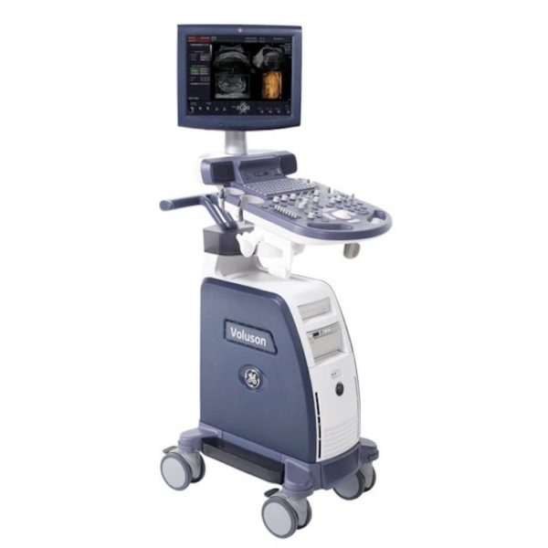
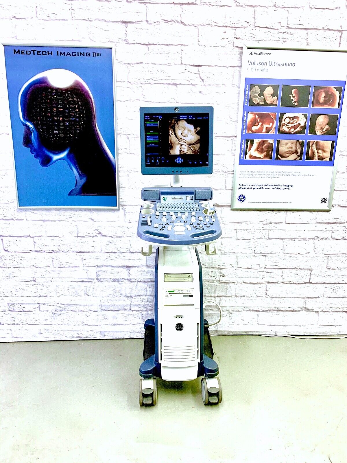
The GE Voluson P8 Ultrasound is an entry-level ultrasound system for women’s healthcare applications. Its automated functions allow physicians in busy practices to maintain high-quality imaging while reducing the time needed to conduct, analyze, and report on exams. The consensus of the GE Voluson P8 is an affordable ultrasound for any medical professional who wants to experience the power of a GE Voluson system for clinical applications. Even though the GE Volsuon P8 system is at a lower price point, it supports in-depth imaging features, such as 3D/4D and HDlive.Dimensions & Weight of the GE Voluson P8Width: 16.5 in (420 mm)
Depth: 626.4 in (70 mm)
Height: 59 in (1495 mm)
Weight: 143 lbs (65 kg)GE Voluson P8 SpecificationsSystem processing channel: Counts up to 161,616 by configuration
Minimum depth of field: 0 – 1 cm (Zoom, probe dependent)
Maximum depth of field: 0 – 30 cm (probe dependent)
Continuous dynamic receive focus/continuous dynamic receive aperture
Adjustable dynamic range: Up to 255 dBGE Voluson P8 Electrical PowerNominal input voltage: 100-240 VAC, frequency 50/60 HzProbes
GE Voluson P8 Probes and Transducers
Convex Probes:
4C-RSLinear Probes:
12L-RSEndocavitary Probes:
E8C-RSPhased Array Probes:
3Sc-RS3D/4D Convex Probes:
RAB2-6-RS3D/4D Endocavitary Probes:
RIC5-9W-RSGE Voluson P8 Features• 17” High Resolution LCD LED Flat Panel Display
• 3 Active Probe Ports
• Integrated HD (500 GB)
• 3 USB Ports for External Peripherals
• 2 USB Ports for On-board Peripherals
• AO(Automatic Optimization)
• CrossXBeam(Spatial Compounding Imaging)
• SRI(Speckle Reduction Imaging)
• B-Steer
• Coded Phased Inversion Harmonic Imaging
• HD-flow
• 3/4D(Real time)
• Virtual Convex
• Patient Information Database
• SonoBiometry
• SonoRender Start
• Raw Data file Standard Voluson P8 Supplies• Aquasonic Ultrasound Gel
• Sono Ultrasound Wipes
• Sony UPP-110HG Thermal Printing Paper
• Sony UPC-21L Color Thermal Printing Package
• Mitsubishi KP95HG Thermal Roll Paper
• Mitsubishi KP65HM-CE High-Density Thermal Paper GE Voluson P8 Ports• 3 USB Ports for External Peripherals
• 2 USB Ports for On-board Peripherals
• RJ45 LAN Port
• 1 HDMI Out Port
• 1 AUDIO Out Port GE Voluson P8 Image Storage Formats & Devices• Storage Formats:– DICOM files (Single- or multi-frame) DCM and DICOM Files with DICOMDIR – Raw Data File (Proprietary format) – Export Data as BMP, TIFF, JPEG, MPEG 4, or MS Video 1
• Storage Devices: USB Memory Stick
• DVD-RW Storage
• HDD Image Storage Options for GE Voluson P8 Ultrasounds• Static 3D Mode
• 3/4D Advanced
• HD Live(3D Only)
• SonoNT/SonoIT
• SonoL&D
• 4D View PC Software
• DICOM 3
• Extended View(XTD)
• Anatomical M-mode
• Report Editor | Samsung Medison SonoAce R3 Ultrasound MachineSonoAce R3 is a portable color ultrasound system with comprehensive diagnostic capabilities. Full Spectrum Imaging (FSI) combines with Speckle Reduction Filter (SRF) to deliver superior quality images, even in the challenging circumstances. The compact and user-friendly design features of the SonoAce R3 ensure portability and manoeuvrability in a lightweight unit. Sporting a 15-inch LCD monitor with Full-Featured Workflow, the SonoAceR3 offers your organisation flexibility, without compromising on precision and efficiency.Slim and Compact DesignThe compact design of SonoAce R3 offers Various-Featured Workflow with a thumbnail menu and shortcut, together with functional key groupings for increased efficiency. The user-friendly design combines a wide-view 15-inch LCD monitor with backlit control keys, customisable measurement package and body markers. The SonoAce R3 features 2 probe ports, 3 USB ports, rapid boot up capability and DICOM 3 compatible image filing. This slim, lightweight unit also has a convenient carrying handle for portability and flexibility.Full Spectrum Imaging (FSI) produces the penetration capabilities associated with the lower frequencies, while still maintaining the fine pixel uniformity associated with the higher frequencies. This combination consistently delivers superior quality images, even in the challenging diagnostic circumstances. The resulting clarity delivers improvements in accuracy and efficiency for greater confidence in your diagnostic procedures. Harmonic Imaging is a technique that records higher frequency information, resulting in greater resolution and fewer artifacts.Synthetic Aperture
The physical radio channel may present some limits to imaging which can be overcome by using Synthetic Aperture Control software. This method uses information from diverse sources to form a more better picture of the area under examination. It creates one single scan line by performing the TX and RX twice to create enhanced penetration and resolution. Synthetic Aperture Control technology delivers a comprehensive picture and to aid accurate diagnosis.QuickScan
QuickScan has been designed to increase workflow efficiency by automatically optimising key imaging parameters at the touch of a button. The resulting increase in efficiency allows your organisation to offer patients an improved service with faster and more accurate diagnoses. This reliable and safe system helps your clinic or hospital to deliver a superior patient experience.Features Samsung Medison SonoAce R3 Ultrasound Machine
Full Spectrum ImagingTM
Quick ScanTM
Color and Power Doppler
Harmonic imaging
Freehand 3D
3D Multi Planar Imaging
Sonoview Image Management
PW Doppler
15″ LCD monitor
Two probe ports
Lightweight | GE Logiq A5 Ultrasound MachineThe lightweight, portable LOGIQ* A5 ultrasound system offers capabilities to help meet your imaging needs — particularly for urological applications.Affordable ultrasound.The LOGIQ A5 puts imaging capabilities at your fingertips in a portable, lightweight design. This system can go from your office to the surgery suite and helps meet a range of general imaging needs. Well-suited for urology, tools include soft tissue image analysis of the kidneys, bladder, penis, testicles, and prostate.Solid imaging performance.
The sleek LOGIQ A5 can help meet your general imaging needs. This black and white ultrasound system has:- B-mode imaging capability and features that allow you to add advancements as your clinical needs grow.
- Auto TGC helps ensure a homogeneous picture from the near-to-far field.
- Virtual Convex provides an expanded field of view that enables you to see more anatomy with linear transducers.
- Color flow Doppler upgrade available to provide dynamic assessment of blood flow in the organs, as well as investigation of hypertension and resulting erectile dysfunction.
Perform imaging with ease.
The LOGIQ A5 has user-friendly features with precision handling.- Automatic Optimization (AO), helps provide improved image quality at the touch of a button for B-mode, spectral and color Doppler imaging.
- Full-size keyboard for optimal ease of use.
- 15-inch adjustable LCD monitor allows a clear view in any position.
- Lightweight package easily travels with you.
Compatible Transducers:- 10L Linear Array
- 11L Wide Band Linear
- 12L Linear Array
- 3.5C Convex
- 3.5CS Convex (2-5 MHz)
- 3CRF Convex
- 3S Phased Array
- 3Sp Wide Band Phased Array Sector
- 4C Convex Array
- 4D3CL Real time 4D Convex
- 4D8C Convex Volume
- 4DE7C Real time 4D Endocavity
- 5CS Convex (2-5 MHz)
- 5S Cardiac Sector
- 5Sp Wide Band Phased Array Sector
- 7S Phased Array
- 8C Convex Array
- 8L Linear (3-7 MHz)
- 9L Wide Band Linear
- BE9C Transrectal
- BE9CS Micro convex Bi-plan
- E8C (4-10 MHz) Wide Band Microconvex
- E8CS Micro-convex (4-11 MHz)
- ERB Brachytherapy Transrectal
- i12L Linear Array
- i739 Intraoperative
- P2D CWD Non-Imaging
- P6D CWD Non-Imaging
- T739 Intra-operative
| Samsung Medison SonoAce R3 Ultrasound Machine
SonoAce R3 is a portable color ultrasound system with comprehensive diagnostic capabilities. Full Spectrum Imaging™ (FSI™) combines with Speckle Reduction Filter™ (SRF™) to deliver superior quality images, even in the challenging circumstances. The compact and user-friendly design features of the SonoAce R3 ensure portability and manoeuvrability in a lightweight unit. Sporting a 15-inch LCD monitor with Full-Featured Workflow, the SonoAceR3 offers your organisation flexibility, without compromising on precision and efficiency.Slim and Compact DesignThe compact design of SonoAce R3 offers Various-Featured Workflow with a thumbnail menu and shortcut, together with functional key groupings for increased efficiency. The user-friendly design combines a wide-view 15-inch LCD monitor with backlit control keys, customisable measurement package and body markers. The SonoAce R3 features 2 probe ports, 3 USB ports, rapid boot up capability and DICOM 3 compatible image filing. This slim, lightweight unit also has a convenient carrying handle for portability and flexibility.Samsung Medison SonoAce R3 Ultrasound MachineFull Spectrum Imaging™ (FSI™) produces the penetration capabilities associated with the lower frequencies, while still maintaining the fine pixel uniformity associated with the higher frequencies. This combination consistently delivers superior quality images, even in the challenging diagnostic circumstances. The resulting clarity delivers improvements in accuracy and efficiency for greater confidence in your diagnostic procedures. Harmonic Imaging is a technique that records higher frequency information, resulting in greater resolution and fewer artifacts.Synthetic Aperture
The physical radio channel may present some limits to imaging which can be overcome by using Synthetic Aperture Control software. This method uses information from diverse sources to form a more better picture of the area under examination. It creates one single scan line by performing the TX and RX twice to create enhanced penetration and resolution. Synthetic Aperture Control technology delivers a comprehensive picture and to aid accurate diagnosis.QuickScan
QuickScan has been designed to increase workflow efficiency by automatically optimising key imaging parameters at the touch of a button. The resulting increase in efficiency allows your organisation to offer patients an improved service with faster and more accurate diagnoses. This reliable and safe system helps your clinic or hospital to deliver a superior patient experience.Features
Full Spectrum ImagingTM
Quick ScanTM
Color and Power Doppler
Harmonic imaging
Freehand 3D
3D Multi Planar Imaging
Sonoview Image Management
PW Doppler
15″ LCD monitor
Two probe ports
Lightweight | GE Voluson E8 Ultrasound Machine15’’ LCD monitor [Rev BT06], 10.4” touchscreen, 3 Active Probe Ports, Floating Keyboard: Interactive back-lighting, Beta-View CrossXBeam, Virtual Convex, Extended View, ATO [Automatic Tissue Optimization], Coded Harmonic Imaging, High Resolution Zoom, Inversion Real-time automatic Doppler, OB/GYN; Vasc. Cardiac; Abd; Small-Parts; Urology; Ped; Ortho; Neuro reporting, Multigestational Calculations, HPRF [High Pulse Repetition Frequency], SonoVCAD [Matrix array volume technology], TruScan [On-board archive], VCI [Volume Contrast Imaging], SonoAVCfollicle [Sonography-based Automated Volume Count follicle], SonoVCADheart [Sonography-based Volume Computer Aided Display], TUI [Tomographic Ultrasound Imaging], CE [Coded Excitation], XTD, SRI III [Speckle reduction imaging], CRI [Compound Resolution Imaging], DICOM, Ethernet port, Video Out, 6 USB ports, Integrated HDD (160 GB) and DVD/CD-RW.
3D/4D, Endocrinology, Family Practice, Gastroenterology, Internal Medicine, Nephrology, Neurology, OB-GYN, Orthopedics, Otorhinolaryngology, Pain Management, Urology, Vascular Probes GE Voluson E8 Ultrasound Machine - 11L-D Linear Array
- 3S-D Cardiac Sector
- 4C-D Convex Array
- 9L-D Linear Array
- AB2-7 Convex
- IC5-9-D Intercavity
- M6C-D Convex
- P2D CWD Non-Imaging
- P6D CWD Non-Imaging
- PA6-8-D Cardiac
- RAB2-5-D 3D/ 4D Convex
- RAB4-8-D 3D/4D Convex
- RIC5-9-D 3D/4D Intracavity
- RIC6-12-D 3D/4D Intracavity
- RNA5-9-D 3D/4D Convex
- RRE6-10-D Endocavitary Micro-Convex
- RSP6-16-D 3D/D Linear
- SP10-16-D Linear
- 19″ monitor
- 4D Real Time
- CFM Doppler Mode
- Coded Contrast Imaging
- CrossBeam
- DICOM 4D
- HD flow
- Power Doppler Mode
- SonoVCAD
- STIC (Spacial-Temporal Image Correlation)
- T.U.I (Tomographic Ulrasound Imaging)
- VOCAL II VCI (Volume Contrast Imaging)
- Black and white printer
- Color printer
- DVD-Recorder
As your patient volume increases and your clinical cases become more complex, you will require an ultrasound system that enables you to deliver truly exceptional care – confidently and efficiently – every time. The Voluson E8 ultrasound system is designed to keep pace with busy practices – handling routine to complex women’s health exams with ease and precision. The premium image quality of the Voluson E8 combined with access to advanced capabilities, delivers the level of performance required by you and your patients. | Acuson Cypress PLUS Ultrasound
Acuson Cypress cardiovascular system PLUS delivers advanced ergonomics and superior performance in a highly portable package. The Cypress System PLUS is a highly miniaturized, all digital, phased array echocardiography system that provides complete studies and outstanding images – even on patients who are difficult to image.The Cypress PLUS transitions easily from your small private office or hospital echo lab to the operating theatre or your patient’s bedside. It’s ultrasound where you need it.TheCypress PLUS delivers high performance in a complete range of cardiovascular capabilities.Designed with advanced ergonomics in mind, the Cypress system PLUS streamlines your workflow and increases your productivity.If you already own a Cypress system, keeping technologically current is simple with the Cypress Upgrade program.The Cypress system is the smallest, lightest, high performance, phased array echo system ever built. Weighing in at just 19 pounds, with digital storage capacity for over 100 studies and network capabilities to send studies from remote locations, clinicians can perform complete echocardiography studies from almost anywhere. The Cypress system was designed for portability, durability and reliability, and to withstand the rigors of constant transportation. Internal system interconnects are minimized. Key components are shock mounted and the entire system sits inside a robust chassis. From hospital to office or from airplane to mountain village, the Cypress system will perform.Full Range of Capabilities
TheCypress system is a complete, full-featured echo cardiography system that provides complete studies and outstanding image quality even on the most difficult to image patients. It offers a full range of features including:– 2D with Harmonics
– Patient Report with audio capture
– M-Mode
– Embedded DICOM conformance
– Color PW and CW Doppler
– High frame rate color flow imaging
– Stress echo
– Network output
– Vascular imaging
– Comprehensive calculations package
– Contrast agent imaging
– USB peripheral support (PC printer connectivity)Physical Characteristics Acuson Cypress PLUS Ultrasound• Single unit houses ultrasound system with control panel, flat panel display and MO drive
• Weight: 8.6 Kg (19 pounds)
• Height: 35.6 cm (14 in)
• Width: 40.6 cm (16 in)
• Depth: 20.3 cm (8 in) – with keyboard folded up 34.3 cm (13.5 in) – with keyboard down
• Heat dissipation: 490 BTU/hour (nominal)User Interface
• Fold-up control panel for portability
• Direct access to system functions through dedicated keyboard controls and multifunctional soft keys
• Easy-access main controls, including multifunctional trackball, enter and select keys, for minimum hand movement while controlling the system
• Adjustable trackball speed
• Functional grouping of keys, rotational knobs, switches and sliding TGC controls for direct access to critical functions
• Tactile feedback on control activation
• Backlighting of control panel labels
• Mode-dependent soft keys
• User-interface tools
• Pre-defined and user-entered labeling textMonitor
• 10.4 inch/26.4 cm flat panel active matrix display screen
• Protective anti-glare, anti-reflective lensHard Drive
• Internal hard drive for image and data storage
• Internal storage of approximately 50 patient studies (25 – 30 loops per study)Transducer Ports
• One transducer connector (type IEC601BF)Operating/Display Modes
• 2-D, fundamental and harmonic imaging
• Color Doppler Velocity (CDV)
• Color Doppler Energy (CDE)
• M-mode, vertical split screen display with 2-D
• Pulsed Wave (PW) Spectral Doppler
• Continuous Wave (CW) Spectral Doppler
• Duplex Doppler (combined 2-D and Spectral Doppler display)2D Calipers – Generic Measurements and Calculations
• Measurements and calculations in 2D, M-mode, color Doppler, PW and CW spectral Doppler modes
• Basic measurements and report calculations
• Comprehensive patient calculations report
• On-screen “mini-reports”
• Complete or configured patient report
• Cardiac package
• Carotid package
• Arterial package
• Venous package
• Abdominal package
• User configurable Sites
• Selectable menu position
• Four cursor types
• Angle correction
• Distance
• Area
• Slope
• Time
• Velocity
• Velocity/pressure
• Mean velocity/pressure gradient
• Velocity Time Integral
• Pressure halftime
• Continuity Equation
• Ejection Fraction
• Left Ventricular Volumes, including End-Systolic and End-Diastolic volumes
• Cardiac output
• Cardiac index
• Fractional area changeCine Review
• 2D Cine memory up to 15 seconds of history
• Color Doppler cine memory up to 15 seconds of history
• M-mode strip cine along with PW and CW Doppler mode
• strip cine memory determined by sweep speed (selectable by the user) 10, 20, 30 or 40 secondsTransducer Technology
• 3V2c: 2-4 MHz wideband phased array
• 7V3c: 3-7 MHz wideband phased array
• 7L3: 5-7 MHz wideband linear array (Vascular)
• 4C1: 2-4 MHz wideband curved linear array (Abdominal)
• V5Ms: 3-6 MHz wideband phased array (micro-pinless (MP) connector) |






Reviews
There are no reviews yet.