| Description | Clinical Applications
• Abdominal
• Gynecology
• Small-Parts
• Vascular
• Pediatrics
• Cardiology
• Urology
• Musculoskeletal (MSK)
• Breast | 3D-4D ultrasound is a technique used during pregnancy that displays three-dimensional images and movement of a fetus (fetal face and other body parts). Other applications include multiplanar imaging of the pelvic organs. This developing technology is also being used in some diagnostic medical procedures such as biopsies and enhanced visualization of cardiac structures. National Ultrasound offers a variety of 3D-4D Ultrasound machines such as GE Voluson i, GE Voluson e, GE Voluson E8, Voluson 730 Expert, Medison Sonoace 8000, Sonoace 9900 or Toshiba with the Xario and Apilo. Our team of expert sales managers will help you find the right 3D-4D ultrasound machine to meet your diagnostic needs while staying within your budget. | The Expert is the new flagship LOGIQ e. The imaging engine comes from GE leading console systems, delivering crisp images in a compact package. Providing ultrasound imaging with precise anatomical detail at a variety of depths. The system includes innovative features that help simplify proceedures. | Acuson sequoia 512 ultrasound click images to enlarge item description acuson sequoia 512 ultrasoundsold as picturedincludes: 2 probes, 4c1 convex & 6l4 linearsony up 895 md printerunit will be cleaned, function checked and certified for electrical safety. Checked, cleaned, certified for electrical safetyguaranteed working when arrives on the guarantee working we need to be notified in 48 hrs. And the seals must be intact. | The Sequoia system’s advanced technologies are designed to make image acquisition easier, reduce exam time and increase overall efficiency. Along with many other included features, the Sequoia system offers Native, a patient specific imaging which adapts both phase and amplitude information of each unique patient’s properties which delivers a consistently better image across a broad range of patients. | Portable Color ultrasound System
This is a portable color ultrasound system that displays good image quality for diverse applications. This small and compact system has various diagnostic capabilities. |
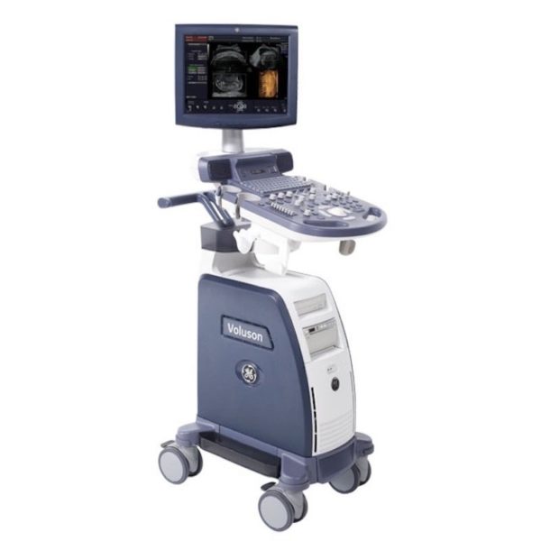
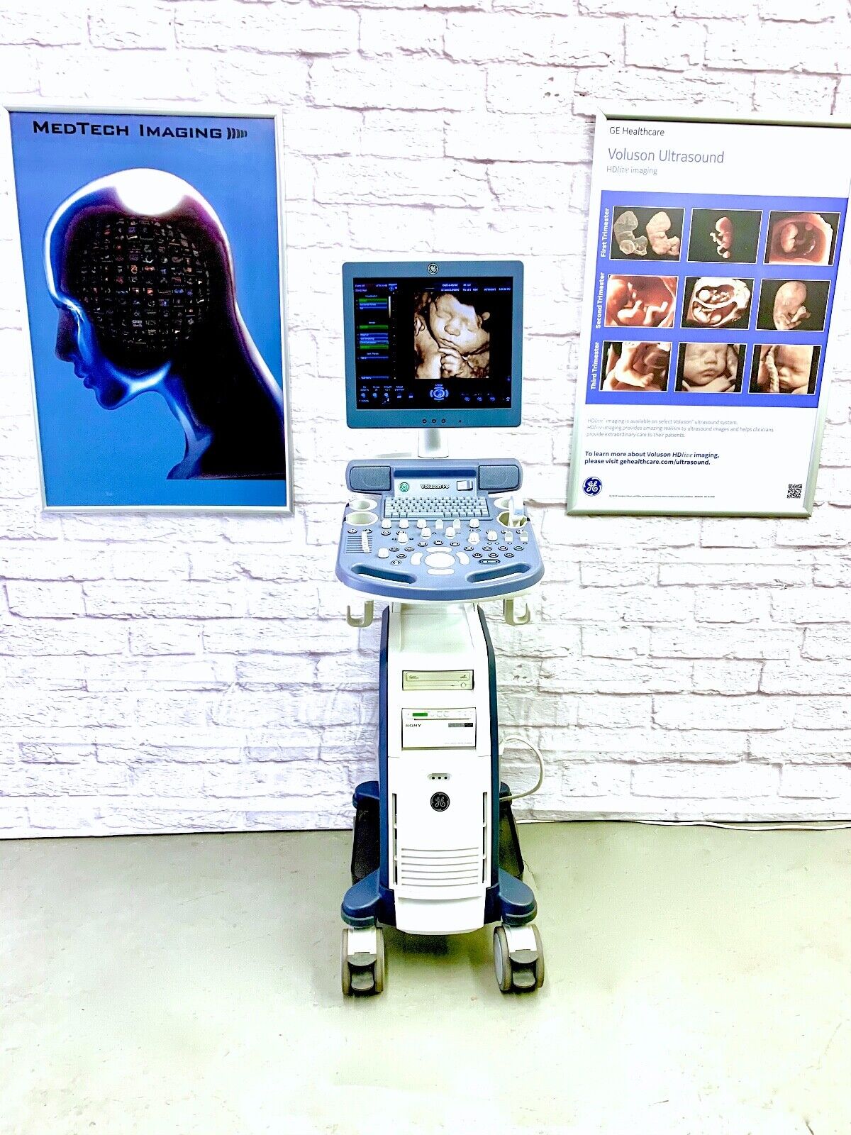
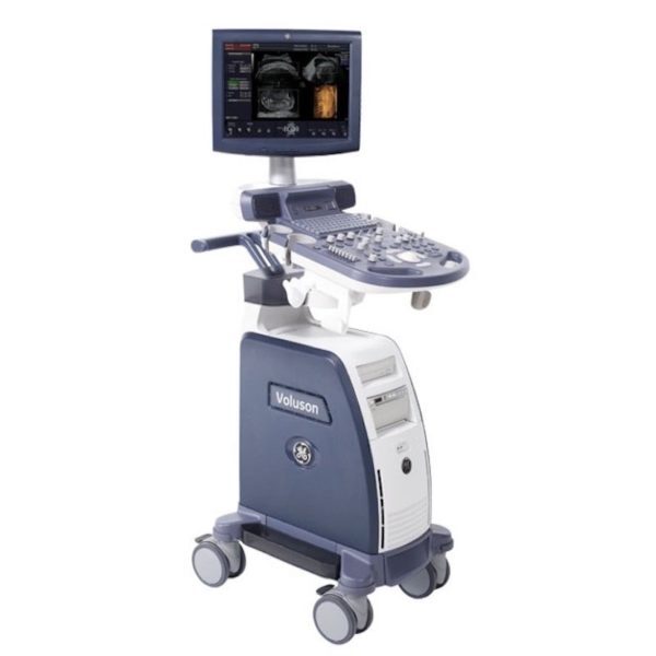
| Content | GE VOLUSON P8 ULTRASOUND
The GE Voluson P8 Ultrasound is an entry-level ultrasound system for women’s healthcare applications. Its automated functions allow physicians in busy practices to maintain high-quality imaging while reducing the time needed to conduct, analyze, and report on exams. The consensus of the GE Voluson P8 is an affordable ultrasound for any medical professional who wants to experience the power of a GE Voluson system for clinical applications. Even though the GE Volsuon P8 system is at a lower price point, it supports in-depth imaging features, such as 3D/4D and HDlive.Dimensions & Weight of the GE Voluson P8Width: 16.5 in (420 mm)
Depth: 626.4 in (70 mm)
Height: 59 in (1495 mm)
Weight: 143 lbs (65 kg)GE Voluson P8 SpecificationsSystem processing channel: Counts up to 161,616 by configuration
Minimum depth of field: 0 – 1 cm (Zoom, probe dependent)
Maximum depth of field: 0 – 30 cm (probe dependent)
Continuous dynamic receive focus/continuous dynamic receive aperture
Adjustable dynamic range: Up to 255 dBGE Voluson P8 Electrical PowerNominal input voltage: 100-240 VAC, frequency 50/60 HzProbes
GE Voluson P8 Probes and Transducers
Convex Probes:
4C-RSLinear Probes:
12L-RSEndocavitary Probes:
E8C-RSPhased Array Probes:
3Sc-RS3D/4D Convex Probes:
RAB2-6-RS3D/4D Endocavitary Probes:
RIC5-9W-RSGE Voluson P8 Features• 17” High Resolution LCD LED Flat Panel Display
• 3 Active Probe Ports
• Integrated HD (500 GB)
• 3 USB Ports for External Peripherals
• 2 USB Ports for On-board Peripherals
• AO(Automatic Optimization)
• CrossXBeam(Spatial Compounding Imaging)
• SRI(Speckle Reduction Imaging)
• B-Steer
• Coded Phased Inversion Harmonic Imaging
• HD-flow
• 3/4D(Real time)
• Virtual Convex
• Patient Information Database
• SonoBiometry
• SonoRender Start
• Raw Data file Standard Voluson P8 Supplies• Aquasonic Ultrasound Gel
• Sono Ultrasound Wipes
• Sony UPP-110HG Thermal Printing Paper
• Sony UPC-21L Color Thermal Printing Package
• Mitsubishi KP95HG Thermal Roll Paper
• Mitsubishi KP65HM-CE High-Density Thermal Paper GE Voluson P8 Ports• 3 USB Ports for External Peripherals
• 2 USB Ports for On-board Peripherals
• RJ45 LAN Port
• 1 HDMI Out Port
• 1 AUDIO Out Port GE Voluson P8 Image Storage Formats & Devices• Storage Formats:– DICOM files (Single- or multi-frame) DCM and DICOM Files with DICOMDIR – Raw Data File (Proprietary format) – Export Data as BMP, TIFF, JPEG, MPEG 4, or MS Video 1
• Storage Devices: USB Memory Stick
• DVD-RW Storage
• HDD Image Storage Options for GE Voluson P8 Ultrasounds• Static 3D Mode
• 3/4D Advanced
• HD Live(3D Only)
• SonoNT/SonoIT
• SonoL&D
• 4D View PC Software
• DICOM 3
• Extended View(XTD)
• Anatomical M-mode
• Report Editor | GE Voluson i Portable 3D/4D UltrasoundFeatures* Lightweight and portable
* Battery operation
* Real-Time 4D
* 3D Multiplanar Display
* 3D Power Doppler
* Automatic Optimization (AO)
* Speckle Reduction Imaging (SRI) and CrossXBeam deliver advanced computational power, allowing for simultaneous processing of CrossXBeam CRI and SRI together – enabling added speckle reduction, contrast resolution and image clarity.
* HD-Flow uses a bi-directional Doppler feature to achieve a more sensitive vascular study and reduce overwriting.
* Volume Contrast Imaging (VCI) allows for image quality in either a single plane or all three planes of a volume acquisition
* Spatio-Temporal Image Correlation (STIC) captures a full virtual fetal heart cycle in real time, and the volume can be saved for offline analysis.
* Tomographic Ultrasound Imaging (TUI) makes analysis and documentation of dynamic studies easier with a simultaneous view of multiple parallel slices of a volume data set.
* Virtual Organ Computer-aided AnaLysis (VOCAL) is a semi-automated volume measurement tool that utilizes computer technology to provide accurate volume calculations.Comes with:GE Voluson i Portable 3D/4D Ultrasound* 1x Voluson i Portable 3D/4D Ultrasound
* 1x RAB2-5RS (2-5MHz) 3/4D Convex Abdominal Probe
* 1x E8C-RS (4-10MHz) Wideband Microconvex Endocavity (Vaginal) Probe
* 1x RIC5-9-RS (5-9MHz) 2D/3D RealTime 4D Endocavitary ProbeThe GE Voluson i ultrasound machine is primarily used for OB-GYN applications, but is versatile and powerful enough to be used for a variety of other applications as well. The Voluson i ultrasound machine carries the power of the GE Voluson line with access to premium 2D and 4D imaging in a portable form factor. The GE Voluson i provides the extraordinary image quality needed to confidently scan for women’s health. The GE Voluson i portable ultrasound machine offers features such as speckle reduction imaging and CrossXBeamCRI, which both lend to the incredible image clarity and quality of this system. The Voluson I portable ultrasound system is known for ease of use, allowing the operator to scan patients faster and more efficiently with better accuracy. | GE Logiq e r7 Portable Ultrasound Machine
The LOGIQ e R7 provides even more impressive veterinary abdominal and cardiac images than the BT12 LOGIQe, combining the high performance of a console system with the portability of a laptop.Highlights of the GE LOGIQ e R7:- Simple. Fast. Precise
- Colour Flow Mode, Power Doppler Imaging (PDI), Pulse Wave Doppler (PWD), Continuous Wave Doppler
- 15” high resolution colour LCD screen
- Integrated solid state hard drive
- Automatic optimisation
- CrossXBeam
- Speckle Reduction Imaging (SRI-HD)
- Virtual Convex / Virtual Apex
- Fine Angle Steer
- HD Zoom (Write Zoom)
- Coded Harmonic Imaging (CHI) – Enhances near field reslolution for improved small parts imaging as well as far field penetration. Diminshes low frequency / amplitude noise and provides clarity to needle, anatomy and motion.
- Raw Data Processing
- Quicksave – Single button push sends single image or entire patient exam to memory stick or network
- On-board manual (Help)
- Loop storage – from live scanning and from memory
- Patient Information Database
- Customisable user interface
- Full M&A calculation package with real time Doppler calculations
- Report writer package – on board reporting package automates report writting
- Portable and battery operated so easy to move from room to room
- Export of stills and clips to USB.
More GE Logiq e r7 Portable Ultrasound MachineThe Expert is the new flagship LOGIQ e. The imaging engine comes from GE leading console systems, delivering crisp images in a compact package. Providing ultrasound imaging with precise anatomical detail at a variety of depths. The system includes innovative features that help simplify proceedures.GE NextGen LOGIQ e R7 Ultrasound System The GE NextGen LOGIQ e R7 Ultrasound System has an imaging engine that delivers crisp images in a compact package. Point-of-care-specific software and transducers help to see the needle to administer a block or perform an aspiration quickly. When productivity is crucial, you can control the console from the transducer, potentially eliminating the need to have a second person assist. Anatomy-specific presets and only displaying the functions that you need helps you to direct your focus on the patient rather than the control panel. Although the system features a large, user-friendly, high-res color display, it is also very durable and compact for easy portability. | Siemens / Acuson Sequoia 512 Ultrasound Machine
The ACUSON Sequoia 512 ultrasound is an ultra-premium system that utilizes revolutionary technology, dedicated to the advancement of diagnostic imaging. It represents some of the most advanced technology in ultrasound imaging. The Acuson Sequoia 512 has unparalleled imaging performance, greater image content, enhanced detail, superior contrast resolution & penetration, and excellent color Doppler sensitivity.One of the best selling ultrasounds in the United States, the Sequois 512 provides superior imaging with Native Patient Specific Imaging that detects, measures and adapts (in real time) to each patient’s individual acoustics.Performance:Reduce image optimization time by up to 80% and allow for an easier, faster and more confident diagnosis:- 2D
- SST Color Doppler
- Color Doppler Energy
- Color Doppler Velocity
- Solo Spectral Doppler
- M-mode and Color Doppler M-mode
- MultiHertz Multiple Frequency Imaging
- DELTA Differential Echo Amplification
- Home Base user interface
- One-Touch Image Control with exam and Image Presets
- Digital acquistion, storage, review and transfer of static images, dynamic clips, and cine
- Digital storage on an integrated hard disk
- Digital transfer via MO Disk
- Integrated Compression engine
- Embedded DICOM conformance
- Patient- oriented file structure
- Native Tissue Harmonic Imaging
- Tissue Equalization™ Technology (Optional)
- Signature II™ (Optional)
- Advanced Imaging package (Optional)
- Auto Doppler (Optional)
- Freestyle Compounding (Optional)
- FreeStyle Extended Imaging (Optional)
- 3D surface Rendering (Optional)
- Cadence™ ADI (Optional)
- Cadence CPS for 512 (Optional)
- Axius™ Edge Assisted EF (Optional)
- Doppler Tissue Imaging (Optional)
- DICOM Print And Store (Optional)
Capabilities:Siemens / Acuson Sequoia 512 Ultrasound MachineGeneral Radiology, Abdominal, Intraoperative Monitoring, OB/GYN, Neonatal, Vascular, Cardiac, Small PartsAvailable Transducers:4V1, 4V2, 8V5, 15L8W, 6L3, 8L5, 6C2, 5C2, 4C1, 3V2C, 5V2C,7V3C, EVC8-4V, EC-10C5, V5M TEE, V7B neo TEE, AcuNav Transducer, 2.0 Non Imaging CW Doppler probe.Acuson sequoia 512 ultrasound click images to enlarge item description acuson sequoia 512 ultrasoundsold as picturedincludes: 2 probes, 4c1 convex & 6l4 linearsony up 895 md printerunit will be cleaned, function checked and certified for electrical safety. Checked, cleaned, certified for electrical safetyguaranteed working when arrives on the guarantee working we need to be notified in 48 hrs. And the seals must be intact. | Siemens Acuson Sequoia 512 LCD Ultrasound Machine18″ Flat panel LCD Monitor–Progressive Media & Display II Package, Cardiac/OB/Vascular Calc. Package, Signature II Option, AUX, CPS Cardiology, CPS General Cadene Trigg Burst, Calc Data to MO, Card Cadence II Contrast Quant, Cornoary Flow reserve, Dicome Store/Print, DTI (Doppler Tissue Imaging), High HR ECG, MIS/MIF, Modality Worklist Native TEQ, Native TEQ, NTHI (Native Tissue Harmonics), Spectral TEQ, TEQ (Tissue Equalization), VVIFeatures- Shared Service
- Native Patient Specific Imaging
- Four Sight TEE view
- Axius quantitative strain rate imaging
- Cadence contrast pulse sequencing technology
- Native Tissue Harmonic Imaging
- Native TEQ ultrasound technology
- Color doppler, Color angio and b-color
- PW/CW Doppler
- M-Mode
- Folding Flat Panel Display
- DICOM capabilities
Applications- Abdominal
- Breast
- OB/GYN
- Vascular
- Pediatrics
- Cardiology
- Musculoskeletal
- Neonatal
Acuson Sequoia 512 Features:- Imaging: Gray-scale 2-D, M-mode, SST Color Doppler, Solo Spectral Doppler, 3-D Surface Rendering, Native Tissue Harmonic Imaging, Auto Doppler, M-Mode, PW Doppler, Color Doppler, Color Power Doppler
- 19" color CRT monitor
- Native tissue harmonic imaging (NTHI)
- DIMAQ: image management
- Advanced vascular analysis
- Advanced spatial compounding
- Native patient specific imaging
- Mitral valve assessment
- CPS contrast pulse sequencing technology
- Clarifying vascular enhancement technology
- High resolution color flow
- Vascular, and General Imaging calculation packages
- Coherent beamformer technology/spatial compounding
- Space/time resolution control
- Cine and zoom/RES
Compatible Ultrasound Probes and Transducers:- Acuson V5Ms TEE Probe
- Acuson 4C1 Curved Array Probe
- Acuson 6C2 Curved Array Probe
- Acuson 8C4 Curved Array Probe
- Acuson 4V1 Vector Array Probe
- Acuson EV-8C4 Endocavitary Probe
- Acuson EC-10C5 Endocavitary Probe
- Acuson 6L3 Linear Array Probe
- Acuson 8L5T Intraoperative Probe
- Acuson 8L5 Linear Array Probe
- Acuson 9L4 Linear Array Probe
- Acuson 15L8 Linear Array Probe
- Acuson 17L5 Linear Array Probe
The Sequoia 512 with LCD ultrasound machine uniquely minimizes automatic image optimization time by up to 80% to make way for an easier and speedier ultrasound test time as well as allow for a more confident diagnosis each time. As a patient-specific imaging ultrasound, it has a unique matched response capacity for accurate image quality, sharp clinical specificity, and optimized diagnostic outcomes that makes infinitesimal pathologies and other anatomical detail in the most difficult-to-image patients becomes more vivid and visible. | Samsung Medison SonoAce R3 Ultrasound MachineSonoAce R3 is a portable color ultrasound system with comprehensive diagnostic capabilities. Full Spectrum Imaging (FSI) combines with Speckle Reduction Filter (SRF) to deliver superior quality images, even in the challenging circumstances. The compact and user-friendly design features of the SonoAce R3 ensure portability and manoeuvrability in a lightweight unit. Sporting a 15-inch LCD monitor with Full-Featured Workflow, the SonoAceR3 offers your organisation flexibility, without compromising on precision and efficiency.Slim and Compact DesignThe compact design of SonoAce R3 offers Various-Featured Workflow with a thumbnail menu and shortcut, together with functional key groupings for increased efficiency. The user-friendly design combines a wide-view 15-inch LCD monitor with backlit control keys, customisable measurement package and body markers. The SonoAce R3 features 2 probe ports, 3 USB ports, rapid boot up capability and DICOM 3 compatible image filing. This slim, lightweight unit also has a convenient carrying handle for portability and flexibility.Full Spectrum Imaging (FSI) produces the penetration capabilities associated with the lower frequencies, while still maintaining the fine pixel uniformity associated with the higher frequencies. This combination consistently delivers superior quality images, even in the challenging diagnostic circumstances. The resulting clarity delivers improvements in accuracy and efficiency for greater confidence in your diagnostic procedures. Harmonic Imaging is a technique that records higher frequency information, resulting in greater resolution and fewer artifacts.Synthetic Aperture
The physical radio channel may present some limits to imaging which can be overcome by using Synthetic Aperture Control software. This method uses information from diverse sources to form a more better picture of the area under examination. It creates one single scan line by performing the TX and RX twice to create enhanced penetration and resolution. Synthetic Aperture Control technology delivers a comprehensive picture and to aid accurate diagnosis.QuickScan
QuickScan has been designed to increase workflow efficiency by automatically optimising key imaging parameters at the touch of a button. The resulting increase in efficiency allows your organisation to offer patients an improved service with faster and more accurate diagnoses. This reliable and safe system helps your clinic or hospital to deliver a superior patient experience.Features Samsung Medison SonoAce R3 Ultrasound Machine
Full Spectrum ImagingTM
Quick ScanTM
Color and Power Doppler
Harmonic imaging
Freehand 3D
3D Multi Planar Imaging
Sonoview Image Management
PW Doppler
15″ LCD monitor
Two probe ports
Lightweight |






Reviews
There are no reviews yet.