| Description | Clinical Applications
• Abdominal
• Gynecology
• Small-Parts
• Vascular
• Pediatrics
• Cardiology
• Urology
• Musculoskeletal (MSK)
• Breast | Function • Gravity Sensor: You can operate the ultrasound system either in transversal layout or vertical layout.• Grid for estimation: With grid on the image area, you can easily estimate the size of the target substance like follicle fertilized egg etc. | Product Features:- Exclusive Raw Data Digital acquisition and storage
- Color Doppler, Spectral Dopler, B, M, Triplex, 3-D
- 150+ Frames per Second TrueDigital imaging
- All digital video clip and still image capture (4,000)
- On-board patient, image and reporting archive
- CD-ROM disk burner included, plus PCMCIA and USB
- Raw Data manipulation on image recall for Post Exam:
- Anatomic M-mode creation and adjustment
- Gain, magnification, and colorization control
- Playback speed, sweep speed, angle, baseline, and more…
- 3-D Digital processing and manipulation o Clip and still measure-annotate-enhance-store new
- DICOM 3.0 capable, plus VetPACS all Digital interfacing
- Angio
- ATO
- TMI
- AMM
- Virtual Convex
- PW/CW Doppler
- ECG/AUX
| M-Turbo is a compact ultrasound imaging system from SonoSite, featuring the image quality and functionality needed to meet your diverse needs. The standard M-Turbo is also still available exclusively from BCF. | The Samsung Medison SonoAce R3 portable ultrasound provides the image quality and features of a premium system at a fraction of the cost. The Samsung Medison SonoAce R3 portable ultrasound is brought to you by Samsung / Medison, a company whose name has been trusted in ultrasound since 1985. The Samsung Medison SonoAce R3 portable ultrasound is intuitive and easy to navigate with sophisticated software to increase workflow. | GE Voluson 730 Expert is among the top-regarded ultrasounds in the industry. With up to 40 volumes per second rate of acquisition and construction of volumetric images in real time. The 730 Expert is loaded with the newest ultrasound technology including: CrossXBeam, Speckle Reduction Imaging, Tomographic Ultrasound Imaging (TUI), Spatio-Temporal Image Correlation (STIC), and much more. |
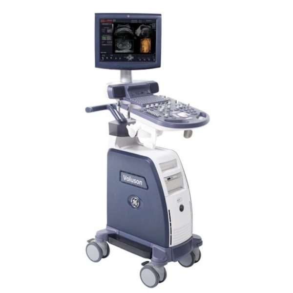
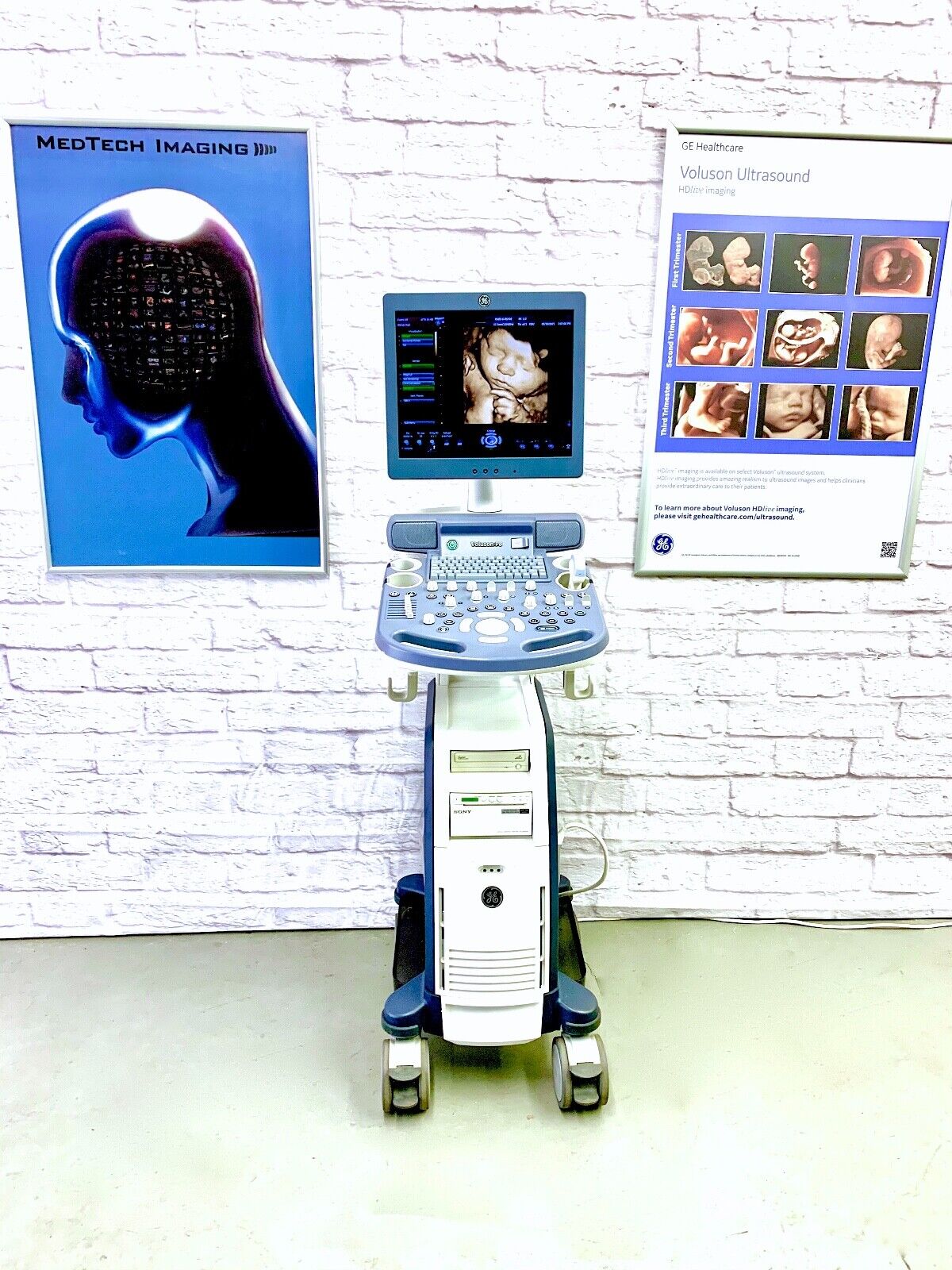
| Content | GE VOLUSON P8 ULTRASOUND
The GE Voluson P8 Ultrasound is an entry-level ultrasound system for women’s healthcare applications. Its automated functions allow physicians in busy practices to maintain high-quality imaging while reducing the time needed to conduct, analyze, and report on exams. The consensus of the GE Voluson P8 is an affordable ultrasound for any medical professional who wants to experience the power of a GE Voluson system for clinical applications. Even though the GE Volsuon P8 system is at a lower price point, it supports in-depth imaging features, such as 3D/4D and HDlive.Dimensions & Weight of the GE Voluson P8Width: 16.5 in (420 mm)
Depth: 626.4 in (70 mm)
Height: 59 in (1495 mm)
Weight: 143 lbs (65 kg)GE Voluson P8 SpecificationsSystem processing channel: Counts up to 161,616 by configuration
Minimum depth of field: 0 – 1 cm (Zoom, probe dependent)
Maximum depth of field: 0 – 30 cm (probe dependent)
Continuous dynamic receive focus/continuous dynamic receive aperture
Adjustable dynamic range: Up to 255 dBGE Voluson P8 Electrical PowerNominal input voltage: 100-240 VAC, frequency 50/60 HzProbes
GE Voluson P8 Probes and Transducers
Convex Probes:
4C-RSLinear Probes:
12L-RSEndocavitary Probes:
E8C-RSPhased Array Probes:
3Sc-RS3D/4D Convex Probes:
RAB2-6-RS3D/4D Endocavitary Probes:
RIC5-9W-RSGE Voluson P8 Features• 17” High Resolution LCD LED Flat Panel Display
• 3 Active Probe Ports
• Integrated HD (500 GB)
• 3 USB Ports for External Peripherals
• 2 USB Ports for On-board Peripherals
• AO(Automatic Optimization)
• CrossXBeam(Spatial Compounding Imaging)
• SRI(Speckle Reduction Imaging)
• B-Steer
• Coded Phased Inversion Harmonic Imaging
• HD-flow
• 3/4D(Real time)
• Virtual Convex
• Patient Information Database
• SonoBiometry
• SonoRender Start
• Raw Data file Standard Voluson P8 Supplies• Aquasonic Ultrasound Gel
• Sono Ultrasound Wipes
• Sony UPP-110HG Thermal Printing Paper
• Sony UPC-21L Color Thermal Printing Package
• Mitsubishi KP95HG Thermal Roll Paper
• Mitsubishi KP65HM-CE High-Density Thermal Paper GE Voluson P8 Ports• 3 USB Ports for External Peripherals
• 2 USB Ports for On-board Peripherals
• RJ45 LAN Port
• 1 HDMI Out Port
• 1 AUDIO Out Port GE Voluson P8 Image Storage Formats & Devices• Storage Formats:– DICOM files (Single- or multi-frame) DCM and DICOM Files with DICOMDIR – Raw Data File (Proprietary format) – Export Data as BMP, TIFF, JPEG, MPEG 4, or MS Video 1
• Storage Devices: USB Memory Stick
• DVD-RW Storage
• HDD Image Storage Options for GE Voluson P8 Ultrasounds• Static 3D Mode
• 3/4D Advanced
• HD Live(3D Only)
• SonoNT/SonoIT
• SonoL&D
• 4D View PC Software
• DICOM 3
• Extended View(XTD)
• Anatomical M-mode
• Report Editor | SIUI CTS-800 Veterinary Ultrasound Machine with Linear Rectal Probe
This small and convenient “Palm-Sized” portable veterinary ultrasound scanner fits in the palm of your hand, and is mostly used for farm animals. Its long battery life and waterproof features makes it the perfect ultrasound scanner for veterinarians on the field.The screen of the SIUI CTS-800V will rotate horizontally or vertically, according to how you are holding it.Gravity Sensor: You can operate the ultrasound system either in transversal layout or vertical layout. It can automatically adapt to two different layouts when you rotate it.Grid for estimation: With grid on the image area you can easily estimate the size of the target substance like follicle fertilized egg etc.SPECIFICATIONS SIUI CTS-800 Veterinary Ultrasound Machine with Linear Rectal ProbeMonitor: 7-inch WVGA LCD monitorWeight: 0.8kg Environmental rating: IP54 (main unit) IP67 (probe head) Battery: 3 hours operation time Display mode: B 2B ZOOM B B/M M Depth: Max to 250mm B gain: 0-100 Image storage: 256 frames Cine loop: 256 frames Probe: L50/7.5 MHz linear rectal probe (standard) L65/5.0 MHz linear rectal probe (optional) R20/5.0 MHz micro convex probe (optional)
– Main unit with 7-inch LCD monitor.
– 1 Probe connector.
– 1 Li-ion battery. 3 hours operation time.
– 1 Adaptor.
– 1 Car Charger.
– Belt and sun shield.
– 1 USB port (Mini).
– Carrying case.PROBES– Rectal Linear L7FVC (6.5/7.1/7.5/8.3/9.0MHz).
– Rectal Linear L5FVC (4.0/4.7/5.0/5.5/6.2MHz).
– Backfat Linear L1FC (150mm, 2.5/3.0/3.5/4.0/5.0MHz).
– Convex C3FC (2.5/3.0/3.5/4.0/5.0MHz).
– Microconvex C5FC (3.5/4.0/5.0/6.5/7.1MHz).CTS-800 is a palm-sized veterinary ultrasound system. Features Weight: 0.8kg Environmental rating: IP54 (main unit) IP67 (probe head) Battery: 3 hours operation time Display mode: B 2B ZOOM B B/M M Depth: Max to 250mm B gain: 0-100 Image storage: 256 frames Cine loop: 256 frames Probe: L50/7.5 MHz linear rectal probe (standard) L65/5.0 MHz linear rectal probe (optional) R20/5.0 MHz micro convex probe (optional) | GE Logiq 5 Ultrasound MachineThe LOGIQ 5 is a mid premium multipurpose color imaging system with high level image quality, computational power and workflow flexibility.Product Features: - Exclusive Raw Data Digital acquisition and storage
- Color Doppler, Spectral Dopler, B, M, Triplex, 3-D
- 150+ Frames per Second TrueDigital imaging
- All digital video clip and still image capture (4,000)
- On-board patient, image and reporting archive
- CD-ROM disk burner included, plus PCMCIA and USB
- Raw Data manipulation on image recall for Post Exam:
- Anatomic M-mode creation and adjustment
- Gain, magnification, and colorization control
- Playback speed, sweep speed, angle, baseline, and more…
- 3-D Digital processing and manipulation o Clip and still measure-annotate-enhance-store new
- DICOM 3.0 capable, plus VetPACS all Digital interfacing
- Angio
- ATO
- TMI
- AMM
- Virtual Convex
- PW/CW Doppler
- ECG/AUX
This unit comes with 2 probes:- Abdominal 3.5C
- Transvaginal E8C
GE Logiq 5 Applications:- Abdominal
- Vascular
- Obstetrics
- Gynecology
- Neonatal
- Urology
- Transcranial
- Cardiology
- Small Parts
GE Logiq P5 BT11 to BT06 Revisions
GE first launched the Logiq P5 in 2006. That first version was designated as BT06. “BT” is an abbreviation of “Break Through” and the number designates the year in which this version was launched. So the GE Logiq P5 premium BT08 was launched in 2008 and was in production till the next version in 2009, the Logiq P5 premium BT09. BT08 and BT09 added CrossXBeam and SRI as standard features and gave access to the 11L linear and E8CS endocavitary probesIn 2011, the Logiq P5 premium BT11 version was released and this is the latest version and is still in production in 2016. BT11 added support for the newer 3Sp adult cardiac sector probe, the 5Sp pediatric cardiac probe, and the 4D8c 4D microconvex probe. New options for Elastography, TVI, Stress Echo, Auto IMT, and SRI-HD were added. The Logiq P5 has always been a reliable ultrasound system, but the BT11 revision is especially stable because of it’s long 5 year history without the need for any further revisions! All this while the Logiq P5 premium became the #1 best selling GE ultrasound unit in production. | Sonosite M-Turbo Ultrasound Machine
M-Turbo is a compact ultrasound imaging system from SonoSite, featuring the image quality and functionality needed to meet your diverse needs.Ideal for the general practitioner looking to do more advanced cardiac and vascular exams.Highlights of the new Sonosite M-Turbo :- Now available the M-Turbo with probe.
- This special version of the M-Turbo comes with a one year warranty, however the probes still have the standard 5 year Sonosite warranty.
- With the M-Turbo you can still add Colour, Pulse Wave and Continuous Wave Doppler.
- The system is now available with DICOM networking.
- Speak to your local Account Manager now to arrange a demonstration.
The standard M-Turbo is also still available exclusively from BCF.Highlights of the Sonosite M-Turbo:- More processing power than MicroMaxx – the next step up in image quality
- 5 year manufacturer’s warranty
- Mains or battery powered so can be used anywhere, anytime
- Able to upgrade from standard black and white system to high quality Colour/Doppler in future
- Large range of probes for all applications
. - Quick start up in just 12 seconds.
More… Sonosite M-Turbo Ultrasound MachinePortable, tough and easy to use. So tough it was originally designed for the American military and comes with a full manufacturer’s 5 year warranty.Despite its size, the M-Turbo produces outstanding cardiac images. It has very high colour Doppler sensitivity.Fully upgradeable to Colour, Pulsed Wave (PW) and Continuous Wave (CW) Doppler and capability to upgrade to DICOM or Wireless DICOM. You can connect it to a PACS system to allow viewing and secure storage of images.Stores still images and movie clips which can be exported via USB in PC friendly formats (jpeg, mpeg and avi).Optional enhanced needle visualisation software (unique to Sonosite) easily highlights needles within the patient allowing for accurate acquisition of a FNA or Tru-Cut biopsy sample.M-Turbo is a compact ultrasound imaging system from SonoSite, featuring the image quality and functionality needed to meet your diverse needs. The standard M-Turbo is also still available exclusively from BCF. | Samsung Medison SonoAce R3 Ultrasound Machine
SonoAce R3 is a portable color ultrasound system with comprehensive diagnostic capabilities. Full Spectrum Imaging™ (FSI™) combines with Speckle Reduction Filter™ (SRF™) to deliver superior quality images, even in the challenging circumstances. The compact and user-friendly design features of the SonoAce R3 ensure portability and manoeuvrability in a lightweight unit. Sporting a 15-inch LCD monitor with Full-Featured Workflow, the SonoAceR3 offers your organisation flexibility, without compromising on precision and efficiency.Slim and Compact DesignThe compact design of SonoAce R3 offers Various-Featured Workflow with a thumbnail menu and shortcut, together with functional key groupings for increased efficiency. The user-friendly design combines a wide-view 15-inch LCD monitor with backlit control keys, customisable measurement package and body markers. The SonoAce R3 features 2 probe ports, 3 USB ports, rapid boot up capability and DICOM 3 compatible image filing. This slim, lightweight unit also has a convenient carrying handle for portability and flexibility.Samsung Medison SonoAce R3 Ultrasound MachineFull Spectrum Imaging™ (FSI™) produces the penetration capabilities associated with the lower frequencies, while still maintaining the fine pixel uniformity associated with the higher frequencies. This combination consistently delivers superior quality images, even in the challenging diagnostic circumstances. The resulting clarity delivers improvements in accuracy and efficiency for greater confidence in your diagnostic procedures. Harmonic Imaging is a technique that records higher frequency information, resulting in greater resolution and fewer artifacts.Synthetic Aperture
The physical radio channel may present some limits to imaging which can be overcome by using Synthetic Aperture Control software. This method uses information from diverse sources to form a more better picture of the area under examination. It creates one single scan line by performing the TX and RX twice to create enhanced penetration and resolution. Synthetic Aperture Control technology delivers a comprehensive picture and to aid accurate diagnosis.QuickScan
QuickScan has been designed to increase workflow efficiency by automatically optimising key imaging parameters at the touch of a button. The resulting increase in efficiency allows your organisation to offer patients an improved service with faster and more accurate diagnoses. This reliable and safe system helps your clinic or hospital to deliver a superior patient experience.Features
Full Spectrum ImagingTM
Quick ScanTM
Color and Power Doppler
Harmonic imaging
Freehand 3D
3D Multi Planar Imaging
Sonoview Image Management
PW Doppler
15″ LCD monitor
Two probe ports
Lightweight | GE Voluson 730 Expert Ultrasound MachineGE Voluson 730 Expert, 3D/4D Realtime Ultrasound System Color Doppler, PW Doppler, Tissue Harmonics, STIC, VOCAL DICOM, M-mode, Angio, ATO, TUI, SonoView II, Crossbeam (CRI) MO Drive, Cineloop, High Resoluotion Interlaced Monitor Probe Options: RAB4-8 4D Convex RIC5-9 4D Transvaginal AB2-7 2D Convex IC5-9 2D Transvaginal SP6-12 2D Linear PA2-5 Cardiac Sector
Two Probes included:- RAB4-8L – Live 4D Abdominal Transducer
- RIC5-9 – Live 4D Transvaginal Transducer
Options:
About the Voluson 730:GE Voluson 730 Expert Ultrasound MachineWith GE Voluson 730, you can now acquire and construct volumetric images in real time – up to 40 volumes per second. The system allows you to explore images in any plane to reveal the smallest details with stunning clarity and apply sophisticated analytical tools to answer virtually any of your clinical questions. Volume Ultrasound not only improves your 3D/4D capabilities, it enhances your 2D imaging as well, for even greater diagnostic confidence in your obstetrical, gynecologic, breast and general imaging studies.The used GE Voluson 730 Expert was among the top-rated and biggest breakthrough ultrasounds in the industry. With up to 40 volumes per second rate of acquisition and construction of volumetric images in real time. The 730 Expert is loaded with the newest ultrasound technology including: CrossXBeam, Speckle Reduction Imaging, Tomographic Ultrasound Imaging (TUI), Spatio-Temporal Image Correlation (STIC), and much more.The Voluson 730 Expert is the more advanced version of the Voluson 730 Pro and was replaced by the GE Voluson e8. Compared to the 730 Pro, the Voluson 730 Expert features a touchscreen interface, higher frame rates in 2D and 4D, and more advanced imaging options such as STIC and TUI.Providian Medical Voluson 730 Expert Review:The Voluson 730 is a workhorse of a 4D ultrasound machine. It remains a popular system in many OB/GYN/Women’s Health offices, as well as 4D “Babyface” studios around the world. It is a durable machine with very good image quality for all women’s health needs. |






Reviews
There are no reviews yet.