| Description | Clinical Applications
• Abdominal
• Gynecology
• Small-Parts
• Vascular
• Pediatrics
• Cardiology
• Urology
• Musculoskeletal (MSK)
• Breast | The New Terason uSmart 3200T Ultrasound System, the first in Terason’s uSmart family of proprietary products. This revolutionary new tablet, deemed “disruptive technology” by industry leaders, will change the way healthcare is delivered in both hospital and outpatient sectors of point-of-care ultrasound. | 3D-4D ultrasound is a technique used during pregnancy that displays three-dimensional images and movement of a fetus (fetal face and other body parts). Other applications include multiplanar imaging of the pelvic organs. This developing technology is also being used in some diagnostic medical procedures such as biopsies and enhanced visualization of cardiac structures. National Ultrasound offers a variety of 3D-4D Ultrasound machines such as GE Voluson i, GE Voluson e, GE Voluson E8, Voluson 730 Expert, Medison Sonoace 8000, Sonoace 9900 or Toshiba with the Xario and Apilo. Our team of expert sales managers will help you find the right 3D-4D ultrasound machine to meet your diagnostic needs while staying within your budget. | GE Voluson 730 Expert is among the top-regarded ultrasounds in the industry. With up to 40 volumes per second rate of acquisition and construction of volumetric images in real time. The 730 Expert is loaded with the newest ultrasound technology including: CrossXBeam, Speckle Reduction Imaging, Tomographic Ultrasound Imaging (TUI), Spatio-Temporal Image Correlation (STIC), and much more. | The more patients you see, the more you need Voluson E8.
The Voluson E8 ultrasound system is designed to keep pace with busy practices that conduct a wide range of Women’s Health exams, from routine scanning to complex assessments.
The imaging excellence of Radiance System Architecture – your diagnostic confidence will be enhanced by the extraordinary image clarity and speed of Radiant System Architecture. | Acuson sequoia 512 ultrasound click images to enlarge item description acuson sequoia 512 ultrasoundsold as picturedincludes: 2 probes, 4c1 convex & 6l4 linearsony up 895 md printerunit will be cleaned, function checked and certified for electrical safety. Checked, cleaned, certified for electrical safetyguaranteed working when arrives on the guarantee working we need to be notified in 48 hrs. And the seals must be intact. |
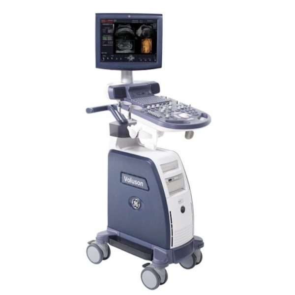
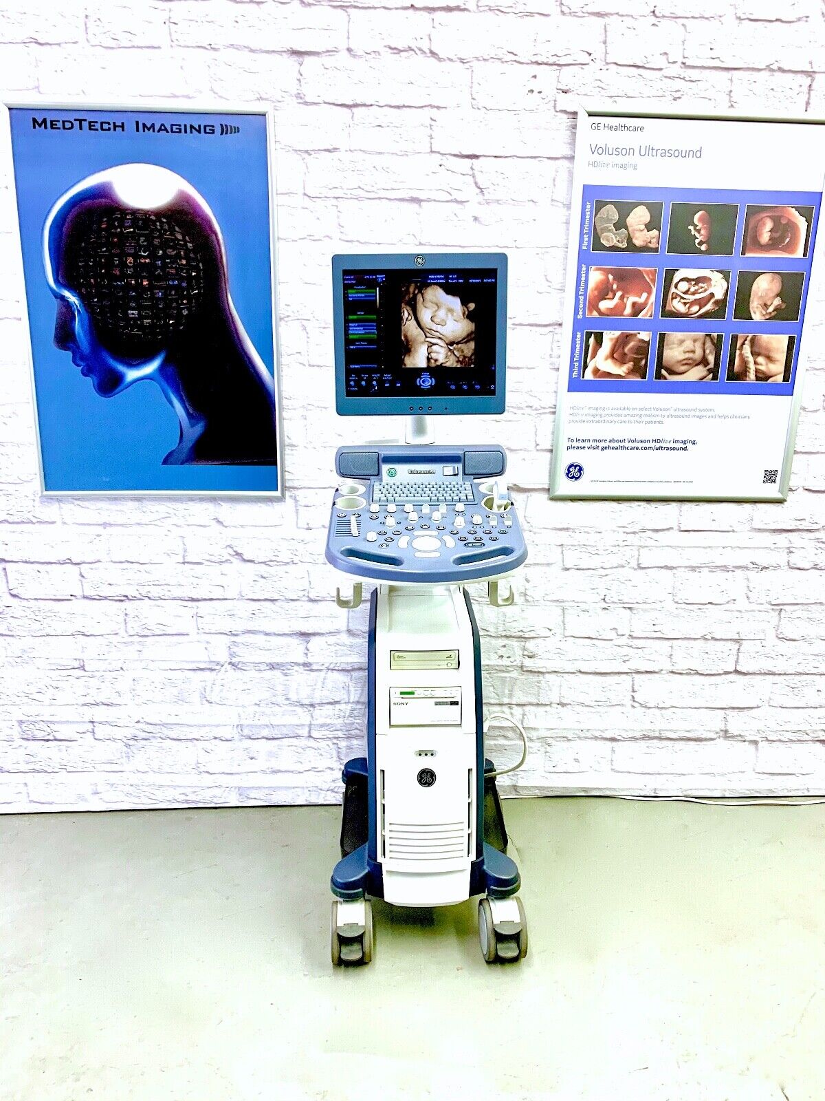
| Content | GE VOLUSON P8 ULTRASOUND
The GE Voluson P8 Ultrasound is an entry-level ultrasound system for women’s healthcare applications. Its automated functions allow physicians in busy practices to maintain high-quality imaging while reducing the time needed to conduct, analyze, and report on exams. The consensus of the GE Voluson P8 is an affordable ultrasound for any medical professional who wants to experience the power of a GE Voluson system for clinical applications. Even though the GE Volsuon P8 system is at a lower price point, it supports in-depth imaging features, such as 3D/4D and HDlive.Dimensions & Weight of the GE Voluson P8Width: 16.5 in (420 mm)
Depth: 626.4 in (70 mm)
Height: 59 in (1495 mm)
Weight: 143 lbs (65 kg)GE Voluson P8 SpecificationsSystem processing channel: Counts up to 161,616 by configuration
Minimum depth of field: 0 – 1 cm (Zoom, probe dependent)
Maximum depth of field: 0 – 30 cm (probe dependent)
Continuous dynamic receive focus/continuous dynamic receive aperture
Adjustable dynamic range: Up to 255 dBGE Voluson P8 Electrical PowerNominal input voltage: 100-240 VAC, frequency 50/60 HzProbes
GE Voluson P8 Probes and Transducers
Convex Probes:
4C-RSLinear Probes:
12L-RSEndocavitary Probes:
E8C-RSPhased Array Probes:
3Sc-RS3D/4D Convex Probes:
RAB2-6-RS3D/4D Endocavitary Probes:
RIC5-9W-RSGE Voluson P8 Features• 17” High Resolution LCD LED Flat Panel Display
• 3 Active Probe Ports
• Integrated HD (500 GB)
• 3 USB Ports for External Peripherals
• 2 USB Ports for On-board Peripherals
• AO(Automatic Optimization)
• CrossXBeam(Spatial Compounding Imaging)
• SRI(Speckle Reduction Imaging)
• B-Steer
• Coded Phased Inversion Harmonic Imaging
• HD-flow
• 3/4D(Real time)
• Virtual Convex
• Patient Information Database
• SonoBiometry
• SonoRender Start
• Raw Data file Standard Voluson P8 Supplies• Aquasonic Ultrasound Gel
• Sono Ultrasound Wipes
• Sony UPP-110HG Thermal Printing Paper
• Sony UPC-21L Color Thermal Printing Package
• Mitsubishi KP95HG Thermal Roll Paper
• Mitsubishi KP65HM-CE High-Density Thermal Paper GE Voluson P8 Ports• 3 USB Ports for External Peripherals
• 2 USB Ports for On-board Peripherals
• RJ45 LAN Port
• 1 HDMI Out Port
• 1 AUDIO Out Port GE Voluson P8 Image Storage Formats & Devices• Storage Formats:– DICOM files (Single- or multi-frame) DCM and DICOM Files with DICOMDIR – Raw Data File (Proprietary format) – Export Data as BMP, TIFF, JPEG, MPEG 4, or MS Video 1
• Storage Devices: USB Memory Stick
• DVD-RW Storage
• HDD Image Storage Options for GE Voluson P8 Ultrasounds• Static 3D Mode
• 3/4D Advanced
• HD Live(3D Only)
• SonoNT/SonoIT
• SonoL&D
• 4D View PC Software
• DICOM 3
• Extended View(XTD)
• Anatomical M-mode
• Report Editor | Terason uSmart 3200t Ultrasound
The new light-weight, power-packed uSmart 3200T Ultrasound System is a fusion of cutting-edge technology with an intuitive interface that defines simplicity. The newest member of the Terason family weighs less than 5 lbs., provides razor-sharp image quality, and is fully equipped with all the features and functionality users have come to expect from Terason products. Developed by clinicians for clinicians, the uSmart 3200T provides the best tools for busy practices, including a 128GB Solid State hard drive, Smart Gestures, an Adaptive Touch Screen, uConnect remote capabilities, and a fast boot-up time.The Terason uSmart 3200T Ultrasound is truly a unique and revolutionary product in the world of portable ultrasound. In our fast-paced technology environment, healthcare providers require products that can keep up with the ever-changing demands of their hospital or practice. We have developed the first tablet-based system where high technology meets superior performanceThe Terason uSmart 3200T is a tablet-style portable color Doppler ultrasound machine designed for the point-of-care bedside ultrasound market.This handheld ultrasound weighs less than 5lbs and uses gestures and touch operation much like a tablet or touchscreen computer. With Terason’s well-known unique beamforming microprocessor technology, the Terason uSmart 3200T Ultrasound provides excellent image quality with an efficient workflow and easy use.Gold Standard, Crystal Clear Images you have to see to believe! As an industry pioneer, Terason developed and is the first to patent color portable ultrasound. Since then, our razor-sharp image quality has earned a reputation for excellence.
The uSmart 3200T captures every detail, subtlety, and nuance that others overlook. The AHVA LCD provides consistent image quality from multiple viewing angles.Grab-And-Go Portability
As part of one of the most comprehensive product families in the industry, the uSmart 3200T weighs in at just under 5 pounds. The touchscreen interface, small footprint an space-saving power dock can go anywhere you need quickly and easily. It’s ready when you are! The Best Tools For Your Practice- Small Gestures
- Adaptive touch screen
- uConnect remote capabilities
- Multi-Function docking station
- Solid State Hard Drive
- Application-Specific presets
- Fast boot-up time
Enhanced Needle Visualization (ENV)
With ENV- AS a leader in ultrasound, Terason is pleased to introduce ENV. This feature provides needle visualization unlike any other. - Frame rate stability
- One-button operation
- Works with all needle gauges
- 15L transducer solution
- Easy to use
Terason uSmart 3200T Ultrasound Specifications- Dimensions: 22.4 cm x 3. 2 cm x 3.21 cm
(8.82″ x 1.26″ x 12.64″)
- 12.5″ x 1.25″ x 8.75″ High-Res AHVA LCD – Touch Screen
- 128 GB Hard Drive
- Input / Output: 3 USB-A, Ethernet & DVI
- Weight: 4.5 pounds
- Export: to USB, Network Folder
- Operates on AC or internal battery power
Imaging Modes:- B- mode
- M-mode
- Color Doppler
- Color Power Doppler
- Directional Color Power Doppler
- PW Doppler
| GE Voluson i Portable 3D/4D UltrasoundFeatures* Lightweight and portable
* Battery operation
* Real-Time 4D
* 3D Multiplanar Display
* 3D Power Doppler
* Automatic Optimization (AO)
* Speckle Reduction Imaging (SRI) and CrossXBeam deliver advanced computational power, allowing for simultaneous processing of CrossXBeam CRI and SRI together – enabling added speckle reduction, contrast resolution and image clarity.
* HD-Flow uses a bi-directional Doppler feature to achieve a more sensitive vascular study and reduce overwriting.
* Volume Contrast Imaging (VCI) allows for image quality in either a single plane or all three planes of a volume acquisition
* Spatio-Temporal Image Correlation (STIC) captures a full virtual fetal heart cycle in real time, and the volume can be saved for offline analysis.
* Tomographic Ultrasound Imaging (TUI) makes analysis and documentation of dynamic studies easier with a simultaneous view of multiple parallel slices of a volume data set.
* Virtual Organ Computer-aided AnaLysis (VOCAL) is a semi-automated volume measurement tool that utilizes computer technology to provide accurate volume calculations.Comes with:GE Voluson i Portable 3D/4D Ultrasound* 1x Voluson i Portable 3D/4D Ultrasound
* 1x RAB2-5RS (2-5MHz) 3/4D Convex Abdominal Probe
* 1x E8C-RS (4-10MHz) Wideband Microconvex Endocavity (Vaginal) Probe
* 1x RIC5-9-RS (5-9MHz) 2D/3D RealTime 4D Endocavitary ProbeThe GE Voluson i ultrasound machine is primarily used for OB-GYN applications, but is versatile and powerful enough to be used for a variety of other applications as well. The Voluson i ultrasound machine carries the power of the GE Voluson line with access to premium 2D and 4D imaging in a portable form factor. The GE Voluson i provides the extraordinary image quality needed to confidently scan for women’s health. The GE Voluson i portable ultrasound machine offers features such as speckle reduction imaging and CrossXBeamCRI, which both lend to the incredible image clarity and quality of this system. The Voluson I portable ultrasound system is known for ease of use, allowing the operator to scan patients faster and more efficiently with better accuracy. | GE Voluson 730 Expert Ultrasound MachineGE Voluson 730 Expert, 3D/4D Realtime Ultrasound System Color Doppler, PW Doppler, Tissue Harmonics, STIC, VOCAL DICOM, M-mode, Angio, ATO, TUI, SonoView II, Crossbeam (CRI) MO Drive, Cineloop, High Resoluotion Interlaced Monitor Probe Options: RAB4-8 4D Convex RIC5-9 4D Transvaginal AB2-7 2D Convex IC5-9 2D Transvaginal SP6-12 2D Linear PA2-5 Cardiac Sector
Two Probes included:- RAB4-8L – Live 4D Abdominal Transducer
- RIC5-9 – Live 4D Transvaginal Transducer
Options:
About the Voluson 730:GE Voluson 730 Expert Ultrasound MachineWith GE Voluson 730, you can now acquire and construct volumetric images in real time – up to 40 volumes per second. The system allows you to explore images in any plane to reveal the smallest details with stunning clarity and apply sophisticated analytical tools to answer virtually any of your clinical questions. Volume Ultrasound not only improves your 3D/4D capabilities, it enhances your 2D imaging as well, for even greater diagnostic confidence in your obstetrical, gynecologic, breast and general imaging studies.The used GE Voluson 730 Expert was among the top-rated and biggest breakthrough ultrasounds in the industry. With up to 40 volumes per second rate of acquisition and construction of volumetric images in real time. The 730 Expert is loaded with the newest ultrasound technology including: CrossXBeam, Speckle Reduction Imaging, Tomographic Ultrasound Imaging (TUI), Spatio-Temporal Image Correlation (STIC), and much more.The Voluson 730 Expert is the more advanced version of the Voluson 730 Pro and was replaced by the GE Voluson e8. Compared to the 730 Pro, the Voluson 730 Expert features a touchscreen interface, higher frame rates in 2D and 4D, and more advanced imaging options such as STIC and TUI.Providian Medical Voluson 730 Expert Review:The Voluson 730 is a workhorse of a 4D ultrasound machine. It remains a popular system in many OB/GYN/Women’s Health offices, as well as 4D “Babyface” studios around the world. It is a durable machine with very good image quality for all women’s health needs. | GE Voluson E8 Ultrasound Machine15’’ LCD monitor [Rev BT06], 10.4” touchscreen, 3 Active Probe Ports, Floating Keyboard: Interactive back-lighting, Beta-View CrossXBeam, Virtual Convex, Extended View, ATO [Automatic Tissue Optimization], Coded Harmonic Imaging, High Resolution Zoom, Inversion Real-time automatic Doppler, OB/GYN; Vasc. Cardiac; Abd; Small-Parts; Urology; Ped; Ortho; Neuro reporting, Multigestational Calculations, HPRF [High Pulse Repetition Frequency], SonoVCAD [Matrix array volume technology], TruScan [On-board archive], VCI [Volume Contrast Imaging], SonoAVCfollicle [Sonography-based Automated Volume Count follicle], SonoVCADheart [Sonography-based Volume Computer Aided Display], TUI [Tomographic Ultrasound Imaging], CE [Coded Excitation], XTD, SRI III [Speckle reduction imaging], CRI [Compound Resolution Imaging], DICOM, Ethernet port, Video Out, 6 USB ports, Integrated HDD (160 GB) and DVD/CD-RW.
3D/4D, Endocrinology, Family Practice, Gastroenterology, Internal Medicine, Nephrology, Neurology, OB-GYN, Orthopedics, Otorhinolaryngology, Pain Management, Urology, Vascular Probes GE Voluson E8 Ultrasound Machine - 11L-D Linear Array
- 3S-D Cardiac Sector
- 4C-D Convex Array
- 9L-D Linear Array
- AB2-7 Convex
- IC5-9-D Intercavity
- M6C-D Convex
- P2D CWD Non-Imaging
- P6D CWD Non-Imaging
- PA6-8-D Cardiac
- RAB2-5-D 3D/ 4D Convex
- RAB4-8-D 3D/4D Convex
- RIC5-9-D 3D/4D Intracavity
- RIC6-12-D 3D/4D Intracavity
- RNA5-9-D 3D/4D Convex
- RRE6-10-D Endocavitary Micro-Convex
- RSP6-16-D 3D/D Linear
- SP10-16-D Linear
- 19″ monitor
- 4D Real Time
- CFM Doppler Mode
- Coded Contrast Imaging
- CrossBeam
- DICOM 4D
- HD flow
- Power Doppler Mode
- SonoVCAD
- STIC (Spacial-Temporal Image Correlation)
- T.U.I (Tomographic Ulrasound Imaging)
- VOCAL II VCI (Volume Contrast Imaging)
- Black and white printer
- Color printer
- DVD-Recorder
As your patient volume increases and your clinical cases become more complex, you will require an ultrasound system that enables you to deliver truly exceptional care – confidently and efficiently – every time. The Voluson E8 ultrasound system is designed to keep pace with busy practices – handling routine to complex women’s health exams with ease and precision. The premium image quality of the Voluson E8 combined with access to advanced capabilities, delivers the level of performance required by you and your patients. | Siemens / Acuson Sequoia 512 Ultrasound Machine
The ACUSON Sequoia 512 ultrasound is an ultra-premium system that utilizes revolutionary technology, dedicated to the advancement of diagnostic imaging. It represents some of the most advanced technology in ultrasound imaging. The Acuson Sequoia 512 has unparalleled imaging performance, greater image content, enhanced detail, superior contrast resolution & penetration, and excellent color Doppler sensitivity.One of the best selling ultrasounds in the United States, the Sequois 512 provides superior imaging with Native Patient Specific Imaging that detects, measures and adapts (in real time) to each patient’s individual acoustics.Performance:Reduce image optimization time by up to 80% and allow for an easier, faster and more confident diagnosis:- 2D
- SST Color Doppler
- Color Doppler Energy
- Color Doppler Velocity
- Solo Spectral Doppler
- M-mode and Color Doppler M-mode
- MultiHertz Multiple Frequency Imaging
- DELTA Differential Echo Amplification
- Home Base user interface
- One-Touch Image Control with exam and Image Presets
- Digital acquistion, storage, review and transfer of static images, dynamic clips, and cine
- Digital storage on an integrated hard disk
- Digital transfer via MO Disk
- Integrated Compression engine
- Embedded DICOM conformance
- Patient- oriented file structure
- Native Tissue Harmonic Imaging
- Tissue Equalization™ Technology (Optional)
- Signature II™ (Optional)
- Advanced Imaging package (Optional)
- Auto Doppler (Optional)
- Freestyle Compounding (Optional)
- FreeStyle Extended Imaging (Optional)
- 3D surface Rendering (Optional)
- Cadence™ ADI (Optional)
- Cadence CPS for 512 (Optional)
- Axius™ Edge Assisted EF (Optional)
- Doppler Tissue Imaging (Optional)
- DICOM Print And Store (Optional)
Capabilities:Siemens / Acuson Sequoia 512 Ultrasound MachineGeneral Radiology, Abdominal, Intraoperative Monitoring, OB/GYN, Neonatal, Vascular, Cardiac, Small PartsAvailable Transducers:4V1, 4V2, 8V5, 15L8W, 6L3, 8L5, 6C2, 5C2, 4C1, 3V2C, 5V2C,7V3C, EVC8-4V, EC-10C5, V5M TEE, V7B neo TEE, AcuNav Transducer, 2.0 Non Imaging CW Doppler probe.Acuson sequoia 512 ultrasound click images to enlarge item description acuson sequoia 512 ultrasoundsold as picturedincludes: 2 probes, 4c1 convex & 6l4 linearsony up 895 md printerunit will be cleaned, function checked and certified for electrical safety. Checked, cleaned, certified for electrical safetyguaranteed working when arrives on the guarantee working we need to be notified in 48 hrs. And the seals must be intact. |






Reviews
There are no reviews yet.