| Description | Clinical Applications
• Abdominal
• Gynecology
• Small-Parts
• Vascular
• Pediatrics
• Cardiology
• Urology
• Musculoskeletal (MSK)
• Breast | Solid imaging performance.
The sleek LOGIQ A5 can help meet your general imaging needs. This black and white ultrasound system has:- B-mode imaging capability and features that allow you to add advancements as your clinical needs grow.
- Auto TGC helps ensure a homogeneous picture from the near-to-far field.
- Virtual Convex provides an expanded field of view that enables you to see more anatomy with linear transducers.
- Color flow Doppler upgrade available to provide dynamic assessment of blood flow in the organs, as well as investigation of hypertension and resulting erectile dysfunction.
| RealTime 4D imaging – continuous, 3D scanning with simultaneous visualization of the A, B and C planes.
The system allows you to explore images in any plane to reveal the smallest details with stunning clarity and apply sophisticated analytical tools to answer virtually any of your clinical questions.
• 3D Static/4D Real-time Imaging (15 FPS) State-of-the-art user interface with on screen menu’s Automatic Tissue Optimization (ATO) 15″ high resolution non-interlaced flat CRT 4 active probe ports Image Archive on CD, MOD, and Hard Drive (40 GB) B-Mode, M-Mode Tissue Harmonic Imaging Digital Beamformer with 512 system processing channel technology CINE memory: up to 256 MB (up to 3000 2D Images) Power Doppler Imaging (PDI) Tissue Doppler Imaging | The Samsung Accuvix XG was the top of the line system from Samsung prior to the release of the newer. The XG was meant as a direct replacement to the popular Accuvix XQ as a shared service system capable of both 4D OBGYN images as well as full cardiac scans. | Ultraformer III’s 7 clip-in transducer cartridges project uniform ultrasound beams directly to multiple layers underneath the skin to promote tighter collagen formation, tissue contraction and reduction in the volume of adipocytes that form bulging areas of tissue.Patients notice an immediate benefit in improved skin tone and texture, with ongoing improvements over 3 to 6 months
Traditionally difficult areas such as eye lifting are made easier with Ultraformer III’s specialised cartridges. It is possible for patients to experience up to 6mm of eyebrow lift, depending on their particular situation. | The Voluson 730 is available in a wide range of system capabilities and affordable price points. The original innovator in 4D OB/GYN imaging provides full diagnostic capabilities and is the perfect for the private practice office regardless of budget. |
| Content | GE VOLUSON P8 ULTRASOUND
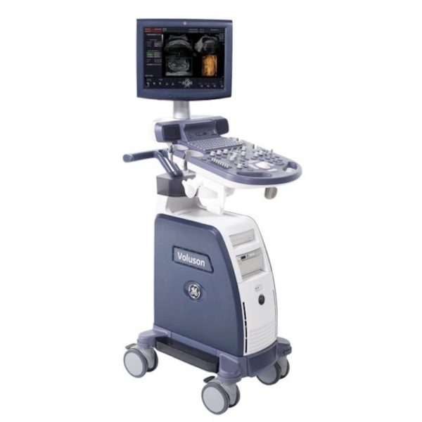
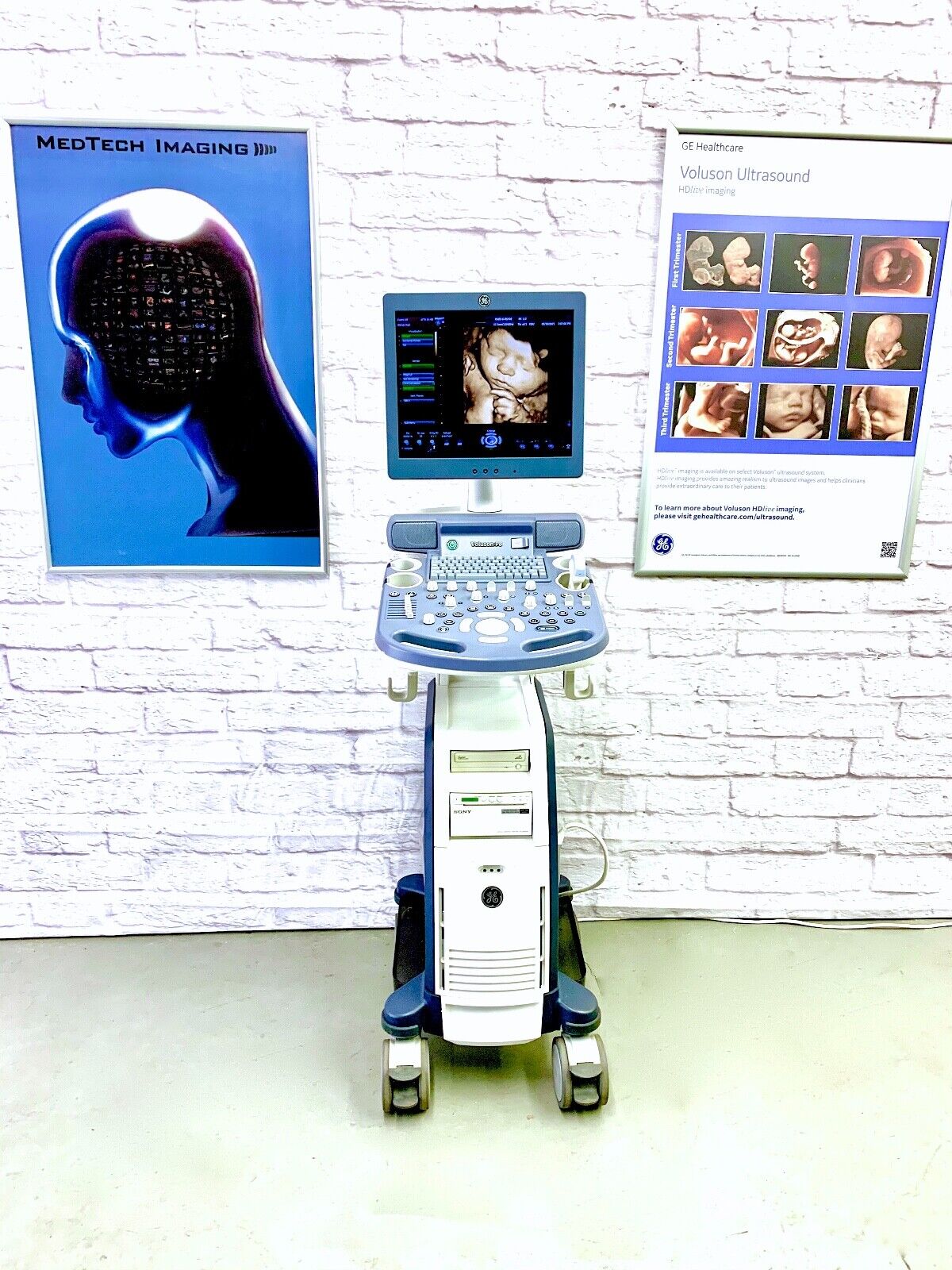
The GE Voluson P8 Ultrasound is an entry-level ultrasound system for women’s healthcare applications. Its automated functions allow physicians in busy practices to maintain high-quality imaging while reducing the time needed to conduct, analyze, and report on exams. The consensus of the GE Voluson P8 is an affordable ultrasound for any medical professional who wants to experience the power of a GE Voluson system for clinical applications. Even though the GE Volsuon P8 system is at a lower price point, it supports in-depth imaging features, such as 3D/4D and HDlive.Dimensions & Weight of the GE Voluson P8Width: 16.5 in (420 mm)
Depth: 626.4 in (70 mm)
Height: 59 in (1495 mm)
Weight: 143 lbs (65 kg)GE Voluson P8 SpecificationsSystem processing channel: Counts up to 161,616 by configuration
Minimum depth of field: 0 – 1 cm (Zoom, probe dependent)
Maximum depth of field: 0 – 30 cm (probe dependent)
Continuous dynamic receive focus/continuous dynamic receive aperture
Adjustable dynamic range: Up to 255 dBGE Voluson P8 Electrical PowerNominal input voltage: 100-240 VAC, frequency 50/60 HzProbes
GE Voluson P8 Probes and Transducers
Convex Probes:
4C-RSLinear Probes:
12L-RSEndocavitary Probes:
E8C-RSPhased Array Probes:
3Sc-RS3D/4D Convex Probes:
RAB2-6-RS3D/4D Endocavitary Probes:
RIC5-9W-RSGE Voluson P8 Features• 17” High Resolution LCD LED Flat Panel Display
• 3 Active Probe Ports
• Integrated HD (500 GB)
• 3 USB Ports for External Peripherals
• 2 USB Ports for On-board Peripherals
• AO(Automatic Optimization)
• CrossXBeam(Spatial Compounding Imaging)
• SRI(Speckle Reduction Imaging)
• B-Steer
• Coded Phased Inversion Harmonic Imaging
• HD-flow
• 3/4D(Real time)
• Virtual Convex
• Patient Information Database
• SonoBiometry
• SonoRender Start
• Raw Data file Standard Voluson P8 Supplies• Aquasonic Ultrasound Gel
• Sono Ultrasound Wipes
• Sony UPP-110HG Thermal Printing Paper
• Sony UPC-21L Color Thermal Printing Package
• Mitsubishi KP95HG Thermal Roll Paper
• Mitsubishi KP65HM-CE High-Density Thermal Paper GE Voluson P8 Ports• 3 USB Ports for External Peripherals
• 2 USB Ports for On-board Peripherals
• RJ45 LAN Port
• 1 HDMI Out Port
• 1 AUDIO Out Port GE Voluson P8 Image Storage Formats & Devices• Storage Formats:– DICOM files (Single- or multi-frame) DCM and DICOM Files with DICOMDIR – Raw Data File (Proprietary format) – Export Data as BMP, TIFF, JPEG, MPEG 4, or MS Video 1
• Storage Devices: USB Memory Stick
• DVD-RW Storage
• HDD Image Storage Options for GE Voluson P8 Ultrasounds• Static 3D Mode
• 3/4D Advanced
• HD Live(3D Only)
• SonoNT/SonoIT
• SonoL&D
• 4D View PC Software
• DICOM 3
• Extended View(XTD)
• Anatomical M-mode
• Report Editor | GE Logiq A5 Ultrasound MachineThe lightweight, portable LOGIQ* A5 ultrasound system offers capabilities to help meet your imaging needs — particularly for urological applications.Affordable ultrasound.The LOGIQ A5 puts imaging capabilities at your fingertips in a portable, lightweight design. This system can go from your office to the surgery suite and helps meet a range of general imaging needs. Well-suited for urology, tools include soft tissue image analysis of the kidneys, bladder, penis, testicles, and prostate.Solid imaging performance.
The sleek LOGIQ A5 can help meet your general imaging needs. This black and white ultrasound system has:- B-mode imaging capability and features that allow you to add advancements as your clinical needs grow.
- Auto TGC helps ensure a homogeneous picture from the near-to-far field.
- Virtual Convex provides an expanded field of view that enables you to see more anatomy with linear transducers.
- Color flow Doppler upgrade available to provide dynamic assessment of blood flow in the organs, as well as investigation of hypertension and resulting erectile dysfunction.
Perform imaging with ease.
The LOGIQ A5 has user-friendly features with precision handling.- Automatic Optimization (AO), helps provide improved image quality at the touch of a button for B-mode, spectral and color Doppler imaging.
- Full-size keyboard for optimal ease of use.
- 15-inch adjustable LCD monitor allows a clear view in any position.
- Lightweight package easily travels with you.
Compatible Transducers:- 10L Linear Array
- 11L Wide Band Linear
- 12L Linear Array
- 3.5C Convex
- 3.5CS Convex (2-5 MHz)
- 3CRF Convex
- 3S Phased Array
- 3Sp Wide Band Phased Array Sector
- 4C Convex Array
- 4D3CL Real time 4D Convex
- 4D8C Convex Volume
- 4DE7C Real time 4D Endocavity
- 5CS Convex (2-5 MHz)
- 5S Cardiac Sector
- 5Sp Wide Band Phased Array Sector
- 7S Phased Array
- 8C Convex Array
- 8L Linear (3-7 MHz)
- 9L Wide Band Linear
- BE9C Transrectal
- BE9CS Micro convex Bi-plan
- E8C (4-10 MHz) Wide Band Microconvex
- E8CS Micro-convex (4-11 MHz)
- ERB Brachytherapy Transrectal
- i12L Linear Array
- i739 Intraoperative
- P2D CWD Non-Imaging
- P6D CWD Non-Imaging
- T739 Intra-operative
| GE Voluson 730 Pro Ultrasound MachineThe Voluson 730 Pro is built on the same platform as GE’s leading Real-Time 4D system, the Voluson 730 Expert. With an easy to use interface and exceptional 2D image quality, in addition to GE’s exclusive 4D imaging technology, the Voluson 730 Pro is ideal for any clinical practice with a variety of patient workloads. Recognized world-wide for its 4D imaging capabilities, the Voluson 730 Pro also features exceptional 2D image quality.RealTime 4D imaging – continuous, 3D scanning with simultaneous visualization of the A, B and C planes.
The system allows you to explore images in any plane to reveal the smallest details with stunning clarity and apply sophisticated analytical tools to answer virtually any of your clinical questions.Performance:GE Voluson 730 Pro Ultrasound Machine
State-of-the-art user interface with high resolution 10.4 inch LCD touch panel
Automatic Tissue Optimization
Tissue Doppler
Coded Harmonic Imaging
Coded Excitation (CE)
HD-Flow
CrossXBeamCRI (Compound Resolution Imaging)
Static 3D Mode
B Mode
Focus & Frequency Composite (FFC)
High Resolution Zoom
Pan Zoom
Steering
Virtual Convex
Beta-View
Patient information database
Image Archive on MOD and hard drive
3D/4D data compression (lossy/lossless)
Inversion
Real-time automatic Doppler calcsCapabilities:
OB/GYN, Vascular, Cardio, Abdominal, Small-Parts, Urology, Pediatrics, Ortho, Neurology, Multigestational Calculations, B-Flow, VCI (Volume Contrast Imaging)Available Probes:
AB2-7 Wide Band Convex Probe
AC2-5 Wide Band Convex Probe
4C-A Wide Band Convex Probe
M7C-H Wide Band Convex Probe
IC5-9H Wide Band Convex Probe
PA2-5P Wide Band Phased Array Probe
PA6-8 Wide Band Phased Array Probe
SP10-16 Wide Band Linear Probe
SP4-10 Wide Band Linear Probe
SP6-12 Wide Band Linear Probe
M12L-H Wide Band Linear Probe (1.25D Array)
RAB2-5L Wide Band Convex Volume Probe
RAB4-8L Wide Band Convex Volume Probe
RIC5-9H Wide Band Convex Volume Probe
RIC5-9W Wide Band Convex Volume Probe
RRE6-10 Wide Band Convex Volume Probe
RSP6-16 Wide Band Linear Volume Probe
RNA5-9 Wide Band Convex Volume Probe
SCW 2.0; non imaging suprasternal CW-Pencil Probe
PCW 4.0; non imaging peripheral CW-Pencil Probe | Medison Accuvix XG Ultrasound System
Samsung Accuvix XG Ultrasound Machine2D Image Features
• Dynamic MR™ / Dynamic MR Plus is designed to enrich gray-scale resolution, as it enhances detection
and contrast resolution while also decreasing speckle echoes. This is particularly useful when evaluating
superficial structures, including thyroid, vessels, pelvic and abdominal anatomy.
• SRF (Speckle Reduction Filter™) enhances image quality by reducing or eliminating the appearance of
speckle echoes from ultrasound images. The degree of speckle reduction implemented is user-selectable.
• Wide Dynamic Range determines the number of gray shades utilized to map the gray-scale image.
It enables display of more details of bright areas and dark areas.
• SCI (Spatial Compounding Image) controls the ultrasound beam electronically by steering, and compounds
many scan lines.
• FSI (Full Spectrum Imaging) incorporates the penetration capabilities associated with lower frequencies,
yet maintains the fine pixel uniformity associated with higher frequencies, to deliver consistently high quality
images even in challenging diagnostic areas.3D Image Features Medison Accuvix XG Ultrasound System
• HDVI gives outstanding image quality and naturally clearer contrast, with excellent tissue differentiation,
edge depiction and speckle reduction, allowing consistent diagnoses with great confidence.
• 3D XI™, comprised of a suite of three innovative imaging applications – Multi-Slice View, Oblique View™ and
Volume CT – offers complete and precise control over 3D/4D volume data manipulation for maximum
diagnostic accuracy.
• 3D MXI is an innovative, cutting-edge 3D image processing technology. Comprising a comprehensive suite of
imaging tools – including Multi Volume Slice, Mirror View, Multi-OVIX, and 3D OH – 3D MXI lets you view,
examine and diagnose 3D volume data with supreme ease, speed and accuracy. • Fully adjustable system
The control panel can be adjusted to the user’s preferred height, for a better working environment and reduced
risk of back pain.
• Wide LED touch-screen
The Accuvix XG’s new LED touch-screen makes it easy to organize and operate.
• 19-inch HD LCD Monitor and articulating monitor arm
A 19-inch LCD monitor enables images to be displayed clearly even with a larger monitor, and the articulating
monitor arm enables easy mobility for a more comfortable and convenient working environment.
• Portability
The Accuvix XG is a lightweight system with 4 swivel wheels that allow easy steering, and a locking function. • Customizable measurement menus
Customizable measurement menus allow access to frequently-used functions, and enable a quicker and
more intuitive workflow.
• User keys and user knob
Accuvix XG offers user keys and a user knob that can map frequently-used functions, enabling the function to be
activated quickly and easily.
• Customized Annotation Menu and body marker
Users can preset up to 360 words of annotations, and body markers for each application, that reduce the time
needed for each examination.
• QuickScan
QuickScan maximizes workflow efficiency by automatically optimizing key imaging parameters with just the
push of a button. • HDVI
HD Volume Imaging(HDVI) gives outstanding image quality and naturally clearer contrast, with excellent tissue
differentiation, edge depiction and speckle reduction, allowing consistent diagnoses with great confidence.
It shows clearer images of subtle lesions, using multi slice image as well as C-plane.
• ElastoScan
ElastoScan™ translates stiff & soft tissue information into gray scale image. This technology defines the alteration
of the stiffness in tissue state more accurately, making it easier for the user to utilize the information in the image. | CLASSYS ULTRAFORMER III UTR3
You’ve heard about it, now see and feel the difference for yourself.Ultraformer III is the leading technology for facial lifting, tightening and contouring. Now with 7 interchangeable cartridges that offer your patients another dimension in non-surgical face and body treatments.Ultraformer III is widely used with over 10 million treatments performed worldwide. It has become the treatment of choice for patients who are not yet indicated or not willing to undergo surgery. This fills a huge gap in the market, and captures an audience of people from 30 years of age all the way to 80.ULTRAFORMER III FEATURESLess discomfort associated with treatments
Patented transducer design for more efficient energy delivery
Dual hand pieces with automatic recognition for ease of use
Faster shot and treatment speed to save time
Easy, user-friendly design and interface
Shot count information and voice assistance
Backed by clinical papersThe CLASSYS ULTRAFORMER III UTR3 advantage with slim transducer designimproves operator visibility
supports very narrow focus and precise treatment
fits facial contoursFast elliptic transducersmore efficient treatment timesUltraformer III’s 7 clip-in transducer cartridges project uniform ultrasound beams directly to multiple layers underneath the skin to promote tighter collagen formation, tissue contraction and reduction in the volume of adipocytes that form bulging areas of tissue.Patients notice an immediate benefit in improved skin tone and texture, with ongoing improvements over 3 to 6 months
Traditionally difficult areas such as eye lifting are made easier with Ultraformer III’s specialised cartridges. It is possible for patients to experience up to 6mm of eyebrow lift, depending on their particular situation.Ultraformer III’s 7 clip-in transducer cartridges project uniform ultrasound beams directly to multiple layers underneath the skin to promote tighter collagen formation, tissue contraction and reduction in the volume of adipocytes that form bulging areas of tissue.Patients notice an immediate benefit in improved skin tone and texture, with ongoing improvements over 3 to 6 months
Traditionally difficult areas such as eye lifting are made easier with Ultraformer III’s specialised cartridges. It is possible for patients to experience up to 6mm of eyebrow lift, depending on their particular situation. | GE Voluson 730 Cardiovascular Ultrasound MachineThe Voluson 730 is available in a wide range of system capabilities and affordable price points. The original innovator in 4D OB/GYN imaging provides full diagnostic capabilities and is the perfect for the private practice office regardless of budget.GE VOLUSON 730 PRO/EXPERT BT08The GE Voluson 730 Pro and the Expert BT08 are among the top-rated ultrasounds, known widely as the workhorses of 4D imaging machines. Especially in today’s fast-paced clinical environments, from routine 2D imaging to powerful 3D/4D imaging capabilities, the GE Voluson 730 Pro and the Expert BT08 ultrasounds allow you to demand more from your machine than ever before.GE Voluson 730 Pro/Expert BT08 ImagingThe GE Voluson 730 Pro ultrasound and the Expert BT08 ultrasound provide the capabilities for acquiring and constructing volumetric images in real-time; in fact, you can achieve up to 40 volumes per second. With this type of imaging technology, users will be able to capture and analyze images of any plane while the ultrasound reveals the smallest details in stunning clarity. With these technologies, the GE Voluson 730 Pro/Expert BT08 ultrasound is highly regarded for:Applications- OB/GYN
- Vascular
- Abdominal
- Small Parts
- MSK
- Urology
- Cardiology
- Neurology
- Orthopedic
- Pediatric
- Neonatal
| Key Standard Features- B, CFM, M, PW, 3D/4D
- Anatomical M-Mode
- 15’’ High-Resolution CRT Monitor
- Automatic Tissue Optimization (ATO)
- Static 3D
- Real Time 4D
- Coded Excitation
- Virtual Convex
- TruScan Architecture (TruAccess, SmartScan, ComfortScan)
- Internal DVD/CD-RW Drive
- 10” Touchscreen User Menu
|
Optional Features- 4D Real Time
- Vocal II
- 4D DiagnoSTIC
- DICOM 3.0 Networking
- Volume Contrast Imaging (VCI)
- B-Flow
- VOCAL
- Volume Contrast Imaging (VCI)
- Speckle Reduction Imaging (SRI)
- Tissue Doppler Imaging (TDI)
- Tomographic Ultrasound Imaging (TUI)
- Coded Contrast Imaging
- Compound Resolution Imaging (CRI)
- DICOM Modality Worklist / Structured Reporting
| Transducers- AB2-7- Convex Array Transducer
- RAB2-5 Volume Convex Array Transducer
- RAB2-5L Volume Convex Array Transducer
- RAB4-8P Volume Convex Array Transducer
- RAB4-8L Volume Convex Array Transducer
- RRE6-10 Volume Convex Array Transducer
- IC5-9 Volume Endocavity Convex Array Transducer
- RIC5-9 Volume Endocavity Convex Array Transducer
- SP10-16 Linear Array Transducer
- SP4-10 Linear Array Transducer
- SP6-12 Linear Array Transducer
|
|






Reviews
There are no reviews yet.