| Description | The Cirrus HD-OCT is a non-contact, high resolution tomographic and biomicroscopic imaging device. It is indicated for in-vivo viewing, axial cross-sectional, and three-dimensional imaging and measurement of anterior and posterior ocular structures, including cornea, retina, retinal nerve fiber layer, ganglion cell plus inner plexiform layer, macula, and optic nerve head. The Cirrus normative databases are quantitative tools for the comparison of retinal nerve fiber layer thickness, macular thickness, ganglion cell plus inner plexiform layer thickness, and optic nerve head measurements to a database of normal subjects. The Cirrus HD-OCT is intended for use as a diagnostic device to aid in the detection and management of ocular diseases including, but not limited to, macular holes, cystoid macular edema, diabetic retinopathy, age-related macular degeneration, and glaucoma. | The Auto “Refkeratometer” RC-800 is the latest in economical, user-friendly, highly accurate auto “refkeratometry”. The RC-800 provides simple, easy measurement by the operator at an economical cost, without compromising accuracy or reliability. | The Tomey Refractometer Keratometer and Topographer only in 1 unit senior citizens and children can also get the inspection with out any trouble because they dont have to move the seat for three measurements. | Experience 6 analyses in 1 device: refraction, keratometry, topography, tonometry, pachymetry and dry eye analysis. | The OPD-Scan III is an automated 5 in 1 instrument that combines topography, wavefront, autorefraction, keratometry, and pupillometry — allowing accurate and reliable analysis of corneal aberration. | The Zeiss Humphrey Acuitus 5015 Auto Refractor Keratometer offers expanded refracting capabilities, allowing a practice to refract children and elderly patients alike. The Zeiss Acuitus 5015 also allows refraction one-day post operative of IOL and refractive surgery patients. The Zeiss Acuitus 5015 includes a flat color screen display which produces clear and concise images. |
| Content | Zeiss Cirrus HD-OCT 4000 OCT Spectral Domain OCT HD
Accessories- The ZEISS Certified Factory Direct Cirrus 4000 HD-OCT also includes the full Accessory Kit
- External Fixation Light
- Test Eye
- Mini Keyboard
- Optical Mouse
- Dust Cover
- Metric 5A/250v Fuse
- 14 Ft. CAT 5e Network Cable
- USB Cable
- IEC 320 to NEMA 12 inch Power Cable
- IEC 320 39in. Power Cable
- Install Disc including Roxio Basic
- Symbol Sheet
- OCT Primer Guide
- Quick Start Guide
HD-OCT Imaging- Methodology: Spectral Domain OCT
- Optical Source: superluminescent diode (SLD), 840 nm
- Optical Power: < 725 μW at the cornea
- Scan Speed: 27,000 A-scans per second
- A-Scan depth: 2.0 mm (in tissue), 1024 points
- Axial resolution: 5 μm (in tissue)
- Transverse resolution: 15 μm (in tissue)
Fundus Imaging- Methodology: Line scanning ophthalmoscope
- Live Fundus Image: During alignment and during OCT scan
- Optical Source: Superluminescent diode (SLD), 750 nm
- Optical Power: < 1.5 mW at the cornea
- Field of View: 36 degrees W x 30 degrees H
- Frame rate: >20 Hz
- Transverse resolution: 25 μm (in tissue)
Iris Imaging- Methodology: CCD Camera
- Resolution: 1280 x 1024
- Live iris image: During alignment
Electrical, Physical and Environmental- Weight: 38kg (83 lbs)
- Dimensions: 65L x 44W x 53H (cm)
- Fixation: Internal, external
- Internal fixation focus adjustment: -20D to +20D (diopters)
- Input devices: Keyboard, mouse
- Electrical rating (115V) Single Phase, 100/120V~ systems:50-60Hz, 5A
- Fuse rating (115V) T 5A 250V
- Electrical rating (230V) Single Phase, 220/240V~ systems:50-60 Hz, 2.5A
- Fuse rating (230V) Fuse rating: T 5A 250V
- Convenience Receptacle output ratings 115V~, 0.5 A Max, 50-60 Hz
- Temperature (transport and storage) -40º to +70º C
- Relative humidity (transport and storage) 10% to 100%, including condensation
- Atmospheric pressure (transport and storage): 500 hPa to 1060 hPa
- Temperature (operation) +10º to +35º C
- Relative humidity (operation) 30% to 75%, excluding condensation
- Atmospheric Pressure (operation) 700 hPa to 1060 hPa
Computer Zeiss Cirrus HD-OCT 4000 OCT Spectral Domain OCT HD- • High performance multi-core processor
- • Internal storage: > 80,000 scans
- • CD-RW, DVD-ROM drive
- • Integrated 15″ color flat panel display.
The Cirrus HD-OCT is a non-contact, high resolution tomographic and biomicroscopic imaging device. It is indicated for in-vivo viewing, axial cross-sectional, and three-dimensional imaging and measurement of anterior and posterior ocular structures, including cornea, retina, retinal nerve fiber layer, ganglion cell plus inner plexiform layer, macula, and optic nerve head. The Cirrus normative databases are quantitative tools for the comparison of retinal nerve fiber layer thickness, macular thickness, ganglion cell plus inner plexiform layer thickness, and optic nerve head measurements to a database of normal subjects. The Cirrus HD-OCT is intended for use as a diagnostic device to aid in the detection and management of ocular diseases including, but not limited to, macular holes, cystoid macular edema, diabetic retinopathy, age-related macular degeneration, and glaucoma. | Tomey RC-800 Auto Refractor Keratometer
Refractometry: Simple handling of the fine adjustment and measurement by yourself. Fogging is applied automatically for each measurement for more accurate results.Keratometry: Short consecutive measurements and time provide reliable data. Measurements at φ3.0mm (R = 8.0mm) give you enough information for contact lens fitting.Diameter Measurement of Cornea and Pupil: Easy measurement by moving the two cursors on the display to the boundaries of Cornea or Pupil.Language Selection: RC-800 has a Multi-language function. You can select your required language from “English”, “Japanese”, “German”, “Chinese”, “Spanish”, “Spanish Latin America”, “Italian”, “Portuguese”, “French” and “Russian”.Features:• Highly Accurate
• Simple to Use
• Diameter Measurement of Cornea and Pupil
• High Speed Printer
• Auto Shot
• Color Touch Screen
• USB connection
• Large Patient Database
• Easy Database Operation
• Multi-Language Operation
• Fourier Refractive Analysis
• Quick Data Reference
• Built-in LCD Alignment
• Keratoconus Screening & Other ApplicationsMeasurement Ranges:Refraction:• Sphere: -25.00D to +22.00D (VD=12.0mm)
• Cylinder: 0D to ±10.00D (VD=12.0mm)
• Axis: 0° to 180°
• Minimum Pupil Dia: Ø2.0mm
• Measurement time: 0.2 second/single eye (data taking time)Keratometry:• Range: 5.00mm to 11.00mm
• Corneal Refraction: 30.68D to 67.50D (n=1.3375)
• Corneal Astigmatism: 0D to 10D (n=1.3375)
• Axis: 0° to 180°
• Measurement area: Ø3mm (at 8.00mm corneal curvature)
• Measurement time: 0.1 second/single eye (data taking time)Other features:Tomey RC-800 Auto Refractor Keratometer• PD Measurement range 50 to 86mm
• Corneal diameter measurement
• Measurement range: 10.0 to 14.0mm
• Display unit: 0.1mm
• Pupil diameter measurement
• Measurement range: 1.0 to 10.0 mm
• Display unit: 0.1mm
• Built-in Printer Thermal printer
• Data output type RS232C / USB-D
• Display 5.7 inch color liquid crystal display
• Dimensions and weight 297(W)x 500(D) x 448(H)mm
• Approx. 17kg
• Power Requirements AC 100V to 240V, 50 / 60 Hz, 80VA to 100VAThe Auto “Refkeratometer” RC-800 is the latest in economical, user-friendly, highly accurate auto “refkeratometry”. The RC-800 provides simple, easy measurement by the operator at an economical cost, without compromising accuracy or reliability. | Tomey RT-7000 Autorefractor Keratometer Topographer
-Three Functions in One Instrument-Easy & Speedy Touch Screen Alignment-6.4 inches color TFT LCD-Dual CCD technology for Refractometry-IOL/ Cataract Mode-New incites for Keratometry KAI, KRI-Auto Alignment / Auto Shot of TopographyCorneal Topography– Auto Alignment / Auto ShotAny one can easily capture measurement with Auto Alignment and Auto Shot. The Measurement variation is significantly reduced no matter what the skill level is of the operator.CL Fitting Simulation-A pseudo-fluorescent pattern of the contact lens selected from the built-in data base can be confirmed and the fitting can be stimulated before the patient wears the trial lens.Corneal Eccentricity Index-The index of CEI (Corneal Eccentricity Index) indicates the ratio of flattering in percentage from the central cornea to the periphery.Three Function in One Instrument– Refractometer Keratometer and Topographer only in 1 unit senior citizens and children can also get the inspection with out any trouble because they don’t have to move the seat for three measurements. Switch from Ref-Kerato Mode to Corneal Topography Mode with only ONE TOUCH. The light come appears with only one button from the measuring head and the Ref-Keratometer is transformed to the topographer.Refractometry– Dual CCD technology for RefractometryTwo CCD cameras are used to capture images for observation and for measurement while providing highly accurate measurement data.Keratometory– New Indices for KeratometryThe Corneal Irregular astigmatism display function is installed to expand the possibility of Keratometer. This is the new function to measure the level of corneal irregular astigmatism which was difficult in the past.SPECIFICATIONSMeasurmentRangesRefraction MeasuremntSphere -25.00D to ±22.00DIVD=12.0mmCylinder 0D to ± 10.00DIVD=12.0mmAxis 0 to 180Corneal Curvature Measurement 5.00mm to 11.00mmMeasurement Zone 3mm R:8.00mmCorneal MappingDisplay Range 9 to 100 D-Normal Mode1.0 to 8.0mm (R:8.00mm)-Special Mode0.9 to 7.0 mm (R:8.00mm)Measurement TimeREF 0.2 SecondsKRT 0.1 SecondsMinimum Pupil Diameter 2.2mmMinimum Measurment Step Refraction MeasurementSphere 0.01DCylinder 0.01DAxis 1Corneal Curvature Measurement 0.01mmPD (Pupil Distance) 50mm to 86mmRecording Built-In Thermal PrinterVertex Distance 0mm, 12.0mm, 13.5mm, 14.0mm, 15.5mm, 16.0mmExterior Output (AN port / 4USB ports)Dimensions Tomey RT-7000 Autorefractor Keratometer TopographerHigh 466mmm or 18 InchesWidth 307mm or 12 InchesDepth 490mm or 19 InchesWeight Approx 20,000Kg / 44.4 PoundsDisplay 6.4 Inches color liquid crystalPower SupplyVoltage AC 100 to 240VFrequency 50/60 HzConsumption Power 120VA to 150VATemperature +10C to + 40CDisplay 30% to 75% | Rodenstock Alino Multifunction Refractometer
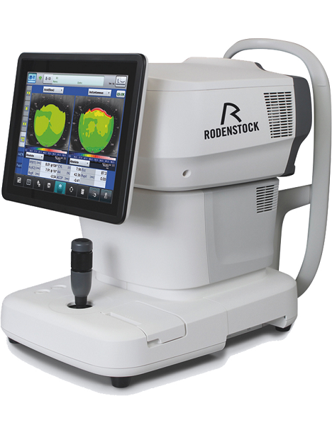
Tune-up your businessExperience 6 analyses in 1 device: Rodenstock Alino Multifunction Refractometer
Are you ready for something that is all-in-one, timesaving, space-saving and suits your business perfectly?Have a look at how powerful ALINO can be.6 in 1 solution01 RefractionHaving a good starting value for subjective refraction is essential. The QUICK REF MODE supports this even for uncooperative patients.02 KeratometryKAI (Kerato-asymmetry index) & KRI (Kerato-regularity index) give the first hint of possible irregularities on the cornea. This supports evaluation of the best vision correction or best-fitting contact lens.03 TonometryThe very gentle air puff created by a new generation of air-flow technology means that patients more readily accept the need for regular check-ups of the IOP measurement.04 PachymetryCorrecting the IOP by measuring the central corneal thickness is essential in interpreting the intraocular pressure.05 TopographyVarious topographic maps support you in carrying out vision screenings, contact lens fittings and in patient education.06 Dry EyeObserve the customers’ eye health and evaluate the long-term tolerability of contact lenses.Take advantage of the combination of tests:Additional servicesTonometry + PachymetryContact lens fitting andtolerance controlKeratometry + Topography + Dry EyeEye healthcareTopography + Tonometry + Pachymetry + Dry EyeSpecifications Rodenstock Alino Multifunction Refractometer
REFRACTIVE POWER MEASUREMENTSpherical refractive power (S) -30.00 D to + 25.00 D (at VD = 12.0 mm)Cylindrical power (C) 0.00 D to ± 12.50 D (at VD = 12.0 mm)Astigmatic axis (A) 0° to 180°KERATOMETRY MEASUREMENTCorneal curvature radius 5.00 mm to 13.00 mmCorneal astigmatic axis 0° to 180°INTRAOCULAR PRESSURE MEASUREMENTMeasurement range 1 mmHg to 60 mmHG (1 hPa to 80 hPa)PACHYMETRY MEASUREMENTMeasurement range 300 μm to 800 μmTOPOGRAPHY MEASUREMENTCorneal curvature radius 5.50 mm to 10.00 mmCorneal astigmatic axis 0° to 180°AUXILIARY FUNCTIONInterpupillary distance Measurement range: 20 mm to 85 mmCorneal diameter and pupil diameter Measurement range: 1.00 mm to 14.00 mmDry eye observation Blinking Analysis, Tear Meniscus height, Hyperemia, Meibomian glandsDATA MANAGEMENTInternal Database Integrated SD CardPrinter Integrated thermal printerData Output Type 3x USB 2.0, 1x LAN, 1x SD Card-slot, 1x WLANExport Format CSV, XML, JPG, PDFDIMENSIONS & ELECTRIC REQUIREMENTSDimensions WDH 312 × 491 × 450 mmWeight Approx. 23 kgVoltage 100 VAC to 240 VACFrequency 50/60 HzPower consumption 110 VA | Nidek OPD-Scan III Corneal AnalyzerComprehensive vision analysis
OPD-Scan III VS is a device that mainly measures corneal shape (topographer) and refractive error (refractometer).
It clarifies causes of vision difficulties through wavefront analysis of information measured over a wide area.
Solutions for face-to-face consultingTablet viewer provides four kinds of easy-to-understand reports for explanation and consulting.
*The tablet is not included or sold with the OPD-Scan III VS.
Basic information reportBasic information to generally judge the patient’s eye conditions.
Topography reportIntuitive maps and numerical data for comprehending the corneal surface.
Simulation reportVisual performance simulations and MTF graphs for a variety of conditions.
Eye diagram image reportEye model to visually understand eye conditions ranging from eye fundus to cornea.
Wide range of display patterns
  Placido image
 Axial map (Additional indication)
 Visual Acuity map (Image charts)
Features Nidek OPD-Scan III Corneal Analyzer- Five-in-One true refractive workstation for all practitioners
- Overview summary for optimal clinical decisions
- A number of summaries
- Wider measurement area
- Greater topography resolution, blue placido rings
The OPD-Scan lll is the Five-in-One true refractive workstation combining - Wavefront Aberrometer
- Topographer
- Auto Refractometer
- Auto Keratometer
- Pupillometer and Pupillographer
- Irregularity helps determine the best strategy for vision correction. Separation into Total, Corneal and Internal components allows determination of the source of the optical pathology.
- PSF images of OPD, Axial, and Internal OPD map simulate objective retinal visual quality from each component of the eye for easy clinical assessment and patient education.
- Corneal Spherical Aberration aids in the selection of aspheric IOLs and contact lenses.
- Color coded Classification Indices help identify post-LASIK corneas and Keratoconus.
- The Astigmatism index aids the implantation of toric IOLs such as incision placement and lens alignment.
- A retroillumination image of cataracts captured during the OPD exam allows better understanding of pupillary effects on vision and in patient education.
| Zeiss Humphrey Acuitus 5015 Autorefractor Keratometer
Autorefractor/Keratometer that
can take a combination of refraction, central and peripheral keratometry. Unit has been tested with a reference eye (not included) and as well with a test subject and
takes accurate OD/OS reading.
Great unit for your optometry practice.
Unit is patient ready. This has just been professionally calibrated by our optical engineer.
Also includes a printed out Acuitus 5015 User manual,power cord.The Zeiss Humphrey Acuitus 5015 Auto Refractor Keratometer offers expanded refracting capabilities, allowing a practice to refract children and elderly patients alike. The Zeiss Acuitus 5015 also allows refraction one-day post operative of IOL and refractive surgery patients. The Zeiss Acuitus 5015 includes a flat color screen display which produces clear and concise images. Additional features on the Zeiss Acuitus 5015 include auto acquisition, auto XYZ, making the measurements easily acquired. Additional benefits on the Zeiss Acuitus 5015 are automatic eye detection, thirty measurements per second, and RS 232 interface. The Zeiss Acuitus 5015 offers a multitude of features in a small compact footprint at an extremely affordable price point. |




Reviews
There are no reviews yet.