| Description | Handheld ultrasound system for Equine and Bovine Gynecology.Supplied with a 9 MHz transrectal probe.
| Function • Gravity Sensor: You can operate the ultrasound system either in transversal layout or vertical layout.• Grid for estimation: With grid on the image area, you can easily estimate the size of the target substance like follicle fertilized egg etc. | Ultraformer III’s 7 clip-in transducer cartridges project uniform ultrasound beams directly to multiple layers underneath the skin to promote tighter collagen formation, tissue contraction and reduction in the volume of adipocytes that form bulging areas of tissue.Patients notice an immediate benefit in improved skin tone and texture, with ongoing improvements over 3 to 6 months
Traditionally difficult areas such as eye lifting are made easier with Ultraformer III’s specialised cartridges. It is possible for patients to experience up to 6mm of eyebrow lift, depending on their particular situation. | Acuson sequoia 512 ultrasound click images to enlarge item description acuson sequoia 512 ultrasoundsold as picturedincludes: 2 probes, 4c1 convex & 6l4 linearsony up 895 md printerunit will be cleaned, function checked and certified for electrical safety. Checked, cleaned, certified for electrical safetyguaranteed working when arrives on the guarantee working we need to be notified in 48 hrs. And the seals must be intact. | The GE Vivid q portable ultrasound machine is a more powerful version of the Vivid i that adds further quantitative features including ICE, Auto EF, AFI, TVI,TT, TSI, Blood Flow and Blood Flow imaging. The Vivid q is the pinnacle of GE’s portable cardiovascular ultrasounds. | The more patients you see, the more you need Voluson E8.
The Voluson E8 ultrasound system is designed to keep pace with busy practices that conduct a wide range of Women’s Health exams, from routine scanning to complex assessments.
The imaging excellence of Radiance System Architecture – your diagnostic confidence will be enhanced by the extraordinary image clarity and speed of Radiant System Architecture. |
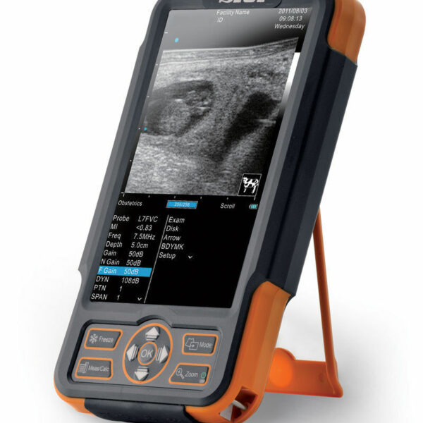
| Content | SIUI CTS-800 Veterinary Ultrasound Machine
The CTS-800 is small, lightweight, and best used for reproduction exams in most farm animals. It features vet software packages for equine, bovine, ovine, canine, feline, and primate.Features SIUI CTS-800 Veterinary Ultrasound Machine- 7.5Mhz Rectal probe with 50mm lens
- 7-inch WVGA LCD monitor
- Weighs approximately 1.8 Lbs. (0.8kg)
- Long lasting battery up to 4.5 hours
- Software & Report for reproductive system
- Gravity sensor for layout change (horizontal/vertical)
- Measurement for distance, area, circumference, volume, angle, heart rate
- Display modes include B, 2B, ZOOM B, B/M, and M mode
Included Accessories- Main unit with rechargeable battery
- Power supply adapter
- Power Cable
- 7.5Mhz Rectal probe with 50mm lens
- Car charger
- USB cable
- Video cable
- Protective case
- Hand straps
- Sunshade
- Storage box
The SIUI CTS 800 vet is a palm-sized portable ultrasound scanner for farm animals. It’s small with a 7-inch LCD monitor. The SIUI CTS-800 has a long battery life and is also waterproof. Another nice feature is a gravity sensor that will rotate the image vertically or horizontally, depending on how you’re holding the ultrasound system.Probes: L50/7.5 MHz linear rectal probe (standard configuration)L65/5.0 MHz linear rectal probe (optional)R20/5.0 MHz micro convex probe (optional)Gravity Sensor: You can operate the ultrasound system either in transversal layout or vertical layout. It can automatically adapt to two different layouts when you rotate it.Grid for estimation: With grid on the image area you can easily estimate the size of the target substance like follicle fertilized egg etc.This is a very popular unit for livestock pregnancy detection. It’s lightweight, durable, waterproof and the battery can last up to 4.5 hours. Its portability and durability plus 2-year warranty as a new system are a great value for the price of this portable veterinary ultrasound machine. | SIUI CTS-800 Veterinary Ultrasound Machine with Linear Rectal Probe
This small and convenient “Palm-Sized” portable veterinary ultrasound scanner fits in the palm of your hand, and is mostly used for farm animals. Its long battery life and waterproof features makes it the perfect ultrasound scanner for veterinarians on the field.The screen of the SIUI CTS-800V will rotate horizontally or vertically, according to how you are holding it.Gravity Sensor: You can operate the ultrasound system either in transversal layout or vertical layout. It can automatically adapt to two different layouts when you rotate it.Grid for estimation: With grid on the image area you can easily estimate the size of the target substance like follicle fertilized egg etc.SPECIFICATIONS SIUI CTS-800 Veterinary Ultrasound Machine with Linear Rectal ProbeMonitor: 7-inch WVGA LCD monitorWeight: 0.8kg Environmental rating: IP54 (main unit) IP67 (probe head) Battery: 3 hours operation time Display mode: B 2B ZOOM B B/M M Depth: Max to 250mm B gain: 0-100 Image storage: 256 frames Cine loop: 256 frames Probe: L50/7.5 MHz linear rectal probe (standard) L65/5.0 MHz linear rectal probe (optional) R20/5.0 MHz micro convex probe (optional)
– Main unit with 7-inch LCD monitor.
– 1 Probe connector.
– 1 Li-ion battery. 3 hours operation time.
– 1 Adaptor.
– 1 Car Charger.
– Belt and sun shield.
– 1 USB port (Mini).
– Carrying case.PROBES– Rectal Linear L7FVC (6.5/7.1/7.5/8.3/9.0MHz).
– Rectal Linear L5FVC (4.0/4.7/5.0/5.5/6.2MHz).
– Backfat Linear L1FC (150mm, 2.5/3.0/3.5/4.0/5.0MHz).
– Convex C3FC (2.5/3.0/3.5/4.0/5.0MHz).
– Microconvex C5FC (3.5/4.0/5.0/6.5/7.1MHz).CTS-800 is a palm-sized veterinary ultrasound system. Features Weight: 0.8kg Environmental rating: IP54 (main unit) IP67 (probe head) Battery: 3 hours operation time Display mode: B 2B ZOOM B B/M M Depth: Max to 250mm B gain: 0-100 Image storage: 256 frames Cine loop: 256 frames Probe: L50/7.5 MHz linear rectal probe (standard) L65/5.0 MHz linear rectal probe (optional) R20/5.0 MHz micro convex probe (optional) | CLASSYS ULTRAFORMER III UTR3
You’ve heard about it, now see and feel the difference for yourself.Ultraformer III is the leading technology for facial lifting, tightening and contouring. Now with 7 interchangeable cartridges that offer your patients another dimension in non-surgical face and body treatments.Ultraformer III is widely used with over 10 million treatments performed worldwide. It has become the treatment of choice for patients who are not yet indicated or not willing to undergo surgery. This fills a huge gap in the market, and captures an audience of people from 30 years of age all the way to 80.ULTRAFORMER III FEATURESLess discomfort associated with treatments
Patented transducer design for more efficient energy delivery
Dual hand pieces with automatic recognition for ease of use
Faster shot and treatment speed to save time
Easy, user-friendly design and interface
Shot count information and voice assistance
Backed by clinical papersThe CLASSYS ULTRAFORMER III UTR3 advantage with slim transducer designimproves operator visibility
supports very narrow focus and precise treatment
fits facial contoursFast elliptic transducersmore efficient treatment timesUltraformer III’s 7 clip-in transducer cartridges project uniform ultrasound beams directly to multiple layers underneath the skin to promote tighter collagen formation, tissue contraction and reduction in the volume of adipocytes that form bulging areas of tissue.Patients notice an immediate benefit in improved skin tone and texture, with ongoing improvements over 3 to 6 months
Traditionally difficult areas such as eye lifting are made easier with Ultraformer III’s specialised cartridges. It is possible for patients to experience up to 6mm of eyebrow lift, depending on their particular situation.Ultraformer III’s 7 clip-in transducer cartridges project uniform ultrasound beams directly to multiple layers underneath the skin to promote tighter collagen formation, tissue contraction and reduction in the volume of adipocytes that form bulging areas of tissue.Patients notice an immediate benefit in improved skin tone and texture, with ongoing improvements over 3 to 6 months
Traditionally difficult areas such as eye lifting are made easier with Ultraformer III’s specialised cartridges. It is possible for patients to experience up to 6mm of eyebrow lift, depending on their particular situation. | Siemens / Acuson Sequoia 512 Ultrasound Machine
The ACUSON Sequoia 512 ultrasound is an ultra-premium system that utilizes revolutionary technology, dedicated to the advancement of diagnostic imaging. It represents some of the most advanced technology in ultrasound imaging. The Acuson Sequoia 512 has unparalleled imaging performance, greater image content, enhanced detail, superior contrast resolution & penetration, and excellent color Doppler sensitivity.One of the best selling ultrasounds in the United States, the Sequois 512 provides superior imaging with Native Patient Specific Imaging that detects, measures and adapts (in real time) to each patient’s individual acoustics.Performance:Reduce image optimization time by up to 80% and allow for an easier, faster and more confident diagnosis:- 2D
- SST Color Doppler
- Color Doppler Energy
- Color Doppler Velocity
- Solo Spectral Doppler
- M-mode and Color Doppler M-mode
- MultiHertz Multiple Frequency Imaging
- DELTA Differential Echo Amplification
- Home Base user interface
- One-Touch Image Control with exam and Image Presets
- Digital acquistion, storage, review and transfer of static images, dynamic clips, and cine
- Digital storage on an integrated hard disk
- Digital transfer via MO Disk
- Integrated Compression engine
- Embedded DICOM conformance
- Patient- oriented file structure
- Native Tissue Harmonic Imaging
- Tissue Equalization™ Technology (Optional)
- Signature II™ (Optional)
- Advanced Imaging package (Optional)
- Auto Doppler (Optional)
- Freestyle Compounding (Optional)
- FreeStyle Extended Imaging (Optional)
- 3D surface Rendering (Optional)
- Cadence™ ADI (Optional)
- Cadence CPS for 512 (Optional)
- Axius™ Edge Assisted EF (Optional)
- Doppler Tissue Imaging (Optional)
- DICOM Print And Store (Optional)
Capabilities:Siemens / Acuson Sequoia 512 Ultrasound MachineGeneral Radiology, Abdominal, Intraoperative Monitoring, OB/GYN, Neonatal, Vascular, Cardiac, Small PartsAvailable Transducers:4V1, 4V2, 8V5, 15L8W, 6L3, 8L5, 6C2, 5C2, 4C1, 3V2C, 5V2C,7V3C, EVC8-4V, EC-10C5, V5M TEE, V7B neo TEE, AcuNav Transducer, 2.0 Non Imaging CW Doppler probe.Acuson sequoia 512 ultrasound click images to enlarge item description acuson sequoia 512 ultrasoundsold as picturedincludes: 2 probes, 4c1 convex & 6l4 linearsony up 895 md printerunit will be cleaned, function checked and certified for electrical safety. Checked, cleaned, certified for electrical safetyguaranteed working when arrives on the guarantee working we need to be notified in 48 hrs. And the seals must be intact. | GE Vivid Q Ultrasound Machine
The powerful GE Vivid Q ultrasound machine is the ultimate lightweight, compact high-performance cardiovascular system for clinicians needing a machine that combines user efficiency and reliable performance. This unique system performs consistently thanks to its TruScan architecture and can support up to 17 probes. The GE Vivid Q is recognized for its premium image quality, advanced image management, and high-performance quantification tools.This advanced GE ultrasound delivers improved patient care and enhanced user productivity with the new M4s-Matrix array technology transducer, which provides greatly enhanced image quality in electronic beamforming, raw data image acquisition and archiving, optimal onboard and post processing technology.This ultrasound system provides high clinical values for quantification and review of stored images, making it one of today’s most sought-after models. Some of its powerful features include compatibility with EchoPac clinical workstation and DICOM networking, speckle tracking strain, integrated stress echo, quantification tools, and reporting capabilities. Also of note is one of the Vivid Q’s best new features, which is its TEE transducer capability.A one-touch button optimization provides premium image quality for workflow efficiency, and maintains AFI- automatic function imaging with complete wallFeatures- Advanced QScan Imaging (incl TSI)
- AdvStress
- ATO
- Auto EF
- Automated Function Imaging
- Blood Flow Imaging
- CardiacApp
- Cardio Lab
- Color Doppler
- CW Doppler
- DICOM Networking
- DICOM: Print
- DICOM: Worklist
- DVD/RW
- ECG
- ECG Cable
- EchoDicom
- EchoPAC
- Evue
- ICE Imaging
- Intima Media Thickness (IMT)
- LVO Contrast
- M Mode
- M4S
- MPEGvue
- PW Doppler
- Small Depth
- TEE
- THI
- Tissue Harmonics Imaging (THI/NTHI)
- Tissue Velocity Imagine & Tissue Tracking
- TSI
- VascAbdContrast
- VascularApp
- VirtualPrinter
- Wide Aperture
GE Vivid Q FeaturesThe GE Vivid Qis a Portable Laptop-style Ultrasound that can be used to help diagnose cardiovascular anatomy. Features and tools based on the previous Vivid 7 and the Vivid E9 have been added to the new lightweight Vivid Q, mainly the M4S-RS transducer that allows for 3D image focusing and enables a higher level of image quality. The Vivid Q ultrasound is versatile and can additionally be used for Cardiac, vascular, Abdominal, Pediatric, Obstetrics, and Emergency medicine. The Vivid Q Ultrasound comes with a 15-inch color LCD monitor that can display a variety of imaging modes including; ICE, Auto EF, AFI, TVI, TT, TSI, and Blood Flow. Additional the Ultrasound system is comparable with 17 different transducers and probes to fit every imaging need. - Auto EF - Automated ejection fraction measurement program designed specifically for the GE Vivid q.
- Tissue Synchronization Imaging (TSI).
- Intracardiac Echo (ICE).
- 16 different compatible probes.
- Coded Octave imaging - enhances image color flow and Doppler imaging.
| GE Voluson E8 Ultrasound Machine15’’ LCD monitor [Rev BT06], 10.4” touchscreen, 3 Active Probe Ports, Floating Keyboard: Interactive back-lighting, Beta-View CrossXBeam, Virtual Convex, Extended View, ATO [Automatic Tissue Optimization], Coded Harmonic Imaging, High Resolution Zoom, Inversion Real-time automatic Doppler, OB/GYN; Vasc. Cardiac; Abd; Small-Parts; Urology; Ped; Ortho; Neuro reporting, Multigestational Calculations, HPRF [High Pulse Repetition Frequency], SonoVCAD [Matrix array volume technology], TruScan [On-board archive], VCI [Volume Contrast Imaging], SonoAVCfollicle [Sonography-based Automated Volume Count follicle], SonoVCADheart [Sonography-based Volume Computer Aided Display], TUI [Tomographic Ultrasound Imaging], CE [Coded Excitation], XTD, SRI III [Speckle reduction imaging], CRI [Compound Resolution Imaging], DICOM, Ethernet port, Video Out, 6 USB ports, Integrated HDD (160 GB) and DVD/CD-RW.
3D/4D, Endocrinology, Family Practice, Gastroenterology, Internal Medicine, Nephrology, Neurology, OB-GYN, Orthopedics, Otorhinolaryngology, Pain Management, Urology, Vascular Probes GE Voluson E8 Ultrasound Machine - 11L-D Linear Array
- 3S-D Cardiac Sector
- 4C-D Convex Array
- 9L-D Linear Array
- AB2-7 Convex
- IC5-9-D Intercavity
- M6C-D Convex
- P2D CWD Non-Imaging
- P6D CWD Non-Imaging
- PA6-8-D Cardiac
- RAB2-5-D 3D/ 4D Convex
- RAB4-8-D 3D/4D Convex
- RIC5-9-D 3D/4D Intracavity
- RIC6-12-D 3D/4D Intracavity
- RNA5-9-D 3D/4D Convex
- RRE6-10-D Endocavitary Micro-Convex
- RSP6-16-D 3D/D Linear
- SP10-16-D Linear
- 19″ monitor
- 4D Real Time
- CFM Doppler Mode
- Coded Contrast Imaging
- CrossBeam
- DICOM 4D
- HD flow
- Power Doppler Mode
- SonoVCAD
- STIC (Spacial-Temporal Image Correlation)
- T.U.I (Tomographic Ulrasound Imaging)
- VOCAL II VCI (Volume Contrast Imaging)
- Black and white printer
- Color printer
- DVD-Recorder
As your patient volume increases and your clinical cases become more complex, you will require an ultrasound system that enables you to deliver truly exceptional care – confidently and efficiently – every time. The Voluson E8 ultrasound system is designed to keep pace with busy practices – handling routine to complex women’s health exams with ease and precision. The premium image quality of the Voluson E8 combined with access to advanced capabilities, delivers the level of performance required by you and your patients. |


Reviews
There are no reviews yet.