| Description | Handheld ultrasound system for Equine and Bovine Gynecology.Supplied with a 9 MHz transrectal probe.
| Acuson sequoia 512 ultrasound click images to enlarge item description acuson sequoia 512 ultrasoundsold as picturedincludes: 2 probes, 4c1 convex & 6l4 linearsony up 895 md printerunit will be cleaned, function checked and certified for electrical safety. Checked, cleaned, certified for electrical safetyguaranteed working when arrives on the guarantee working we need to be notified in 48 hrs. And the seals must be intact. | RealTime 4D imaging – continuous, 3D scanning with simultaneous visualization of the A, B and C planes.
The system allows you to explore images in any plane to reveal the smallest details with stunning clarity and apply sophisticated analytical tools to answer virtually any of your clinical questions.
• 3D Static/4D Real-time Imaging (15 FPS) State-of-the-art user interface with on screen menu’s Automatic Tissue Optimization (ATO) 15″ high resolution non-interlaced flat CRT 4 active probe ports Image Archive on CD, MOD, and Hard Drive (40 GB) B-Mode, M-Mode Tissue Harmonic Imaging Digital Beamformer with 512 system processing channel technology CINE memory: up to 256 MB (up to 3000 2D Images) Power Doppler Imaging (PDI) Tissue Doppler Imaging | 15″ LCD monitor, 2 Hot-swappable Probe ports, Keyboard with programmable keys, HPRF (High Pulse Repetition Frequency), MBP (Multi-beam Processing), Trapezoidal Imaging, Panoramic Imaging, Advanced THI (Tissue Harmonic Imaging), TDI (Tissue Doppler Imaging) ,MicroScan (Noise & Artefact Reduction), Multi Parameter Compound Imaging, M Tuning (One Touch Image Optimization), Steerable M mode, uScan, Bi-Plane Transrectal Imaging, Multi-Plane Adult and Paediatric TEE/TOE, Endocavitary Imaging with Extended Field-of-View, Stress Echo, anatomic M mode, PDF report, Automatic flow volume analysis, IMT, HQNF (High Q Noise Filter), 2 USB ports, Video in/out ports, Li-ion battery. | Acuson Cypress cardiovascular system PLUS delivers advanced ergonomics and superior performance in a highly portable package. TheCypress System PLUSis a highly miniaturized, all digital, phased array echocardiography system that provides complete studies and outstanding images – even on patients who are difficult to image. | Ultraformer III’s 7 clip-in transducer cartridges project uniform ultrasound beams directly to multiple layers underneath the skin to promote tighter collagen formation, tissue contraction and reduction in the volume of adipocytes that form bulging areas of tissue.Patients notice an immediate benefit in improved skin tone and texture, with ongoing improvements over 3 to 6 months
Traditionally difficult areas such as eye lifting are made easier with Ultraformer III’s specialised cartridges. It is possible for patients to experience up to 6mm of eyebrow lift, depending on their particular situation. |
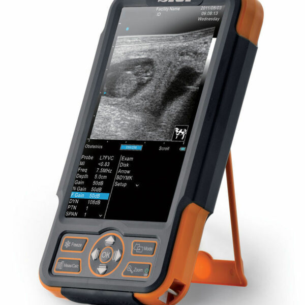
| Content | SIUI CTS-800 Veterinary Ultrasound Machine
The CTS-800 is small, lightweight, and best used for reproduction exams in most farm animals. It features vet software packages for equine, bovine, ovine, canine, feline, and primate.Features SIUI CTS-800 Veterinary Ultrasound Machine- 7.5Mhz Rectal probe with 50mm lens
- 7-inch WVGA LCD monitor
- Weighs approximately 1.8 Lbs. (0.8kg)
- Long lasting battery up to 4.5 hours
- Software & Report for reproductive system
- Gravity sensor for layout change (horizontal/vertical)
- Measurement for distance, area, circumference, volume, angle, heart rate
- Display modes include B, 2B, ZOOM B, B/M, and M mode
Included Accessories- Main unit with rechargeable battery
- Power supply adapter
- Power Cable
- 7.5Mhz Rectal probe with 50mm lens
- Car charger
- USB cable
- Video cable
- Protective case
- Hand straps
- Sunshade
- Storage box
The SIUI CTS 800 vet is a palm-sized portable ultrasound scanner for farm animals. It’s small with a 7-inch LCD monitor. The SIUI CTS-800 has a long battery life and is also waterproof. Another nice feature is a gravity sensor that will rotate the image vertically or horizontally, depending on how you’re holding the ultrasound system.Probes: L50/7.5 MHz linear rectal probe (standard configuration)L65/5.0 MHz linear rectal probe (optional)R20/5.0 MHz micro convex probe (optional)Gravity Sensor: You can operate the ultrasound system either in transversal layout or vertical layout. It can automatically adapt to two different layouts when you rotate it.Grid for estimation: With grid on the image area you can easily estimate the size of the target substance like follicle fertilized egg etc.This is a very popular unit for livestock pregnancy detection. It’s lightweight, durable, waterproof and the battery can last up to 4.5 hours. Its portability and durability plus 2-year warranty as a new system are a great value for the price of this portable veterinary ultrasound machine. | Siemens / Acuson Sequoia 512 Ultrasound Machine
The ACUSON Sequoia 512 ultrasound is an ultra-premium system that utilizes revolutionary technology, dedicated to the advancement of diagnostic imaging. It represents some of the most advanced technology in ultrasound imaging. The Acuson Sequoia 512 has unparalleled imaging performance, greater image content, enhanced detail, superior contrast resolution & penetration, and excellent color Doppler sensitivity.One of the best selling ultrasounds in the United States, the Sequois 512 provides superior imaging with Native Patient Specific Imaging that detects, measures and adapts (in real time) to each patient’s individual acoustics.Performance:Reduce image optimization time by up to 80% and allow for an easier, faster and more confident diagnosis:- 2D
- SST Color Doppler
- Color Doppler Energy
- Color Doppler Velocity
- Solo Spectral Doppler
- M-mode and Color Doppler M-mode
- MultiHertz Multiple Frequency Imaging
- DELTA Differential Echo Amplification
- Home Base user interface
- One-Touch Image Control with exam and Image Presets
- Digital acquistion, storage, review and transfer of static images, dynamic clips, and cine
- Digital storage on an integrated hard disk
- Digital transfer via MO Disk
- Integrated Compression engine
- Embedded DICOM conformance
- Patient- oriented file structure
- Native Tissue Harmonic Imaging
- Tissue Equalization™ Technology (Optional)
- Signature II™ (Optional)
- Advanced Imaging package (Optional)
- Auto Doppler (Optional)
- Freestyle Compounding (Optional)
- FreeStyle Extended Imaging (Optional)
- 3D surface Rendering (Optional)
- Cadence™ ADI (Optional)
- Cadence CPS for 512 (Optional)
- Axius™ Edge Assisted EF (Optional)
- Doppler Tissue Imaging (Optional)
- DICOM Print And Store (Optional)
Capabilities:Siemens / Acuson Sequoia 512 Ultrasound MachineGeneral Radiology, Abdominal, Intraoperative Monitoring, OB/GYN, Neonatal, Vascular, Cardiac, Small PartsAvailable Transducers:4V1, 4V2, 8V5, 15L8W, 6L3, 8L5, 6C2, 5C2, 4C1, 3V2C, 5V2C,7V3C, EVC8-4V, EC-10C5, V5M TEE, V7B neo TEE, AcuNav Transducer, 2.0 Non Imaging CW Doppler probe.Acuson sequoia 512 ultrasound click images to enlarge item description acuson sequoia 512 ultrasoundsold as picturedincludes: 2 probes, 4c1 convex & 6l4 linearsony up 895 md printerunit will be cleaned, function checked and certified for electrical safety. Checked, cleaned, certified for electrical safetyguaranteed working when arrives on the guarantee working we need to be notified in 48 hrs. And the seals must be intact. | GE Voluson 730 Pro Ultrasound MachineThe Voluson 730 Pro is built on the same platform as GE’s leading Real-Time 4D system, the Voluson 730 Expert. With an easy to use interface and exceptional 2D image quality, in addition to GE’s exclusive 4D imaging technology, the Voluson 730 Pro is ideal for any clinical practice with a variety of patient workloads. Recognized world-wide for its 4D imaging capabilities, the Voluson 730 Pro also features exceptional 2D image quality.RealTime 4D imaging – continuous, 3D scanning with simultaneous visualization of the A, B and C planes.
The system allows you to explore images in any plane to reveal the smallest details with stunning clarity and apply sophisticated analytical tools to answer virtually any of your clinical questions.Performance:GE Voluson 730 Pro Ultrasound Machine
State-of-the-art user interface with high resolution 10.4 inch LCD touch panel
Automatic Tissue Optimization
Tissue Doppler
Coded Harmonic Imaging
Coded Excitation (CE)
HD-Flow
CrossXBeamCRI (Compound Resolution Imaging)
Static 3D Mode
B Mode
Focus & Frequency Composite (FFC)
High Resolution Zoom
Pan Zoom
Steering
Virtual Convex
Beta-View
Patient information database
Image Archive on MOD and hard drive
3D/4D data compression (lossy/lossless)
Inversion
Real-time automatic Doppler calcsCapabilities:
OB/GYN, Vascular, Cardio, Abdominal, Small-Parts, Urology, Pediatrics, Ortho, Neurology, Multigestational Calculations, B-Flow, VCI (Volume Contrast Imaging)Available Probes:
AB2-7 Wide Band Convex Probe
AC2-5 Wide Band Convex Probe
4C-A Wide Band Convex Probe
M7C-H Wide Band Convex Probe
IC5-9H Wide Band Convex Probe
PA2-5P Wide Band Phased Array Probe
PA6-8 Wide Band Phased Array Probe
SP10-16 Wide Band Linear Probe
SP4-10 Wide Band Linear Probe
SP6-12 Wide Band Linear Probe
M12L-H Wide Band Linear Probe (1.25D Array)
RAB2-5L Wide Band Convex Volume Probe
RAB4-8L Wide Band Convex Volume Probe
RIC5-9H Wide Band Convex Volume Probe
RIC5-9W Wide Band Convex Volume Probe
RRE6-10 Wide Band Convex Volume Probe
RSP6-16 Wide Band Linear Volume Probe
RNA5-9 Wide Band Convex Volume Probe
SCW 2.0; non imaging suprasternal CW-Pencil Probe
PCW 4.0; non imaging peripheral CW-Pencil Probe | SonoScape S8 Portable UltrasoundSystem Description: The SonoScape S8 portable ultrasound is an award winning portable ultrasound system with outstanding image quality in all modalities. SonoScape has designed a fully featured, shared service ultrasound system producing image quality one would expect from a much more expensive console system. Frost & Sullivan “Product Quality Leadership” Award:
“The S8 is versatile in performance,” notes Frost & Sullivan Research Analyst Shriram Shanmugham. “It uses a 15 MHz linear transducer, 512 super high density transducers, and is the only echocardiography system that also offers 4D imaging capability. None of the company’s competitors offers such a combination of 4D and high-end echocardiography technology affording portability.” “The high frame rate and high definition image quality provide users better diagnostic experience than ever” Application:- Shared Service!
- Ob/Gyn
- Anesthesia
- Urology
- Vascular / Vascular Access
- MSK
- Breast Imaging
- Abdominal
- Small Parts
- Cardiac
Features:- 15” LCD Monitor
- 2 Hot Swap Probe Ports
- High Pulse Repetition Frequency [HPRF]
- Multi Beam Processing [MBP]
- Trapezoidal Imaging
- Panoramic
- Imaging
- Advanced Tissue Harmonic Imaging
- M-Tuning [One touch image optimization]
- uScan
- Multi Plane Adult & Pediatric TEE
- Intimae Media Thickness [IMT]
- High Q Nosie Filter [HQNF]
- B-Mode
- M-Mode
- Color Doppler
- Power Doppler
- Spectral Steered Doppler
- Real time Triplex: B, Flow & PW Doppler / B, Flow & Color M
- ECG
- 4D
- Tissue Doppler Imaging
- B-Color
- Anatomic M-Mode
- DICOM 3.0
- 2 USB Ports
- Video In/Out
| | Options:- Mobile Trolley
- DVD/CD-RW
- Printer
- Stress Echo
Transducers:- C344, 5-2MHz/R40mm, Abdominal Convex Probe
- C362, 5-2MHz/R60mm, Abdominal Convex probe
- C542, 7-4MHz/R40mm, Abdominal & Pediatric Probe
- C311, 4-2MHz/R15mm, Micro-convex Cardiac Probe
- C611, 8-4MHz/R11mm, Micro-convex Pediatric Cardiac Probe
- L741, 10-5MHz/46mm, Vascular Small part Linear Probe
- L742, 15-5MHz/38mm, Vascular Small part linear probe
- L743, 15-5MHz/46mm, Vascular Small part linear probe
- L541, 7-4MHz/38mm, Vascular Linear probe
- 10L1, 12-6MHz/36mm, Vascular Small Parts Probe
- 10I2, 15-5MHz/25mm, Linear Surgical Probe
- 6V1, 8-4MHz/R11mm, Endovaginal Probe (135 degree)
- 6V3, 9-5MHz/R10mm, Endovaginal probe (200 degree)
- EC9-5, 9-5MHz/R8mm, micro-convex Endocavity probe (150 degree)
- 2P1, 4-2MHz, Phased Array Cardiac Probe
- 5P1, 7-4MHz, Phased Array Pediatric Cardiac Probe
- MPTEE, 7-4MHz, Multi-plane Phased Array TEE Probe
- MPTEE Mini, 7-4MHz Pediatric Multi-plane Phased Array TEE Probe
- VC6-2, 6-2MHz/R40mm, Curved volumetric probe for Live 3D
Biopsy Guides:- C344, C362, L741, L742, L743, 10LI, L541, 6V1, 6V3, EC9-5, 2P1, 7U2
|
| Acuson Cypress PLUS UltrasoundAcuson Cypress cardiovascular system PLUS delivers advanced ergonomics and superior performance in a highly portable package. TheCypress System PLUSis a highly miniaturized, all digital, phased array echocardiography system that provides complete studies and outstanding images – even on patients who are difficult to image.The Cypress PLUS transitions easily from your small private office or hospital echo lab to the operating theatre or your patient’s bedside. It’s ultrasound where you need it.The Cypress PLUS delivers high performance in a complete range of cardiovascular capabilities.Designed with advanced ergonomics in mind, the Cypress system PLUS streamlines your workflow and increases your productivity.If you already own a Cypress system, keeping technologically current is simple with the Cypress Upgrade program.The Cypress system is the smallest, lightest, high performance, phased array echo system ever built. Weighing in at just 19 pounds, with digital storage capacity for over 100 studies and network capabilities to send studies from remote locations, clinicians can perform complete echocardiography studies from almost anywhere. TheCypress system was designed for portability, durability and reliability, and to withstand the rigors of constant transportation. Internal system interconnects are minimized. Key components are shock mounted and the entire system sits inside a robust chassis. From hospital to office or from airplane to mountain village, the Cypress system will perform.Full Range of Capabilities
The Cypress system is a complete, full-featured echo cardiography system that provides complete studies and outstanding image quality even on the most difficult to image patients. It offers a full range of features including:Acuson Cypress PLUS Ultrasound– 2D with Harmonics
– Patient Report with audio capture
– M-Mode
– Embedded DICOM conformance
– Color PW and CW Doppler
– High frame rate color flow imaging
– Stress echo
– Network output
– Vascular imaging
– Comprehensive calculations package
– Contrast agent imaging
– USB peripheral support (PC printer connectivity)Physical Characteristics
• Single unit houses ultrasound system with control panel, flat panel display and MO drive
• Weight: 8.6 Kg (19 pounds)
• Height: 35.6 cm (14 in)
• Width: 40.6 cm (16 in)
• Depth: 20.3 cm (8 in) – with keyboard folded up 34.3 cm (13.5 in) – with keyboard down
• Heat dissipation: 490 BTU/hour (nominal)User Interface
• Fold-up control panel for portability
• Direct access to system functions through dedicated keyboard controls and multifunctional soft keys
• Easy-access main controls, including multifunctional trackball, enter and select keys, for minimum hand movement while controlling the system
• Adjustable trackball speed
• Functional grouping of keys, rotational knobs, switches and sliding TGC controls for direct access to critical functions
• Tactile feedback on control activation
• Backlighting of control panel labels
• Mode-dependent soft keys
• User-interface tools
• Pre-defined and user-entered labeling textMonitor
• 10.4 inch/26.4 cm flat panel active matrix display screen
• Protective anti-glare, anti-reflective lensHard Drive
• Internal hard drive for image and data storage
• Internal storage of approximately 50 patient studies (25 – 30 loops per study)Transducer Ports
• One transducer connector (type IEC601BF)Operating/Display Modes
• 2-D, fundamental and harmonic imaging
• Color Doppler Velocity (CDV)
• Color Doppler Energy (CDE)
• M-mode, vertical split screen display with 2-D
• Pulsed Wave (PW) Spectral Doppler
• Continuous Wave (CW) Spectral Doppler
• Duplex Doppler (combined 2-D and Spectral Doppler display)2D Calipers – Generic Measurements and Calculations
• Measurements and calculations in 2D, M-mode, color Doppler, PW and CW spectral Doppler modes
• Basic measurements and report calculations
• Comprehensive patient calculations report
• On-screen “mini-reports”
• Complete or configured patient report
• Cardiac package
• Carotid package
• Arterial package
• Venous package
• Abdominal package
• User configurable Sites
• Selectable menu position
• Four cursor types
• Angle correction
• Distance
• Area
• Slope
• Time
• Velocity
• Velocity/pressure
• Mean velocity/pressure gradient
• Velocity Time Integral
• Pressure halftime
• Continuity Equation
• Ejection Fraction
• Left Ventricular Volumes, including End-Systolic and End-Diastolic volumes
• Cardiac output
• Cardiac index
• Fractional area changeCine Review
• 2D Cine memory up to 15 seconds of history
• Color Doppler cine memory up to 15 seconds of history
• M-mode strip cine along with PW and CW Doppler mode
• strip cine memory determined by sweep speed (selectable by the user) 10, 20, 30 or 40 secondsTransducer Technology
• 3V2c: 2-4 MHz wideband phased array
• 7V3c: 3-7 MHz wideband phased array
• 7L3: 5-7 MHz wideband linear array (Vascular)
• 4C1: 2-4 MHz wideband curved linear array (Abdominal)
• V5Ms: 3-6 MHz wideband phased array (micro-pinless (MP) connector) | CLASSYS ULTRAFORMER III UTR3
You’ve heard about it, now see and feel the difference for yourself.Ultraformer III is the leading technology for facial lifting, tightening and contouring. Now with 7 interchangeable cartridges that offer your patients another dimension in non-surgical face and body treatments.Ultraformer III is widely used with over 10 million treatments performed worldwide. It has become the treatment of choice for patients who are not yet indicated or not willing to undergo surgery. This fills a huge gap in the market, and captures an audience of people from 30 years of age all the way to 80.ULTRAFORMER III FEATURESLess discomfort associated with treatments
Patented transducer design for more efficient energy delivery
Dual hand pieces with automatic recognition for ease of use
Faster shot and treatment speed to save time
Easy, user-friendly design and interface
Shot count information and voice assistance
Backed by clinical papersThe CLASSYS ULTRAFORMER III UTR3 advantage with slim transducer designimproves operator visibility
supports very narrow focus and precise treatment
fits facial contoursFast elliptic transducersmore efficient treatment timesUltraformer III’s 7 clip-in transducer cartridges project uniform ultrasound beams directly to multiple layers underneath the skin to promote tighter collagen formation, tissue contraction and reduction in the volume of adipocytes that form bulging areas of tissue.Patients notice an immediate benefit in improved skin tone and texture, with ongoing improvements over 3 to 6 months
Traditionally difficult areas such as eye lifting are made easier with Ultraformer III’s specialised cartridges. It is possible for patients to experience up to 6mm of eyebrow lift, depending on their particular situation.Ultraformer III’s 7 clip-in transducer cartridges project uniform ultrasound beams directly to multiple layers underneath the skin to promote tighter collagen formation, tissue contraction and reduction in the volume of adipocytes that form bulging areas of tissue.Patients notice an immediate benefit in improved skin tone and texture, with ongoing improvements over 3 to 6 months
Traditionally difficult areas such as eye lifting are made easier with Ultraformer III’s specialised cartridges. It is possible for patients to experience up to 6mm of eyebrow lift, depending on their particular situation. |




Reviews
There are no reviews yet.