| Description | Handheld ultrasound system for Equine and Bovine Gynecology.Supplied with a 9 MHz transrectal probe.
| The Sequoia system’s advanced technologies are designed to make image acquisition easier, reduce exam time and increase overall efficiency. Along with many other included features, the Sequoia system offers Native, a patient specific imaging which adapts both phase and amplitude information of each unique patient’s properties which delivers a consistently better image across a broad range of patients. | The GE Vivid q portable ultrasound machine is a more powerful version of the Vivid i that adds further quantitative features including ICE, Auto EF, AFI, TVI,TT, TSI, Blood Flow and Blood Flow imaging. The Vivid q is the pinnacle of GE’s portable cardiovascular ultrasounds. | The Voluson 730 is available in a wide range of system capabilities and affordable price points. The original innovator in 4D OB/GYN imaging provides full diagnostic capabilities and is the perfect for the private practice office regardless of budget. | GE Voluson 730 Expert is among the top-regarded ultrasounds in the industry. With up to 40 volumes per second rate of acquisition and construction of volumetric images in real time. The 730 Expert is loaded with the newest ultrasound technology including: CrossXBeam, Speckle Reduction Imaging, Tomographic Ultrasound Imaging (TUI), Spatio-Temporal Image Correlation (STIC), and much more. | The Expert is the new flagship LOGIQ e. The imaging engine comes from GE leading console systems, delivering crisp images in a compact package. Providing ultrasound imaging with precise anatomical detail at a variety of depths. The system includes innovative features that help simplify proceedures. |
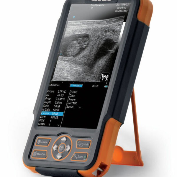
| Content | SIUI CTS-800 Veterinary Ultrasound Machine
The CTS-800 is small, lightweight, and best used for reproduction exams in most farm animals. It features vet software packages for equine, bovine, ovine, canine, feline, and primate.Features SIUI CTS-800 Veterinary Ultrasound Machine- 7.5Mhz Rectal probe with 50mm lens
- 7-inch WVGA LCD monitor
- Weighs approximately 1.8 Lbs. (0.8kg)
- Long lasting battery up to 4.5 hours
- Software & Report for reproductive system
- Gravity sensor for layout change (horizontal/vertical)
- Measurement for distance, area, circumference, volume, angle, heart rate
- Display modes include B, 2B, ZOOM B, B/M, and M mode
Included Accessories- Main unit with rechargeable battery
- Power supply adapter
- Power Cable
- 7.5Mhz Rectal probe with 50mm lens
- Car charger
- USB cable
- Video cable
- Protective case
- Hand straps
- Sunshade
- Storage box
The SIUI CTS 800 vet is a palm-sized portable ultrasound scanner for farm animals. It’s small with a 7-inch LCD monitor. The SIUI CTS-800 has a long battery life and is also waterproof. Another nice feature is a gravity sensor that will rotate the image vertically or horizontally, depending on how you’re holding the ultrasound system.Probes: L50/7.5 MHz linear rectal probe (standard configuration)L65/5.0 MHz linear rectal probe (optional)R20/5.0 MHz micro convex probe (optional)Gravity Sensor: You can operate the ultrasound system either in transversal layout or vertical layout. It can automatically adapt to two different layouts when you rotate it.Grid for estimation: With grid on the image area you can easily estimate the size of the target substance like follicle fertilized egg etc.This is a very popular unit for livestock pregnancy detection. It’s lightweight, durable, waterproof and the battery can last up to 4.5 hours. Its portability and durability plus 2-year warranty as a new system are a great value for the price of this portable veterinary ultrasound machine. | Siemens Acuson Sequoia 512 LCD Ultrasound Machine18″ Flat panel LCD Monitor–Progressive Media & Display II Package, Cardiac/OB/Vascular Calc. Package, Signature II Option, AUX, CPS Cardiology, CPS General Cadene Trigg Burst, Calc Data to MO, Card Cadence II Contrast Quant, Cornoary Flow reserve, Dicome Store/Print, DTI (Doppler Tissue Imaging), High HR ECG, MIS/MIF, Modality Worklist Native TEQ, Native TEQ, NTHI (Native Tissue Harmonics), Spectral TEQ, TEQ (Tissue Equalization), VVIFeatures- Shared Service
- Native Patient Specific Imaging
- Four Sight TEE view
- Axius quantitative strain rate imaging
- Cadence contrast pulse sequencing technology
- Native Tissue Harmonic Imaging
- Native TEQ ultrasound technology
- Color doppler, Color angio and b-color
- PW/CW Doppler
- M-Mode
- Folding Flat Panel Display
- DICOM capabilities
Applications- Abdominal
- Breast
- OB/GYN
- Vascular
- Pediatrics
- Cardiology
- Musculoskeletal
- Neonatal
Acuson Sequoia 512 Features:- Imaging: Gray-scale 2-D, M-mode, SST Color Doppler, Solo Spectral Doppler, 3-D Surface Rendering, Native Tissue Harmonic Imaging, Auto Doppler, M-Mode, PW Doppler, Color Doppler, Color Power Doppler
- 19" color CRT monitor
- Native tissue harmonic imaging (NTHI)
- DIMAQ: image management
- Advanced vascular analysis
- Advanced spatial compounding
- Native patient specific imaging
- Mitral valve assessment
- CPS contrast pulse sequencing technology
- Clarifying vascular enhancement technology
- High resolution color flow
- Vascular, and General Imaging calculation packages
- Coherent beamformer technology/spatial compounding
- Space/time resolution control
- Cine and zoom/RES
Compatible Ultrasound Probes and Transducers:- Acuson V5Ms TEE Probe
- Acuson 4C1 Curved Array Probe
- Acuson 6C2 Curved Array Probe
- Acuson 8C4 Curved Array Probe
- Acuson 4V1 Vector Array Probe
- Acuson EV-8C4 Endocavitary Probe
- Acuson EC-10C5 Endocavitary Probe
- Acuson 6L3 Linear Array Probe
- Acuson 8L5T Intraoperative Probe
- Acuson 8L5 Linear Array Probe
- Acuson 9L4 Linear Array Probe
- Acuson 15L8 Linear Array Probe
- Acuson 17L5 Linear Array Probe
The Sequoia 512 with LCD ultrasound machine uniquely minimizes automatic image optimization time by up to 80% to make way for an easier and speedier ultrasound test time as well as allow for a more confident diagnosis each time. As a patient-specific imaging ultrasound, it has a unique matched response capacity for accurate image quality, sharp clinical specificity, and optimized diagnostic outcomes that makes infinitesimal pathologies and other anatomical detail in the most difficult-to-image patients becomes more vivid and visible. | GE Vivid Q Ultrasound Machine
The powerful GE Vivid Q ultrasound machine is the ultimate lightweight, compact high-performance cardiovascular system for clinicians needing a machine that combines user efficiency and reliable performance. This unique system performs consistently thanks to its TruScan architecture and can support up to 17 probes. The GE Vivid Q is recognized for its premium image quality, advanced image management, and high-performance quantification tools.This advanced GE ultrasound delivers improved patient care and enhanced user productivity with the new M4s-Matrix array technology transducer, which provides greatly enhanced image quality in electronic beamforming, raw data image acquisition and archiving, optimal onboard and post processing technology.This ultrasound system provides high clinical values for quantification and review of stored images, making it one of today’s most sought-after models. Some of its powerful features include compatibility with EchoPac clinical workstation and DICOM networking, speckle tracking strain, integrated stress echo, quantification tools, and reporting capabilities. Also of note is one of the Vivid Q’s best new features, which is its TEE transducer capability.A one-touch button optimization provides premium image quality for workflow efficiency, and maintains AFI- automatic function imaging with complete wallFeatures- Advanced QScan Imaging (incl TSI)
- AdvStress
- ATO
- Auto EF
- Automated Function Imaging
- Blood Flow Imaging
- CardiacApp
- Cardio Lab
- Color Doppler
- CW Doppler
- DICOM Networking
- DICOM: Print
- DICOM: Worklist
- DVD/RW
- ECG
- ECG Cable
- EchoDicom
- EchoPAC
- Evue
- ICE Imaging
- Intima Media Thickness (IMT)
- LVO Contrast
- M Mode
- M4S
- MPEGvue
- PW Doppler
- Small Depth
- TEE
- THI
- Tissue Harmonics Imaging (THI/NTHI)
- Tissue Velocity Imagine & Tissue Tracking
- TSI
- VascAbdContrast
- VascularApp
- VirtualPrinter
- Wide Aperture
GE Vivid Q FeaturesThe GE Vivid Qis a Portable Laptop-style Ultrasound that can be used to help diagnose cardiovascular anatomy. Features and tools based on the previous Vivid 7 and the Vivid E9 have been added to the new lightweight Vivid Q, mainly the M4S-RS transducer that allows for 3D image focusing and enables a higher level of image quality. The Vivid Q ultrasound is versatile and can additionally be used for Cardiac, vascular, Abdominal, Pediatric, Obstetrics, and Emergency medicine. The Vivid Q Ultrasound comes with a 15-inch color LCD monitor that can display a variety of imaging modes including; ICE, Auto EF, AFI, TVI, TT, TSI, and Blood Flow. Additional the Ultrasound system is comparable with 17 different transducers and probes to fit every imaging need. - Auto EF - Automated ejection fraction measurement program designed specifically for the GE Vivid q.
- Tissue Synchronization Imaging (TSI).
- Intracardiac Echo (ICE).
- 16 different compatible probes.
- Coded Octave imaging - enhances image color flow and Doppler imaging.
| GE Voluson 730 Cardiovascular Ultrasound MachineThe Voluson 730 is available in a wide range of system capabilities and affordable price points. The original innovator in 4D OB/GYN imaging provides full diagnostic capabilities and is the perfect for the private practice office regardless of budget.GE VOLUSON 730 PRO/EXPERT BT08The GE Voluson 730 Pro and the Expert BT08 are among the top-rated ultrasounds, known widely as the workhorses of 4D imaging machines. Especially in today’s fast-paced clinical environments, from routine 2D imaging to powerful 3D/4D imaging capabilities, the GE Voluson 730 Pro and the Expert BT08 ultrasounds allow you to demand more from your machine than ever before.GE Voluson 730 Pro/Expert BT08 ImagingThe GE Voluson 730 Pro ultrasound and the Expert BT08 ultrasound provide the capabilities for acquiring and constructing volumetric images in real-time; in fact, you can achieve up to 40 volumes per second. With this type of imaging technology, users will be able to capture and analyze images of any plane while the ultrasound reveals the smallest details in stunning clarity. With these technologies, the GE Voluson 730 Pro/Expert BT08 ultrasound is highly regarded for:Applications- OB/GYN
- Vascular
- Abdominal
- Small Parts
- MSK
- Urology
- Cardiology
- Neurology
- Orthopedic
- Pediatric
- Neonatal
| Key Standard Features- B, CFM, M, PW, 3D/4D
- Anatomical M-Mode
- 15’’ High-Resolution CRT Monitor
- Automatic Tissue Optimization (ATO)
- Static 3D
- Real Time 4D
- Coded Excitation
- Virtual Convex
- TruScan Architecture (TruAccess, SmartScan, ComfortScan)
- Internal DVD/CD-RW Drive
- 10” Touchscreen User Menu
|
Optional Features- 4D Real Time
- Vocal II
- 4D DiagnoSTIC
- DICOM 3.0 Networking
- Volume Contrast Imaging (VCI)
- B-Flow
- VOCAL
- Volume Contrast Imaging (VCI)
- Speckle Reduction Imaging (SRI)
- Tissue Doppler Imaging (TDI)
- Tomographic Ultrasound Imaging (TUI)
- Coded Contrast Imaging
- Compound Resolution Imaging (CRI)
- DICOM Modality Worklist / Structured Reporting
| Transducers- AB2-7- Convex Array Transducer
- RAB2-5 Volume Convex Array Transducer
- RAB2-5L Volume Convex Array Transducer
- RAB4-8P Volume Convex Array Transducer
- RAB4-8L Volume Convex Array Transducer
- RRE6-10 Volume Convex Array Transducer
- IC5-9 Volume Endocavity Convex Array Transducer
- RIC5-9 Volume Endocavity Convex Array Transducer
- SP10-16 Linear Array Transducer
- SP4-10 Linear Array Transducer
- SP6-12 Linear Array Transducer
|
| GE Voluson 730 Expert Ultrasound MachineGE Voluson 730 Expert, 3D/4D Realtime Ultrasound System Color Doppler, PW Doppler, Tissue Harmonics, STIC, VOCAL DICOM, M-mode, Angio, ATO, TUI, SonoView II, Crossbeam (CRI) MO Drive, Cineloop, High Resoluotion Interlaced Monitor Probe Options: RAB4-8 4D Convex RIC5-9 4D Transvaginal AB2-7 2D Convex IC5-9 2D Transvaginal SP6-12 2D Linear PA2-5 Cardiac Sector
Two Probes included:- RAB4-8L – Live 4D Abdominal Transducer
- RIC5-9 – Live 4D Transvaginal Transducer
Options:
About the Voluson 730:GE Voluson 730 Expert Ultrasound MachineWith GE Voluson 730, you can now acquire and construct volumetric images in real time – up to 40 volumes per second. The system allows you to explore images in any plane to reveal the smallest details with stunning clarity and apply sophisticated analytical tools to answer virtually any of your clinical questions. Volume Ultrasound not only improves your 3D/4D capabilities, it enhances your 2D imaging as well, for even greater diagnostic confidence in your obstetrical, gynecologic, breast and general imaging studies.The used GE Voluson 730 Expert was among the top-rated and biggest breakthrough ultrasounds in the industry. With up to 40 volumes per second rate of acquisition and construction of volumetric images in real time. The 730 Expert is loaded with the newest ultrasound technology including: CrossXBeam, Speckle Reduction Imaging, Tomographic Ultrasound Imaging (TUI), Spatio-Temporal Image Correlation (STIC), and much more.The Voluson 730 Expert is the more advanced version of the Voluson 730 Pro and was replaced by the GE Voluson e8. Compared to the 730 Pro, the Voluson 730 Expert features a touchscreen interface, higher frame rates in 2D and 4D, and more advanced imaging options such as STIC and TUI.Providian Medical Voluson 730 Expert Review:The Voluson 730 is a workhorse of a 4D ultrasound machine. It remains a popular system in many OB/GYN/Women’s Health offices, as well as 4D “Babyface” studios around the world. It is a durable machine with very good image quality for all women’s health needs. | GE Logiq e r7 Portable Ultrasound Machine
The LOGIQ e R7 provides even more impressive veterinary abdominal and cardiac images than the BT12 LOGIQe, combining the high performance of a console system with the portability of a laptop.Highlights of the GE LOGIQ e R7:- Simple. Fast. Precise
- Colour Flow Mode, Power Doppler Imaging (PDI), Pulse Wave Doppler (PWD), Continuous Wave Doppler
- 15” high resolution colour LCD screen
- Integrated solid state hard drive
- Automatic optimisation
- CrossXBeam
- Speckle Reduction Imaging (SRI-HD)
- Virtual Convex / Virtual Apex
- Fine Angle Steer
- HD Zoom (Write Zoom)
- Coded Harmonic Imaging (CHI) – Enhances near field reslolution for improved small parts imaging as well as far field penetration. Diminshes low frequency / amplitude noise and provides clarity to needle, anatomy and motion.
- Raw Data Processing
- Quicksave – Single button push sends single image or entire patient exam to memory stick or network
- On-board manual (Help)
- Loop storage – from live scanning and from memory
- Patient Information Database
- Customisable user interface
- Full M&A calculation package with real time Doppler calculations
- Report writer package – on board reporting package automates report writting
- Portable and battery operated so easy to move from room to room
- Export of stills and clips to USB.
More GE Logiq e r7 Portable Ultrasound MachineThe Expert is the new flagship LOGIQ e. The imaging engine comes from GE leading console systems, delivering crisp images in a compact package. Providing ultrasound imaging with precise anatomical detail at a variety of depths. The system includes innovative features that help simplify proceedures.GE NextGen LOGIQ e R7 Ultrasound System The GE NextGen LOGIQ e R7 Ultrasound System has an imaging engine that delivers crisp images in a compact package. Point-of-care-specific software and transducers help to see the needle to administer a block or perform an aspiration quickly. When productivity is crucial, you can control the console from the transducer, potentially eliminating the need to have a second person assist. Anatomy-specific presets and only displaying the functions that you need helps you to direct your focus on the patient rather than the control panel. Although the system features a large, user-friendly, high-res color display, it is also very durable and compact for easy portability. |


Reviews
There are no reviews yet.