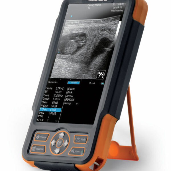
| Content | SIUI CTS-800 Veterinary Ultrasound Machine
The CTS-800 is small, lightweight, and best used for reproduction exams in most farm animals. It features vet software packages for equine, bovine, ovine, canine, feline, and primate.Features SIUI CTS-800 Veterinary Ultrasound Machine- 7.5Mhz Rectal probe with 50mm lens
- 7-inch WVGA LCD monitor
- Weighs approximately 1.8 Lbs. (0.8kg)
- Long lasting battery up to 4.5 hours
- Software & Report for reproductive system
- Gravity sensor for layout change (horizontal/vertical)
- Measurement for distance, area, circumference, volume, angle, heart rate
- Display modes include B, 2B, ZOOM B, B/M, and M mode
Included Accessories- Main unit with rechargeable battery
- Power supply adapter
- Power Cable
- 7.5Mhz Rectal probe with 50mm lens
- Car charger
- USB cable
- Video cable
- Protective case
- Hand straps
- Sunshade
- Storage box
The SIUI CTS 800 vet is a palm-sized portable ultrasound scanner for farm animals. It’s small with a 7-inch LCD monitor. The SIUI CTS-800 has a long battery life and is also waterproof. Another nice feature is a gravity sensor that will rotate the image vertically or horizontally, depending on how you’re holding the ultrasound system.Probes: L50/7.5 MHz linear rectal probe (standard configuration)L65/5.0 MHz linear rectal probe (optional)R20/5.0 MHz micro convex probe (optional)Gravity Sensor: You can operate the ultrasound system either in transversal layout or vertical layout. It can automatically adapt to two different layouts when you rotate it.Grid for estimation: With grid on the image area you can easily estimate the size of the target substance like follicle fertilized egg etc.This is a very popular unit for livestock pregnancy detection. It’s lightweight, durable, waterproof and the battery can last up to 4.5 hours. Its portability and durability plus 2-year warranty as a new system are a great value for the price of this portable veterinary ultrasound machine. | Medison Accuvix XG Ultrasound System
Samsung Accuvix XG Ultrasound Machine2D Image Features
• Dynamic MR™ / Dynamic MR Plus is designed to enrich gray-scale resolution, as it enhances detection
and contrast resolution while also decreasing speckle echoes. This is particularly useful when evaluating
superficial structures, including thyroid, vessels, pelvic and abdominal anatomy.
• SRF (Speckle Reduction Filter™) enhances image quality by reducing or eliminating the appearance of
speckle echoes from ultrasound images. The degree of speckle reduction implemented is user-selectable.
• Wide Dynamic Range determines the number of gray shades utilized to map the gray-scale image.
It enables display of more details of bright areas and dark areas.
• SCI (Spatial Compounding Image) controls the ultrasound beam electronically by steering, and compounds
many scan lines.
• FSI (Full Spectrum Imaging) incorporates the penetration capabilities associated with lower frequencies,
yet maintains the fine pixel uniformity associated with higher frequencies, to deliver consistently high quality
images even in challenging diagnostic areas.3D Image Features Medison Accuvix XG Ultrasound System
• HDVI gives outstanding image quality and naturally clearer contrast, with excellent tissue differentiation,
edge depiction and speckle reduction, allowing consistent diagnoses with great confidence.
• 3D XI™, comprised of a suite of three innovative imaging applications – Multi-Slice View, Oblique View™ and
Volume CT – offers complete and precise control over 3D/4D volume data manipulation for maximum
diagnostic accuracy.
• 3D MXI is an innovative, cutting-edge 3D image processing technology. Comprising a comprehensive suite of
imaging tools – including Multi Volume Slice, Mirror View, Multi-OVIX, and 3D OH – 3D MXI lets you view,
examine and diagnose 3D volume data with supreme ease, speed and accuracy. • Fully adjustable system
The control panel can be adjusted to the user’s preferred height, for a better working environment and reduced
risk of back pain.
• Wide LED touch-screen
The Accuvix XG’s new LED touch-screen makes it easy to organize and operate.
• 19-inch HD LCD Monitor and articulating monitor arm
A 19-inch LCD monitor enables images to be displayed clearly even with a larger monitor, and the articulating
monitor arm enables easy mobility for a more comfortable and convenient working environment.
• Portability
The Accuvix XG is a lightweight system with 4 swivel wheels that allow easy steering, and a locking function. • Customizable measurement menus
Customizable measurement menus allow access to frequently-used functions, and enable a quicker and
more intuitive workflow.
• User keys and user knob
Accuvix XG offers user keys and a user knob that can map frequently-used functions, enabling the function to be
activated quickly and easily.
• Customized Annotation Menu and body marker
Users can preset up to 360 words of annotations, and body markers for each application, that reduce the time
needed for each examination.
• QuickScan
QuickScan maximizes workflow efficiency by automatically optimizing key imaging parameters with just the
push of a button. • HDVI
HD Volume Imaging(HDVI) gives outstanding image quality and naturally clearer contrast, with excellent tissue
differentiation, edge depiction and speckle reduction, allowing consistent diagnoses with great confidence.
It shows clearer images of subtle lesions, using multi slice image as well as C-plane.
• ElastoScan
ElastoScan™ translates stiff & soft tissue information into gray scale image. This technology defines the alteration
of the stiffness in tissue state more accurately, making it easier for the user to utilize the information in the image. | GE Logiq A5 Ultrasound MachineThe lightweight, portable LOGIQ* A5 ultrasound system offers capabilities to help meet your imaging needs — particularly for urological applications.Affordable ultrasound.The LOGIQ A5 puts imaging capabilities at your fingertips in a portable, lightweight design. This system can go from your office to the surgery suite and helps meet a range of general imaging needs. Well-suited for urology, tools include soft tissue image analysis of the kidneys, bladder, penis, testicles, and prostate.Solid imaging performance.
The sleek LOGIQ A5 can help meet your general imaging needs. This black and white ultrasound system has:- B-mode imaging capability and features that allow you to add advancements as your clinical needs grow.
- Auto TGC helps ensure a homogeneous picture from the near-to-far field.
- Virtual Convex provides an expanded field of view that enables you to see more anatomy with linear transducers.
- Color flow Doppler upgrade available to provide dynamic assessment of blood flow in the organs, as well as investigation of hypertension and resulting erectile dysfunction.
Perform imaging with ease.
The LOGIQ A5 has user-friendly features with precision handling.- Automatic Optimization (AO), helps provide improved image quality at the touch of a button for B-mode, spectral and color Doppler imaging.
- Full-size keyboard for optimal ease of use.
- 15-inch adjustable LCD monitor allows a clear view in any position.
- Lightweight package easily travels with you.
Compatible Transducers:- 10L Linear Array
- 11L Wide Band Linear
- 12L Linear Array
- 3.5C Convex
- 3.5CS Convex (2-5 MHz)
- 3CRF Convex
- 3S Phased Array
- 3Sp Wide Band Phased Array Sector
- 4C Convex Array
- 4D3CL Real time 4D Convex
- 4D8C Convex Volume
- 4DE7C Real time 4D Endocavity
- 5CS Convex (2-5 MHz)
- 5S Cardiac Sector
- 5Sp Wide Band Phased Array Sector
- 7S Phased Array
- 8C Convex Array
- 8L Linear (3-7 MHz)
- 9L Wide Band Linear
- BE9C Transrectal
- BE9CS Micro convex Bi-plan
- E8C (4-10 MHz) Wide Band Microconvex
- E8CS Micro-convex (4-11 MHz)
- ERB Brachytherapy Transrectal
- i12L Linear Array
- i739 Intraoperative
- P2D CWD Non-Imaging
- P6D CWD Non-Imaging
- T739 Intra-operative
| Terason uSmart 3200t Ultrasound
The new light-weight, power-packed uSmart 3200T Ultrasound System is a fusion of cutting-edge technology with an intuitive interface that defines simplicity. The newest member of the Terason family weighs less than 5 lbs., provides razor-sharp image quality, and is fully equipped with all the features and functionality users have come to expect from Terason products. Developed by clinicians for clinicians, the uSmart 3200T provides the best tools for busy practices, including a 128GB Solid State hard drive, Smart Gestures, an Adaptive Touch Screen, uConnect remote capabilities, and a fast boot-up time.The Terason uSmart 3200T Ultrasound is truly a unique and revolutionary product in the world of portable ultrasound. In our fast-paced technology environment, healthcare providers require products that can keep up with the ever-changing demands of their hospital or practice. We have developed the first tablet-based system where high technology meets superior performanceThe Terason uSmart 3200T is a tablet-style portable color Doppler ultrasound machine designed for the point-of-care bedside ultrasound market.This handheld ultrasound weighs less than 5lbs and uses gestures and touch operation much like a tablet or touchscreen computer. With Terason’s well-known unique beamforming microprocessor technology, the Terason uSmart 3200T Ultrasound provides excellent image quality with an efficient workflow and easy use.Gold Standard, Crystal Clear Images you have to see to believe! As an industry pioneer, Terason developed and is the first to patent color portable ultrasound. Since then, our razor-sharp image quality has earned a reputation for excellence.
The uSmart 3200T captures every detail, subtlety, and nuance that others overlook. The AHVA LCD provides consistent image quality from multiple viewing angles.Grab-And-Go Portability
As part of one of the most comprehensive product families in the industry, the uSmart 3200T weighs in at just under 5 pounds. The touchscreen interface, small footprint an space-saving power dock can go anywhere you need quickly and easily. It’s ready when you are! The Best Tools For Your Practice- Small Gestures
- Adaptive touch screen
- uConnect remote capabilities
- Multi-Function docking station
- Solid State Hard Drive
- Application-Specific presets
- Fast boot-up time
Enhanced Needle Visualization (ENV)
With ENV- AS a leader in ultrasound, Terason is pleased to introduce ENV. This feature provides needle visualization unlike any other. - Frame rate stability
- One-button operation
- Works with all needle gauges
- 15L transducer solution
- Easy to use
Terason uSmart 3200T Ultrasound Specifications- Dimensions: 22.4 cm x 3. 2 cm x 3.21 cm
(8.82″ x 1.26″ x 12.64″)
- 12.5″ x 1.25″ x 8.75″ High-Res AHVA LCD – Touch Screen
- 128 GB Hard Drive
- Input / Output: 3 USB-A, Ethernet & DVI
- Weight: 4.5 pounds
- Export: to USB, Network Folder
- Operates on AC or internal battery power
Imaging Modes:- B- mode
- M-mode
- Color Doppler
- Color Power Doppler
- Directional Color Power Doppler
- PW Doppler
| Siemens Acuson Sequoia 512 LCD Ultrasound Machine18″ Flat panel LCD Monitor–Progressive Media & Display II Package, Cardiac/OB/Vascular Calc. Package, Signature II Option, AUX, CPS Cardiology, CPS General Cadene Trigg Burst, Calc Data to MO, Card Cadence II Contrast Quant, Cornoary Flow reserve, Dicome Store/Print, DTI (Doppler Tissue Imaging), High HR ECG, MIS/MIF, Modality Worklist Native TEQ, Native TEQ, NTHI (Native Tissue Harmonics), Spectral TEQ, TEQ (Tissue Equalization), VVIFeatures- Shared Service
- Native Patient Specific Imaging
- Four Sight TEE view
- Axius quantitative strain rate imaging
- Cadence contrast pulse sequencing technology
- Native Tissue Harmonic Imaging
- Native TEQ ultrasound technology
- Color doppler, Color angio and b-color
- PW/CW Doppler
- M-Mode
- Folding Flat Panel Display
- DICOM capabilities
Applications- Abdominal
- Breast
- OB/GYN
- Vascular
- Pediatrics
- Cardiology
- Musculoskeletal
- Neonatal
Acuson Sequoia 512 Features:- Imaging: Gray-scale 2-D, M-mode, SST Color Doppler, Solo Spectral Doppler, 3-D Surface Rendering, Native Tissue Harmonic Imaging, Auto Doppler, M-Mode, PW Doppler, Color Doppler, Color Power Doppler
- 19" color CRT monitor
- Native tissue harmonic imaging (NTHI)
- DIMAQ: image management
- Advanced vascular analysis
- Advanced spatial compounding
- Native patient specific imaging
- Mitral valve assessment
- CPS contrast pulse sequencing technology
- Clarifying vascular enhancement technology
- High resolution color flow
- Vascular, and General Imaging calculation packages
- Coherent beamformer technology/spatial compounding
- Space/time resolution control
- Cine and zoom/RES
Compatible Ultrasound Probes and Transducers:- Acuson V5Ms TEE Probe
- Acuson 4C1 Curved Array Probe
- Acuson 6C2 Curved Array Probe
- Acuson 8C4 Curved Array Probe
- Acuson 4V1 Vector Array Probe
- Acuson EV-8C4 Endocavitary Probe
- Acuson EC-10C5 Endocavitary Probe
- Acuson 6L3 Linear Array Probe
- Acuson 8L5T Intraoperative Probe
- Acuson 8L5 Linear Array Probe
- Acuson 9L4 Linear Array Probe
- Acuson 15L8 Linear Array Probe
- Acuson 17L5 Linear Array Probe
The Sequoia 512 with LCD ultrasound machine uniquely minimizes automatic image optimization time by up to 80% to make way for an easier and speedier ultrasound test time as well as allow for a more confident diagnosis each time. As a patient-specific imaging ultrasound, it has a unique matched response capacity for accurate image quality, sharp clinical specificity, and optimized diagnostic outcomes that makes infinitesimal pathologies and other anatomical detail in the most difficult-to-image patients becomes more vivid and visible. | SonoSite Nanomaxx Ultrasound MachineThe SonoSite NanoMaxx ultrasound system combines premium functionality and affordability with the versatility and portability that makes SonoSite a top competitor in the portable ultrasound market. The SonoSite NanoMaxx comes in a sleek, hand-held package that never compromises performance, with applications ranging from emergency medicine and anesthesia to vascular.MedCorp offers the refurbished SonoSite NanoMaxx for purchase. Used SonoSite NanoMaxx from MedCorp offer everything you’d expect from a newer machine, including PC/MAC export configurations, 2D, Color and Color Power Doppler modes, and 2GB of internal memory that can hold 50 images each for 40 patients, all in an 8” touch-screen interface. However, because this is a refurbished SonoSite NanoMaxx, the price is lower than the new models. - 8″ Touch-screen interface
- 2D, Color and Color Power Doppler modes
- 2GB internal memory that can hold 50 images each for 40 patients
- 2 hours of battery power
- PC/MAC export configuration
- Four preset pictograph screen positions
- Drop resistant magnesium case
System Description:SonoSite Nanomaxx Ultrasound Machine
The NanoMaxx ultrasound system offers excellent performance at an affordable price. It is easy to use and offers top of the line diagnostic imaging, color power Doppler and color-flow velocity. The NanoMaxx can aid users in making clinical decisions or guide interventional procedures. The unit uses proprietary technology which allows the optimization of many system settings by simply pressing a button. Application:
Abdominal, Cardiology, Gynecology, Nerve, Obstetrics, CIMT, Musculoskeletal, Small Parts, Superficial, Vascular, Venous |


Reviews
There are no reviews yet.