| Description | Applications:
• Fetal/Obstetrics
• Abdominal (includes renal, GYN/Pelvic)
• Pediatric
• Small Organ (breast, testes, thyroid)
• Neonatal Cephalic
• Adult Cephalic
• Cardiac (adult and pediatric)
• Peripheral Vascular
• Musculo-skeletal Conventional and Superficial
• Urology (including prostate)
• Transrectal
• Transvaginal
• Transesophageal | The Expert is the new flagship LOGIQ e. The imaging engine comes from GE leading console systems, delivering crisp images in a compact package. Providing ultrasound imaging with precise anatomical detail at a variety of depths. The system includes innovative features that help simplify proceedures. | 15″ LCD monitor, 2 Hot-swappable Probe ports, Keyboard with programmable keys, HPRF (High Pulse Repetition Frequency), MBP (Multi-beam Processing), Trapezoidal Imaging, Panoramic Imaging, Advanced THI (Tissue Harmonic Imaging), TDI (Tissue Doppler Imaging) ,MicroScan (Noise & Artefact Reduction), Multi Parameter Compound Imaging, M Tuning (One Touch Image Optimization), Steerable M mode, uScan, Bi-Plane Transrectal Imaging, Multi-Plane Adult and Paediatric TEE/TOE, Endocavitary Imaging with Extended Field-of-View, Stress Echo, anatomic M mode, PDF report, Automatic flow volume analysis, IMT, HQNF (High Q Noise Filter), 2 USB ports, Video in/out ports, Li-ion battery. | M-Turbo is a compact ultrasound imaging system from SonoSite, featuring the image quality and functionality needed to meet your diverse needs. The standard M-Turbo is also still available exclusively from BCF. | The Sequoia system’s advanced technologies are designed to make image acquisition easier, reduce exam time and increase overall efficiency. Along with many other included features, the Sequoia system offers Native, a patient specific imaging which adapts both phase and amplitude information of each unique patient’s properties which delivers a consistently better image across a broad range of patients. | Function • Gravity Sensor: You can operate the ultrasound system either in transversal layout or vertical layout.• Grid for estimation: With grid on the image area, you can easily estimate the size of the target substance like follicle fertilized egg etc. |
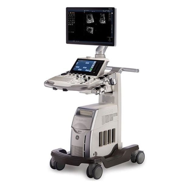
| Content | GE LOGIQ S7 WITH XDCLEAR ULTRASOUND
The GE Logiq S7 with XDclear Ultrasound is a mid range multi-purpose ultrasound machine. The GE Logiq S7 with XDClear has been fully upgraded, 23″ large LCD monitor and 10.1″ LCD touch screen. It becomes very reliable and trustworthy while reaching the revision 3.0, and GE incorporated single crystal transducers, so called XDClear transducers such as C1-6-D and C3-10-D. With one of the market most demanding technologies, XDClear, the user can enjoy premium image resolution, penetration, and wide bandwidth from two XDClear options. The GE Logiq S7 with XDClear built with the same system platform of GE Logiq S8 with XDClear, S-agile architecture, but just some of the features and transducer are not included or more economic options and transducers are integrated. Although the GE Logiq S7 with XDClear is classified as a mid range system, it offers a great benefit to users with advanced automated features such as Scan Assistant, OB Measure Assistant, and Breast Measure Assistant. Also, Automated Function Imaging(AFI), XDClear probes, large 23″ LCD monitor and 10.1″ touch screen are very unique features in mid-range.GE Logiq S7 With XDClear Dimensions and Weight:• Height: 1,210mm (Minimum), 1,760mm (Max.)
• Width: 530mm (Caster), 520mm (Keyboard), 565mm (Monitor)
• Depth: 865mm (Max.), 795mm (Caster)
• Weight: 90 kg (198 lb.) GE Logiq S7 With XDClear Specifications:• Digital beamformer
• Displayed Imaging Depth: 0 – 33 cm
• Minimum Depth of Field: 0 – 2 cm (Zoom) (probe dependent)
• Maximum Depth of Field: 0 – 33 cm (probe dependent)
• Continuous dynamic receive focus/continuous dynamic receive aperture GE Logiq S7 With XDClear Electrical power:• Voltage:100-120 Vac or 220-240 Vac
• Frequency: 50/60 Hz
• Power: Consumption maximum of 900 VA with peripherals GE Logiq S7 XDClear Features:• High resolution 23″ wide LCD monitor
• High resolution 10.1″ wide LCD touch screen
• 4 active probe ports and 1 parking probe port
• Integrated HDD
• Integrated DVD Multi Drive
• Integrated speakers
• Locking mechanism that provides rolling lock and caster swivel lock
• Integrated cable management
• Front and rear handles
• Easily removable air filters
• Automatic Optimization
• CrossXBeam
• Speckle Reduction Imaging (SRI-HD)
• Fine Angle Steer
• Coded Harmonic Imaging
• Virtual Convex
• Advanced 3D
• Patient information database
• Image Archive on integrated CD/DVD and hard disk drive
• Raw Data Analysis
• Real-time automatic Doppler calcs
• OB Calcs
• Fetal Trending
• Multi gestational Calcs
• Hip Dysplasia Calcs
• Gynecological Calcs
• Vascular Calcs
• Urological Calcs
• Renal Calcs
• Cardiac Calcs
• inSite ExC Capability
• On-board electronic documentation
• MPEGVue
• Key Macro
• Network Storage
• Quick Save
• Quick Patient Entry
• Email2MMS Accessories GE Logiq S7 with XDclear Ultrasound GE Logiq S7 With XDClear Peripheral Options:• Sony Digital UP-D711 Termal Printer
• Sony Fixture Kit for Digital UP-D711 Thermal Printer
• Sony Digital UP-D25 Color Thermal Printer
• Sony Digital UP-D897 BW Thermal Printer
• Mitsubishi P93W/E Thermal Printer
• Mitsubishi P95DW Thermal Printer
• Printer installation kit
• External USB printer connection
• Printing using Bluetooth connectivity (Inkjet printer option)
• Wireless LAN
• HDMI output available for compatible devices
• Footswitch with programmable functionality
• Universal Video Converter
• Power Assistant
• Battery Pack (for Power Assistant)
• Isolation Transformer
• Isolated 1Gb Ethernet connection
• Isolated USB connector
• EMI filter (PWR supply noise filter)
• RS-DLP Probe Adapter for TEE Probe
• Side Cabinet (Japan only)
• Drawer
• Small Probe Holder (Probe Holder Adapter for Small Probe)
• Multi Purpose Holder (Vertical Endocavitary Probe Holder)
• Optional Probe Holder (Side Probe Holder)
• Probe cable hanger
• ECG + AHA/IEC Cables
• USB 3 Pedal Foot Switch GE Logiq S7 With XDClear Supplies:Aquasonic ultrasound gel
Sono ultrasound wipes
Sony UPP-110HG thermal printing paper
Sony UPC-21L color thermal printing pack
Mitsubishi KP95HG thermal roll paper (new)
Mitsubishi KP65HM-CE High density thermal paper GE Logiq S7 With XDClear ports:• User accessible USB x 5 ports
• HDMI connector
• Ethernet 1000/100/10BaseT
• Audio Line Out (1.5mm pin jack) GE Logiq S7 With XDClear image storage:• On-board database of patient information from past exams
• Storage Formats:
• DICOM – compressed/ uncompressed, single/ multiframe, with/ without Raw Data
• Export JPEG, JPEG2000, WMV (MPEG 4), and AVI formats
• Storage Devices: – USB Flash Device: 64MB to 64GB (for exporting individual images/clips) – CD-R storage: 700MB – DVD storage: R (4.7GB) – Hard Drive Image Storage: ~300GB – Compare previous exam images with current exam images – Reload of archived data sets – Network Storage support for Import, Export, DICOM Read, SaveAs, SaveAs GE Logiq S7 with XDClear Options:• Auto IMT
• Auto EF
• Automated Volume Calculation (VOCAL II)
• Automated Function Imaging (AFI)
• Breast Measure Assistant
• OB Measure Assistant
• B-Flow/B-Flow Color
• B Steer+
• Breast Productivity Package
• Coded Contrast Imaging
• HRES Contrast
• Compare Assistant
• CW Doppler
• DICOM 3.0 connectivity
• Digital Video Recording Software (Software DVR)
• Elastography
• Elastography Quantification
• Flow Quantification
• LOGIQView
• Real Time 4D
• Report Writer
• Scan Assistant
• Stress Echo
• Thyroid Productivity Package
• Tissue Velocity Imaging (TVI)
• Tomographic Ultrasound Imaging (TUI)
• Volume Contrast Imaging (VCI) Static
• OmniView
• STIC
• Advanced Probes (XDclear)
• Cabinet: High/Mid/Low GE Logiq S7 with XDClear Applications:• Fetal/Obstetrics
• Abdominal (includes renal, GYN/Pelvic)
• Pediatric
• Small Organ (breast, testes, thyroid)
• Neonatal Cephalic
• Adult Cephalic
• Cardiac (adult and pediatric)
• Peripheral Vascular
• Musculo-skeletal Conventional and Superficial
• Urology (including prostate)
• Transrectal
• Transvaginal
• Transesophageal | GE Logiq e r7 Portable Ultrasound Machine
The LOGIQ e R7 provides even more impressive veterinary abdominal and cardiac images than the BT12 LOGIQe, combining the high performance of a console system with the portability of a laptop.Highlights of the GE LOGIQ e R7:- Simple. Fast. Precise
- Colour Flow Mode, Power Doppler Imaging (PDI), Pulse Wave Doppler (PWD), Continuous Wave Doppler
- 15” high resolution colour LCD screen
- Integrated solid state hard drive
- Automatic optimisation
- CrossXBeam
- Speckle Reduction Imaging (SRI-HD)
- Virtual Convex / Virtual Apex
- Fine Angle Steer
- HD Zoom (Write Zoom)
- Coded Harmonic Imaging (CHI) – Enhances near field reslolution for improved small parts imaging as well as far field penetration. Diminshes low frequency / amplitude noise and provides clarity to needle, anatomy and motion.
- Raw Data Processing
- Quicksave – Single button push sends single image or entire patient exam to memory stick or network
- On-board manual (Help)
- Loop storage – from live scanning and from memory
- Patient Information Database
- Customisable user interface
- Full M&A calculation package with real time Doppler calculations
- Report writer package – on board reporting package automates report writting
- Portable and battery operated so easy to move from room to room
- Export of stills and clips to USB.
More GE Logiq e r7 Portable Ultrasound MachineThe Expert is the new flagship LOGIQ e. The imaging engine comes from GE leading console systems, delivering crisp images in a compact package. Providing ultrasound imaging with precise anatomical detail at a variety of depths. The system includes innovative features that help simplify proceedures.GE NextGen LOGIQ e R7 Ultrasound System The GE NextGen LOGIQ e R7 Ultrasound System has an imaging engine that delivers crisp images in a compact package. Point-of-care-specific software and transducers help to see the needle to administer a block or perform an aspiration quickly. When productivity is crucial, you can control the console from the transducer, potentially eliminating the need to have a second person assist. Anatomy-specific presets and only displaying the functions that you need helps you to direct your focus on the patient rather than the control panel. Although the system features a large, user-friendly, high-res color display, it is also very durable and compact for easy portability. | SonoScape S8 Portable UltrasoundSystem Description: The SonoScape S8 portable ultrasound is an award winning portable ultrasound system with outstanding image quality in all modalities. SonoScape has designed a fully featured, shared service ultrasound system producing image quality one would expect from a much more expensive console system. Frost & Sullivan “Product Quality Leadership” Award:
“The S8 is versatile in performance,” notes Frost & Sullivan Research Analyst Shriram Shanmugham. “It uses a 15 MHz linear transducer, 512 super high density transducers, and is the only echocardiography system that also offers 4D imaging capability. None of the company’s competitors offers such a combination of 4D and high-end echocardiography technology affording portability.” “The high frame rate and high definition image quality provide users better diagnostic experience than ever” Application:- Shared Service!
- Ob/Gyn
- Anesthesia
- Urology
- Vascular / Vascular Access
- MSK
- Breast Imaging
- Abdominal
- Small Parts
- Cardiac
Features:- 15” LCD Monitor
- 2 Hot Swap Probe Ports
- High Pulse Repetition Frequency [HPRF]
- Multi Beam Processing [MBP]
- Trapezoidal Imaging
- Panoramic
- Imaging
- Advanced Tissue Harmonic Imaging
- M-Tuning [One touch image optimization]
- uScan
- Multi Plane Adult & Pediatric TEE
- Intimae Media Thickness [IMT]
- High Q Nosie Filter [HQNF]
- B-Mode
- M-Mode
- Color Doppler
- Power Doppler
- Spectral Steered Doppler
- Real time Triplex: B, Flow & PW Doppler / B, Flow & Color M
- ECG
- 4D
- Tissue Doppler Imaging
- B-Color
- Anatomic M-Mode
- DICOM 3.0
- 2 USB Ports
- Video In/Out
| | Options:- Mobile Trolley
- DVD/CD-RW
- Printer
- Stress Echo
Transducers:- C344, 5-2MHz/R40mm, Abdominal Convex Probe
- C362, 5-2MHz/R60mm, Abdominal Convex probe
- C542, 7-4MHz/R40mm, Abdominal & Pediatric Probe
- C311, 4-2MHz/R15mm, Micro-convex Cardiac Probe
- C611, 8-4MHz/R11mm, Micro-convex Pediatric Cardiac Probe
- L741, 10-5MHz/46mm, Vascular Small part Linear Probe
- L742, 15-5MHz/38mm, Vascular Small part linear probe
- L743, 15-5MHz/46mm, Vascular Small part linear probe
- L541, 7-4MHz/38mm, Vascular Linear probe
- 10L1, 12-6MHz/36mm, Vascular Small Parts Probe
- 10I2, 15-5MHz/25mm, Linear Surgical Probe
- 6V1, 8-4MHz/R11mm, Endovaginal Probe (135 degree)
- 6V3, 9-5MHz/R10mm, Endovaginal probe (200 degree)
- EC9-5, 9-5MHz/R8mm, micro-convex Endocavity probe (150 degree)
- 2P1, 4-2MHz, Phased Array Cardiac Probe
- 5P1, 7-4MHz, Phased Array Pediatric Cardiac Probe
- MPTEE, 7-4MHz, Multi-plane Phased Array TEE Probe
- MPTEE Mini, 7-4MHz Pediatric Multi-plane Phased Array TEE Probe
- VC6-2, 6-2MHz/R40mm, Curved volumetric probe for Live 3D
Biopsy Guides:- C344, C362, L741, L742, L743, 10LI, L541, 6V1, 6V3, EC9-5, 2P1, 7U2
|
| Sonosite M-Turbo Ultrasound Machine
M-Turbo is a compact ultrasound imaging system from SonoSite, featuring the image quality and functionality needed to meet your diverse needs.Ideal for the general practitioner looking to do more advanced cardiac and vascular exams.Highlights of the new Sonosite M-Turbo :- Now available the M-Turbo with probe.
- This special version of the M-Turbo comes with a one year warranty, however the probes still have the standard 5 year Sonosite warranty.
- With the M-Turbo you can still add Colour, Pulse Wave and Continuous Wave Doppler.
- The system is now available with DICOM networking.
- Speak to your local Account Manager now to arrange a demonstration.
The standard M-Turbo is also still available exclusively from BCF.Highlights of the Sonosite M-Turbo:- More processing power than MicroMaxx – the next step up in image quality
- 5 year manufacturer’s warranty
- Mains or battery powered so can be used anywhere, anytime
- Able to upgrade from standard black and white system to high quality Colour/Doppler in future
- Large range of probes for all applications
. - Quick start up in just 12 seconds.
More… Sonosite M-Turbo Ultrasound MachinePortable, tough and easy to use. So tough it was originally designed for the American military and comes with a full manufacturer’s 5 year warranty.Despite its size, the M-Turbo produces outstanding cardiac images. It has very high colour Doppler sensitivity.Fully upgradeable to Colour, Pulsed Wave (PW) and Continuous Wave (CW) Doppler and capability to upgrade to DICOM or Wireless DICOM. You can connect it to a PACS system to allow viewing and secure storage of images.Stores still images and movie clips which can be exported via USB in PC friendly formats (jpeg, mpeg and avi).Optional enhanced needle visualisation software (unique to Sonosite) easily highlights needles within the patient allowing for accurate acquisition of a FNA or Tru-Cut biopsy sample.M-Turbo is a compact ultrasound imaging system from SonoSite, featuring the image quality and functionality needed to meet your diverse needs. The standard M-Turbo is also still available exclusively from BCF. | Siemens Acuson Sequoia 512 LCD Ultrasound Machine18″ Flat panel LCD Monitor–Progressive Media & Display II Package, Cardiac/OB/Vascular Calc. Package, Signature II Option, AUX, CPS Cardiology, CPS General Cadene Trigg Burst, Calc Data to MO, Card Cadence II Contrast Quant, Cornoary Flow reserve, Dicome Store/Print, DTI (Doppler Tissue Imaging), High HR ECG, MIS/MIF, Modality Worklist Native TEQ, Native TEQ, NTHI (Native Tissue Harmonics), Spectral TEQ, TEQ (Tissue Equalization), VVIFeatures- Shared Service
- Native Patient Specific Imaging
- Four Sight TEE view
- Axius quantitative strain rate imaging
- Cadence contrast pulse sequencing technology
- Native Tissue Harmonic Imaging
- Native TEQ ultrasound technology
- Color doppler, Color angio and b-color
- PW/CW Doppler
- M-Mode
- Folding Flat Panel Display
- DICOM capabilities
Applications- Abdominal
- Breast
- OB/GYN
- Vascular
- Pediatrics
- Cardiology
- Musculoskeletal
- Neonatal
Acuson Sequoia 512 Features:- Imaging: Gray-scale 2-D, M-mode, SST Color Doppler, Solo Spectral Doppler, 3-D Surface Rendering, Native Tissue Harmonic Imaging, Auto Doppler, M-Mode, PW Doppler, Color Doppler, Color Power Doppler
- 19" color CRT monitor
- Native tissue harmonic imaging (NTHI)
- DIMAQ: image management
- Advanced vascular analysis
- Advanced spatial compounding
- Native patient specific imaging
- Mitral valve assessment
- CPS contrast pulse sequencing technology
- Clarifying vascular enhancement technology
- High resolution color flow
- Vascular, and General Imaging calculation packages
- Coherent beamformer technology/spatial compounding
- Space/time resolution control
- Cine and zoom/RES
Compatible Ultrasound Probes and Transducers:- Acuson V5Ms TEE Probe
- Acuson 4C1 Curved Array Probe
- Acuson 6C2 Curved Array Probe
- Acuson 8C4 Curved Array Probe
- Acuson 4V1 Vector Array Probe
- Acuson EV-8C4 Endocavitary Probe
- Acuson EC-10C5 Endocavitary Probe
- Acuson 6L3 Linear Array Probe
- Acuson 8L5T Intraoperative Probe
- Acuson 8L5 Linear Array Probe
- Acuson 9L4 Linear Array Probe
- Acuson 15L8 Linear Array Probe
- Acuson 17L5 Linear Array Probe
The Sequoia 512 with LCD ultrasound machine uniquely minimizes automatic image optimization time by up to 80% to make way for an easier and speedier ultrasound test time as well as allow for a more confident diagnosis each time. As a patient-specific imaging ultrasound, it has a unique matched response capacity for accurate image quality, sharp clinical specificity, and optimized diagnostic outcomes that makes infinitesimal pathologies and other anatomical detail in the most difficult-to-image patients becomes more vivid and visible. | SIUI CTS-800 Veterinary Ultrasound Machine with Linear Rectal Probe
This small and convenient “Palm-Sized” portable veterinary ultrasound scanner fits in the palm of your hand, and is mostly used for farm animals. Its long battery life and waterproof features makes it the perfect ultrasound scanner for veterinarians on the field.The screen of the SIUI CTS-800V will rotate horizontally or vertically, according to how you are holding it.Gravity Sensor: You can operate the ultrasound system either in transversal layout or vertical layout. It can automatically adapt to two different layouts when you rotate it.Grid for estimation: With grid on the image area you can easily estimate the size of the target substance like follicle fertilized egg etc.SPECIFICATIONS SIUI CTS-800 Veterinary Ultrasound Machine with Linear Rectal ProbeMonitor: 7-inch WVGA LCD monitorWeight: 0.8kg Environmental rating: IP54 (main unit) IP67 (probe head) Battery: 3 hours operation time Display mode: B 2B ZOOM B B/M M Depth: Max to 250mm B gain: 0-100 Image storage: 256 frames Cine loop: 256 frames Probe: L50/7.5 MHz linear rectal probe (standard) L65/5.0 MHz linear rectal probe (optional) R20/5.0 MHz micro convex probe (optional)
– Main unit with 7-inch LCD monitor.
– 1 Probe connector.
– 1 Li-ion battery. 3 hours operation time.
– 1 Adaptor.
– 1 Car Charger.
– Belt and sun shield.
– 1 USB port (Mini).
– Carrying case.PROBES– Rectal Linear L7FVC (6.5/7.1/7.5/8.3/9.0MHz).
– Rectal Linear L5FVC (4.0/4.7/5.0/5.5/6.2MHz).
– Backfat Linear L1FC (150mm, 2.5/3.0/3.5/4.0/5.0MHz).
– Convex C3FC (2.5/3.0/3.5/4.0/5.0MHz).
– Microconvex C5FC (3.5/4.0/5.0/6.5/7.1MHz).CTS-800 is a palm-sized veterinary ultrasound system. Features Weight: 0.8kg Environmental rating: IP54 (main unit) IP67 (probe head) Battery: 3 hours operation time Display mode: B 2B ZOOM B B/M M Depth: Max to 250mm B gain: 0-100 Image storage: 256 frames Cine loop: 256 frames Probe: L50/7.5 MHz linear rectal probe (standard) L65/5.0 MHz linear rectal probe (optional) R20/5.0 MHz micro convex probe (optional) |


Reviews
There are no reviews yet.