| Description | Applications:
• Fetal/Obstetrics
• Abdominal (includes renal, GYN/Pelvic)
• Pediatric
• Small Organ (breast, testes, thyroid)
• Neonatal Cephalic
• Adult Cephalic
• Cardiac (adult and pediatric)
• Peripheral Vascular
• Musculo-skeletal Conventional and Superficial
• Urology (including prostate)
• Transrectal
• Transvaginal
• Transesophageal | Solid imaging performance.
The sleek LOGIQ A5 can help meet your general imaging needs. This black and white ultrasound system has:- B-mode imaging capability and features that allow you to add advancements as your clinical needs grow.
- Auto TGC helps ensure a homogeneous picture from the near-to-far field.
- Virtual Convex provides an expanded field of view that enables you to see more anatomy with linear transducers.
- Color flow Doppler upgrade available to provide dynamic assessment of blood flow in the organs, as well as investigation of hypertension and resulting erectile dysfunction.
| The GE Vivid q portable ultrasound machine is a more powerful version of the Vivid i that adds further quantitative features including ICE, Auto EF, AFI, TVI,TT, TSI, Blood Flow and Blood Flow imaging. The Vivid q is the pinnacle of GE’s portable cardiovascular ultrasounds. | The Expert is the new flagship LOGIQ e. The imaging engine comes from GE leading console systems, delivering crisp images in a compact package. Providing ultrasound imaging with precise anatomical detail at a variety of depths. The system includes innovative features that help simplify proceedures. | MedCorp is a trusted source for new and refurbished ultrasound systems with a combined 50 years experience in the industry and a detailed process for ensuring that each system they sell is as good as the day it was manufactured. | RealTime 4D imaging – continuous, 3D scanning with simultaneous visualization of the A, B and C planes.
The system allows you to explore images in any plane to reveal the smallest details with stunning clarity and apply sophisticated analytical tools to answer virtually any of your clinical questions.
• 3D Static/4D Real-time Imaging (15 FPS) State-of-the-art user interface with on screen menu’s Automatic Tissue Optimization (ATO) 15″ high resolution non-interlaced flat CRT 4 active probe ports Image Archive on CD, MOD, and Hard Drive (40 GB) B-Mode, M-Mode Tissue Harmonic Imaging Digital Beamformer with 512 system processing channel technology CINE memory: up to 256 MB (up to 3000 2D Images) Power Doppler Imaging (PDI) Tissue Doppler Imaging |
| Content | GE LOGIQ S7 WITH XDCLEAR ULTRASOUND
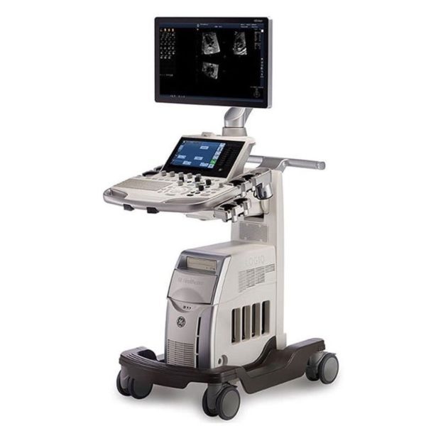
The GE Logiq S7 with XDclear Ultrasound is a mid range multi-purpose ultrasound machine. The GE Logiq S7 with XDClear has been fully upgraded, 23″ large LCD monitor and 10.1″ LCD touch screen. It becomes very reliable and trustworthy while reaching the revision 3.0, and GE incorporated single crystal transducers, so called XDClear transducers such as C1-6-D and C3-10-D. With one of the market most demanding technologies, XDClear, the user can enjoy premium image resolution, penetration, and wide bandwidth from two XDClear options. The GE Logiq S7 with XDClear built with the same system platform of GE Logiq S8 with XDClear, S-agile architecture, but just some of the features and transducer are not included or more economic options and transducers are integrated. Although the GE Logiq S7 with XDClear is classified as a mid range system, it offers a great benefit to users with advanced automated features such as Scan Assistant, OB Measure Assistant, and Breast Measure Assistant. Also, Automated Function Imaging(AFI), XDClear probes, large 23″ LCD monitor and 10.1″ touch screen are very unique features in mid-range.GE Logiq S7 With XDClear Dimensions and Weight:• Height: 1,210mm (Minimum), 1,760mm (Max.)
• Width: 530mm (Caster), 520mm (Keyboard), 565mm (Monitor)
• Depth: 865mm (Max.), 795mm (Caster)
• Weight: 90 kg (198 lb.) GE Logiq S7 With XDClear Specifications:• Digital beamformer
• Displayed Imaging Depth: 0 – 33 cm
• Minimum Depth of Field: 0 – 2 cm (Zoom) (probe dependent)
• Maximum Depth of Field: 0 – 33 cm (probe dependent)
• Continuous dynamic receive focus/continuous dynamic receive aperture GE Logiq S7 With XDClear Electrical power:• Voltage:100-120 Vac or 220-240 Vac
• Frequency: 50/60 Hz
• Power: Consumption maximum of 900 VA with peripherals GE Logiq S7 XDClear Features:• High resolution 23″ wide LCD monitor
• High resolution 10.1″ wide LCD touch screen
• 4 active probe ports and 1 parking probe port
• Integrated HDD
• Integrated DVD Multi Drive
• Integrated speakers
• Locking mechanism that provides rolling lock and caster swivel lock
• Integrated cable management
• Front and rear handles
• Easily removable air filters
• Automatic Optimization
• CrossXBeam
• Speckle Reduction Imaging (SRI-HD)
• Fine Angle Steer
• Coded Harmonic Imaging
• Virtual Convex
• Advanced 3D
• Patient information database
• Image Archive on integrated CD/DVD and hard disk drive
• Raw Data Analysis
• Real-time automatic Doppler calcs
• OB Calcs
• Fetal Trending
• Multi gestational Calcs
• Hip Dysplasia Calcs
• Gynecological Calcs
• Vascular Calcs
• Urological Calcs
• Renal Calcs
• Cardiac Calcs
• inSite ExC Capability
• On-board electronic documentation
• MPEGVue
• Key Macro
• Network Storage
• Quick Save
• Quick Patient Entry
• Email2MMS Accessories GE Logiq S7 with XDclear Ultrasound GE Logiq S7 With XDClear Peripheral Options:• Sony Digital UP-D711 Termal Printer
• Sony Fixture Kit for Digital UP-D711 Thermal Printer
• Sony Digital UP-D25 Color Thermal Printer
• Sony Digital UP-D897 BW Thermal Printer
• Mitsubishi P93W/E Thermal Printer
• Mitsubishi P95DW Thermal Printer
• Printer installation kit
• External USB printer connection
• Printing using Bluetooth connectivity (Inkjet printer option)
• Wireless LAN
• HDMI output available for compatible devices
• Footswitch with programmable functionality
• Universal Video Converter
• Power Assistant
• Battery Pack (for Power Assistant)
• Isolation Transformer
• Isolated 1Gb Ethernet connection
• Isolated USB connector
• EMI filter (PWR supply noise filter)
• RS-DLP Probe Adapter for TEE Probe
• Side Cabinet (Japan only)
• Drawer
• Small Probe Holder (Probe Holder Adapter for Small Probe)
• Multi Purpose Holder (Vertical Endocavitary Probe Holder)
• Optional Probe Holder (Side Probe Holder)
• Probe cable hanger
• ECG + AHA/IEC Cables
• USB 3 Pedal Foot Switch GE Logiq S7 With XDClear Supplies:Aquasonic ultrasound gel
Sono ultrasound wipes
Sony UPP-110HG thermal printing paper
Sony UPC-21L color thermal printing pack
Mitsubishi KP95HG thermal roll paper (new)
Mitsubishi KP65HM-CE High density thermal paper GE Logiq S7 With XDClear ports:• User accessible USB x 5 ports
• HDMI connector
• Ethernet 1000/100/10BaseT
• Audio Line Out (1.5mm pin jack) GE Logiq S7 With XDClear image storage:• On-board database of patient information from past exams
• Storage Formats:
• DICOM – compressed/ uncompressed, single/ multiframe, with/ without Raw Data
• Export JPEG, JPEG2000, WMV (MPEG 4), and AVI formats
• Storage Devices: – USB Flash Device: 64MB to 64GB (for exporting individual images/clips) – CD-R storage: 700MB – DVD storage: R (4.7GB) – Hard Drive Image Storage: ~300GB – Compare previous exam images with current exam images – Reload of archived data sets – Network Storage support for Import, Export, DICOM Read, SaveAs, SaveAs GE Logiq S7 with XDClear Options:• Auto IMT
• Auto EF
• Automated Volume Calculation (VOCAL II)
• Automated Function Imaging (AFI)
• Breast Measure Assistant
• OB Measure Assistant
• B-Flow/B-Flow Color
• B Steer+
• Breast Productivity Package
• Coded Contrast Imaging
• HRES Contrast
• Compare Assistant
• CW Doppler
• DICOM 3.0 connectivity
• Digital Video Recording Software (Software DVR)
• Elastography
• Elastography Quantification
• Flow Quantification
• LOGIQView
• Real Time 4D
• Report Writer
• Scan Assistant
• Stress Echo
• Thyroid Productivity Package
• Tissue Velocity Imaging (TVI)
• Tomographic Ultrasound Imaging (TUI)
• Volume Contrast Imaging (VCI) Static
• OmniView
• STIC
• Advanced Probes (XDclear)
• Cabinet: High/Mid/Low GE Logiq S7 with XDClear Applications:• Fetal/Obstetrics
• Abdominal (includes renal, GYN/Pelvic)
• Pediatric
• Small Organ (breast, testes, thyroid)
• Neonatal Cephalic
• Adult Cephalic
• Cardiac (adult and pediatric)
• Peripheral Vascular
• Musculo-skeletal Conventional and Superficial
• Urology (including prostate)
• Transrectal
• Transvaginal
• Transesophageal | GE Logiq A5 Ultrasound MachineThe lightweight, portable LOGIQ* A5 ultrasound system offers capabilities to help meet your imaging needs — particularly for urological applications.Affordable ultrasound.The LOGIQ A5 puts imaging capabilities at your fingertips in a portable, lightweight design. This system can go from your office to the surgery suite and helps meet a range of general imaging needs. Well-suited for urology, tools include soft tissue image analysis of the kidneys, bladder, penis, testicles, and prostate.Solid imaging performance.
The sleek LOGIQ A5 can help meet your general imaging needs. This black and white ultrasound system has:- B-mode imaging capability and features that allow you to add advancements as your clinical needs grow.
- Auto TGC helps ensure a homogeneous picture from the near-to-far field.
- Virtual Convex provides an expanded field of view that enables you to see more anatomy with linear transducers.
- Color flow Doppler upgrade available to provide dynamic assessment of blood flow in the organs, as well as investigation of hypertension and resulting erectile dysfunction.
Perform imaging with ease.
The LOGIQ A5 has user-friendly features with precision handling.- Automatic Optimization (AO), helps provide improved image quality at the touch of a button for B-mode, spectral and color Doppler imaging.
- Full-size keyboard for optimal ease of use.
- 15-inch adjustable LCD monitor allows a clear view in any position.
- Lightweight package easily travels with you.
Compatible Transducers:- 10L Linear Array
- 11L Wide Band Linear
- 12L Linear Array
- 3.5C Convex
- 3.5CS Convex (2-5 MHz)
- 3CRF Convex
- 3S Phased Array
- 3Sp Wide Band Phased Array Sector
- 4C Convex Array
- 4D3CL Real time 4D Convex
- 4D8C Convex Volume
- 4DE7C Real time 4D Endocavity
- 5CS Convex (2-5 MHz)
- 5S Cardiac Sector
- 5Sp Wide Band Phased Array Sector
- 7S Phased Array
- 8C Convex Array
- 8L Linear (3-7 MHz)
- 9L Wide Band Linear
- BE9C Transrectal
- BE9CS Micro convex Bi-plan
- E8C (4-10 MHz) Wide Band Microconvex
- E8CS Micro-convex (4-11 MHz)
- ERB Brachytherapy Transrectal
- i12L Linear Array
- i739 Intraoperative
- P2D CWD Non-Imaging
- P6D CWD Non-Imaging
- T739 Intra-operative
| GE Vivid Q Ultrasound Machine
The powerful GE Vivid Q ultrasound machine is the ultimate lightweight, compact high-performance cardiovascular system for clinicians needing a machine that combines user efficiency and reliable performance. This unique system performs consistently thanks to its TruScan architecture and can support up to 17 probes. The GE Vivid Q is recognized for its premium image quality, advanced image management, and high-performance quantification tools.This advanced GE ultrasound delivers improved patient care and enhanced user productivity with the new M4s-Matrix array technology transducer, which provides greatly enhanced image quality in electronic beamforming, raw data image acquisition and archiving, optimal onboard and post processing technology.This ultrasound system provides high clinical values for quantification and review of stored images, making it one of today’s most sought-after models. Some of its powerful features include compatibility with EchoPac clinical workstation and DICOM networking, speckle tracking strain, integrated stress echo, quantification tools, and reporting capabilities. Also of note is one of the Vivid Q’s best new features, which is its TEE transducer capability.A one-touch button optimization provides premium image quality for workflow efficiency, and maintains AFI- automatic function imaging with complete wallFeatures- Advanced QScan Imaging (incl TSI)
- AdvStress
- ATO
- Auto EF
- Automated Function Imaging
- Blood Flow Imaging
- CardiacApp
- Cardio Lab
- Color Doppler
- CW Doppler
- DICOM Networking
- DICOM: Print
- DICOM: Worklist
- DVD/RW
- ECG
- ECG Cable
- EchoDicom
- EchoPAC
- Evue
- ICE Imaging
- Intima Media Thickness (IMT)
- LVO Contrast
- M Mode
- M4S
- MPEGvue
- PW Doppler
- Small Depth
- TEE
- THI
- Tissue Harmonics Imaging (THI/NTHI)
- Tissue Velocity Imagine & Tissue Tracking
- TSI
- VascAbdContrast
- VascularApp
- VirtualPrinter
- Wide Aperture
GE Vivid Q FeaturesThe GE Vivid Qis a Portable Laptop-style Ultrasound that can be used to help diagnose cardiovascular anatomy. Features and tools based on the previous Vivid 7 and the Vivid E9 have been added to the new lightweight Vivid Q, mainly the M4S-RS transducer that allows for 3D image focusing and enables a higher level of image quality. The Vivid Q ultrasound is versatile and can additionally be used for Cardiac, vascular, Abdominal, Pediatric, Obstetrics, and Emergency medicine. The Vivid Q Ultrasound comes with a 15-inch color LCD monitor that can display a variety of imaging modes including; ICE, Auto EF, AFI, TVI, TT, TSI, and Blood Flow. Additional the Ultrasound system is comparable with 17 different transducers and probes to fit every imaging need. - Auto EF - Automated ejection fraction measurement program designed specifically for the GE Vivid q.
- Tissue Synchronization Imaging (TSI).
- Intracardiac Echo (ICE).
- 16 different compatible probes.
- Coded Octave imaging - enhances image color flow and Doppler imaging.
| GE Logiq e r7 Portable Ultrasound Machine
The LOGIQ e R7 provides even more impressive veterinary abdominal and cardiac images than the BT12 LOGIQe, combining the high performance of a console system with the portability of a laptop.Highlights of the GE LOGIQ e R7:- Simple. Fast. Precise
- Colour Flow Mode, Power Doppler Imaging (PDI), Pulse Wave Doppler (PWD), Continuous Wave Doppler
- 15” high resolution colour LCD screen
- Integrated solid state hard drive
- Automatic optimisation
- CrossXBeam
- Speckle Reduction Imaging (SRI-HD)
- Virtual Convex / Virtual Apex
- Fine Angle Steer
- HD Zoom (Write Zoom)
- Coded Harmonic Imaging (CHI) – Enhances near field reslolution for improved small parts imaging as well as far field penetration. Diminshes low frequency / amplitude noise and provides clarity to needle, anatomy and motion.
- Raw Data Processing
- Quicksave – Single button push sends single image or entire patient exam to memory stick or network
- On-board manual (Help)
- Loop storage – from live scanning and from memory
- Patient Information Database
- Customisable user interface
- Full M&A calculation package with real time Doppler calculations
- Report writer package – on board reporting package automates report writting
- Portable and battery operated so easy to move from room to room
- Export of stills and clips to USB.
More GE Logiq e r7 Portable Ultrasound MachineThe Expert is the new flagship LOGIQ e. The imaging engine comes from GE leading console systems, delivering crisp images in a compact package. Providing ultrasound imaging with precise anatomical detail at a variety of depths. The system includes innovative features that help simplify proceedures.GE NextGen LOGIQ e R7 Ultrasound System The GE NextGen LOGIQ e R7 Ultrasound System has an imaging engine that delivers crisp images in a compact package. Point-of-care-specific software and transducers help to see the needle to administer a block or perform an aspiration quickly. When productivity is crucial, you can control the console from the transducer, potentially eliminating the need to have a second person assist. Anatomy-specific presets and only displaying the functions that you need helps you to direct your focus on the patient rather than the control panel. Although the system features a large, user-friendly, high-res color display, it is also very durable and compact for easy portability. | SonoSite Nanomaxx Ultrasound MachineThe SonoSite NanoMaxx ultrasound system combines premium functionality and affordability with the versatility and portability that makes SonoSite a top competitor in the portable ultrasound market. The SonoSite NanoMaxx comes in a sleek, hand-held package that never compromises performance, with applications ranging from emergency medicine and anesthesia to vascular.MedCorp offers the refurbished SonoSite NanoMaxx for purchase. Used SonoSite NanoMaxx from MedCorp offer everything you’d expect from a newer machine, including PC/MAC export configurations, 2D, Color and Color Power Doppler modes, and 2GB of internal memory that can hold 50 images each for 40 patients, all in an 8” touch-screen interface. However, because this is a refurbished SonoSite NanoMaxx, the price is lower than the new models. - 8″ Touch-screen interface
- 2D, Color and Color Power Doppler modes
- 2GB internal memory that can hold 50 images each for 40 patients
- 2 hours of battery power
- PC/MAC export configuration
- Four preset pictograph screen positions
- Drop resistant magnesium case
System Description:SonoSite Nanomaxx Ultrasound Machine
The NanoMaxx ultrasound system offers excellent performance at an affordable price. It is easy to use and offers top of the line diagnostic imaging, color power Doppler and color-flow velocity. The NanoMaxx can aid users in making clinical decisions or guide interventional procedures. The unit uses proprietary technology which allows the optimization of many system settings by simply pressing a button. Application:
Abdominal, Cardiology, Gynecology, Nerve, Obstetrics, CIMT, Musculoskeletal, Small Parts, Superficial, Vascular, Venous | GE Voluson 730 Pro Ultrasound MachineThe Voluson 730 Pro is built on the same platform as GE’s leading Real-Time 4D system, the Voluson 730 Expert. With an easy to use interface and exceptional 2D image quality, in addition to GE’s exclusive 4D imaging technology, the Voluson 730 Pro is ideal for any clinical practice with a variety of patient workloads. Recognized world-wide for its 4D imaging capabilities, the Voluson 730 Pro also features exceptional 2D image quality.RealTime 4D imaging – continuous, 3D scanning with simultaneous visualization of the A, B and C planes.
The system allows you to explore images in any plane to reveal the smallest details with stunning clarity and apply sophisticated analytical tools to answer virtually any of your clinical questions.Performance:GE Voluson 730 Pro Ultrasound Machine
State-of-the-art user interface with high resolution 10.4 inch LCD touch panel
Automatic Tissue Optimization
Tissue Doppler
Coded Harmonic Imaging
Coded Excitation (CE)
HD-Flow
CrossXBeamCRI (Compound Resolution Imaging)
Static 3D Mode
B Mode
Focus & Frequency Composite (FFC)
High Resolution Zoom
Pan Zoom
Steering
Virtual Convex
Beta-View
Patient information database
Image Archive on MOD and hard drive
3D/4D data compression (lossy/lossless)
Inversion
Real-time automatic Doppler calcsCapabilities:
OB/GYN, Vascular, Cardio, Abdominal, Small-Parts, Urology, Pediatrics, Ortho, Neurology, Multigestational Calculations, B-Flow, VCI (Volume Contrast Imaging)Available Probes:
AB2-7 Wide Band Convex Probe
AC2-5 Wide Band Convex Probe
4C-A Wide Band Convex Probe
M7C-H Wide Band Convex Probe
IC5-9H Wide Band Convex Probe
PA2-5P Wide Band Phased Array Probe
PA6-8 Wide Band Phased Array Probe
SP10-16 Wide Band Linear Probe
SP4-10 Wide Band Linear Probe
SP6-12 Wide Band Linear Probe
M12L-H Wide Band Linear Probe (1.25D Array)
RAB2-5L Wide Band Convex Volume Probe
RAB4-8L Wide Band Convex Volume Probe
RIC5-9H Wide Band Convex Volume Probe
RIC5-9W Wide Band Convex Volume Probe
RRE6-10 Wide Band Convex Volume Probe
RSP6-16 Wide Band Linear Volume Probe
RNA5-9 Wide Band Convex Volume Probe
SCW 2.0; non imaging suprasternal CW-Pencil Probe
PCW 4.0; non imaging peripheral CW-Pencil Probe |




Reviews
There are no reviews yet.