| Description | Applications:
• Fetal/Obstetrics
• Abdominal (includes renal, GYN/Pelvic)
• Pediatric
• Small Organ (breast, testes, thyroid)
• Neonatal Cephalic
• Adult Cephalic
• Cardiac (adult and pediatric)
• Peripheral Vascular
• Musculo-skeletal Conventional and Superficial
• Urology (including prostate)
• Transrectal
• Transvaginal
• Transesophageal | Designed to make your job easier, faster and more accurate, the MicroMaxx ultrasound tool makes point-of-care ultrasound an affordable reality. It offers impressive image quality. Wireless data transfer. Extreme durability. The Sonosite Micromaxx also features SonoCalc IMT, edge detection software for cardiology and Tissue Harmonic Imaging | Acuson sequoia 512 ultrasound click images to enlarge item description acuson sequoia 512 ultrasoundsold as picturedincludes: 2 probes, 4c1 convex & 6l4 linearsony up 895 md printerunit will be cleaned, function checked and certified for electrical safety. Checked, cleaned, certified for electrical safetyguaranteed working when arrives on the guarantee working we need to be notified in 48 hrs. And the seals must be intact. | Ultraformer III’s 7 clip-in transducer cartridges project uniform ultrasound beams directly to multiple layers underneath the skin to promote tighter collagen formation, tissue contraction and reduction in the volume of adipocytes that form bulging areas of tissue.Patients notice an immediate benefit in improved skin tone and texture, with ongoing improvements over 3 to 6 months
Traditionally difficult areas such as eye lifting are made easier with Ultraformer III’s specialised cartridges. It is possible for patients to experience up to 6mm of eyebrow lift, depending on their particular situation. | The SonoSite Titan is the earliest version of the PoC dedicated portable ultrasound machine from Sonosite. Though its form factor is compact, it provides high resolution imaging, rapid responsive time, and supports a broad range of applications including cardiac. | Solid imaging performance.
The sleek LOGIQ A5 can help meet your general imaging needs. This black and white ultrasound system has:- B-mode imaging capability and features that allow you to add advancements as your clinical needs grow.
- Auto TGC helps ensure a homogeneous picture from the near-to-far field.
- Virtual Convex provides an expanded field of view that enables you to see more anatomy with linear transducers.
- Color flow Doppler upgrade available to provide dynamic assessment of blood flow in the organs, as well as investigation of hypertension and resulting erectile dysfunction.
|
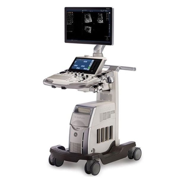
| Content | GE LOGIQ S7 WITH XDCLEAR ULTRASOUND
The GE Logiq S7 with XDclear Ultrasound is a mid range multi-purpose ultrasound machine. The GE Logiq S7 with XDClear has been fully upgraded, 23″ large LCD monitor and 10.1″ LCD touch screen. It becomes very reliable and trustworthy while reaching the revision 3.0, and GE incorporated single crystal transducers, so called XDClear transducers such as C1-6-D and C3-10-D. With one of the market most demanding technologies, XDClear, the user can enjoy premium image resolution, penetration, and wide bandwidth from two XDClear options. The GE Logiq S7 with XDClear built with the same system platform of GE Logiq S8 with XDClear, S-agile architecture, but just some of the features and transducer are not included or more economic options and transducers are integrated. Although the GE Logiq S7 with XDClear is classified as a mid range system, it offers a great benefit to users with advanced automated features such as Scan Assistant, OB Measure Assistant, and Breast Measure Assistant. Also, Automated Function Imaging(AFI), XDClear probes, large 23″ LCD monitor and 10.1″ touch screen are very unique features in mid-range.GE Logiq S7 With XDClear Dimensions and Weight:• Height: 1,210mm (Minimum), 1,760mm (Max.)
• Width: 530mm (Caster), 520mm (Keyboard), 565mm (Monitor)
• Depth: 865mm (Max.), 795mm (Caster)
• Weight: 90 kg (198 lb.) GE Logiq S7 With XDClear Specifications:• Digital beamformer
• Displayed Imaging Depth: 0 – 33 cm
• Minimum Depth of Field: 0 – 2 cm (Zoom) (probe dependent)
• Maximum Depth of Field: 0 – 33 cm (probe dependent)
• Continuous dynamic receive focus/continuous dynamic receive aperture GE Logiq S7 With XDClear Electrical power:• Voltage:100-120 Vac or 220-240 Vac
• Frequency: 50/60 Hz
• Power: Consumption maximum of 900 VA with peripherals GE Logiq S7 XDClear Features:• High resolution 23″ wide LCD monitor
• High resolution 10.1″ wide LCD touch screen
• 4 active probe ports and 1 parking probe port
• Integrated HDD
• Integrated DVD Multi Drive
• Integrated speakers
• Locking mechanism that provides rolling lock and caster swivel lock
• Integrated cable management
• Front and rear handles
• Easily removable air filters
• Automatic Optimization
• CrossXBeam
• Speckle Reduction Imaging (SRI-HD)
• Fine Angle Steer
• Coded Harmonic Imaging
• Virtual Convex
• Advanced 3D
• Patient information database
• Image Archive on integrated CD/DVD and hard disk drive
• Raw Data Analysis
• Real-time automatic Doppler calcs
• OB Calcs
• Fetal Trending
• Multi gestational Calcs
• Hip Dysplasia Calcs
• Gynecological Calcs
• Vascular Calcs
• Urological Calcs
• Renal Calcs
• Cardiac Calcs
• inSite ExC Capability
• On-board electronic documentation
• MPEGVue
• Key Macro
• Network Storage
• Quick Save
• Quick Patient Entry
• Email2MMS Accessories GE Logiq S7 with XDclear Ultrasound GE Logiq S7 With XDClear Peripheral Options:• Sony Digital UP-D711 Termal Printer
• Sony Fixture Kit for Digital UP-D711 Thermal Printer
• Sony Digital UP-D25 Color Thermal Printer
• Sony Digital UP-D897 BW Thermal Printer
• Mitsubishi P93W/E Thermal Printer
• Mitsubishi P95DW Thermal Printer
• Printer installation kit
• External USB printer connection
• Printing using Bluetooth connectivity (Inkjet printer option)
• Wireless LAN
• HDMI output available for compatible devices
• Footswitch with programmable functionality
• Universal Video Converter
• Power Assistant
• Battery Pack (for Power Assistant)
• Isolation Transformer
• Isolated 1Gb Ethernet connection
• Isolated USB connector
• EMI filter (PWR supply noise filter)
• RS-DLP Probe Adapter for TEE Probe
• Side Cabinet (Japan only)
• Drawer
• Small Probe Holder (Probe Holder Adapter for Small Probe)
• Multi Purpose Holder (Vertical Endocavitary Probe Holder)
• Optional Probe Holder (Side Probe Holder)
• Probe cable hanger
• ECG + AHA/IEC Cables
• USB 3 Pedal Foot Switch GE Logiq S7 With XDClear Supplies:Aquasonic ultrasound gel
Sono ultrasound wipes
Sony UPP-110HG thermal printing paper
Sony UPC-21L color thermal printing pack
Mitsubishi KP95HG thermal roll paper (new)
Mitsubishi KP65HM-CE High density thermal paper GE Logiq S7 With XDClear ports:• User accessible USB x 5 ports
• HDMI connector
• Ethernet 1000/100/10BaseT
• Audio Line Out (1.5mm pin jack) GE Logiq S7 With XDClear image storage:• On-board database of patient information from past exams
• Storage Formats:
• DICOM – compressed/ uncompressed, single/ multiframe, with/ without Raw Data
• Export JPEG, JPEG2000, WMV (MPEG 4), and AVI formats
• Storage Devices: – USB Flash Device: 64MB to 64GB (for exporting individual images/clips) – CD-R storage: 700MB – DVD storage: R (4.7GB) – Hard Drive Image Storage: ~300GB – Compare previous exam images with current exam images – Reload of archived data sets – Network Storage support for Import, Export, DICOM Read, SaveAs, SaveAs GE Logiq S7 with XDClear Options:• Auto IMT
• Auto EF
• Automated Volume Calculation (VOCAL II)
• Automated Function Imaging (AFI)
• Breast Measure Assistant
• OB Measure Assistant
• B-Flow/B-Flow Color
• B Steer+
• Breast Productivity Package
• Coded Contrast Imaging
• HRES Contrast
• Compare Assistant
• CW Doppler
• DICOM 3.0 connectivity
• Digital Video Recording Software (Software DVR)
• Elastography
• Elastography Quantification
• Flow Quantification
• LOGIQView
• Real Time 4D
• Report Writer
• Scan Assistant
• Stress Echo
• Thyroid Productivity Package
• Tissue Velocity Imaging (TVI)
• Tomographic Ultrasound Imaging (TUI)
• Volume Contrast Imaging (VCI) Static
• OmniView
• STIC
• Advanced Probes (XDclear)
• Cabinet: High/Mid/Low GE Logiq S7 with XDClear Applications:• Fetal/Obstetrics
• Abdominal (includes renal, GYN/Pelvic)
• Pediatric
• Small Organ (breast, testes, thyroid)
• Neonatal Cephalic
• Adult Cephalic
• Cardiac (adult and pediatric)
• Peripheral Vascular
• Musculo-skeletal Conventional and Superficial
• Urology (including prostate)
• Transrectal
• Transvaginal
• Transesophageal | SonoSite MicroMaxx Ultrasound Machine
The SonoSite MicroMaxx Portable Ultrasound is the third portable system to emerge from SonoSite. With improved image quality, a larger screen and additional imaging features, this system is the upgrade to the SonoSite Titan. The SonoSite MicroMaxx portable ultrasound is a well suited portable ultrasound, and is capable of performing the duties of a shared service system.The portable SonoSite MicroMaxx Portable Ultrasound system has purpose-built software and proprietary ASIC design yielding high-quality images with speed and reliability while efficiently operating on either AC or battery power. From busy offices and big-city hospitals to critical-care situations where seconds count, the handheld SonoSite MicroMaxx ultrasound system, boots up quickly, from off to scanning in under 15 seconds. Sonosite Micromaxx portable ultrasound transducers, probes, service and pricing are available through UDS Portable Ultrasound.Big machine performance in a hand-carried unitMicroMaxx’s purpose-built software and proprietary ASIC design yields high-quality images with speed and reliability while efficiently operating on either AC or battery power.MicroMaxx goes anywhere you need itFrom busy offices and big-city hospitals to critical-care situations where seconds count, the SonoSite MicroMaxx&trade system boots up quickly, from “off” to scanning in under 15 seconds. This easily carried device gets medical professionals into-and out of-tight spaces fast.Embedded software means solid reliabilityThe MicroMaxx system software is hard-wired and purpose-built for faster boot.Chip Fusion TechnologySonoSite’s proprietary Chip Fusion™ Technology enables advanced, cart-based image resolution in a hand-carried unit by integrating digital signal processing and multiple ultrasound functions into a custom ASIC microchip to deliver unprecedented performance without the size, weight and cost of conventional systems.Clinical utilityThe MicroMaxx system comes with a wide range of transducers for multiple clinical applications. With MicroMaxx, SonoSite is extending the capabilities of hand-carried systems by adding high-performance features.Durability SonoSite MicroMaxx Ultrasound MachineSonoSite products and technology deliver proven reliability and durability in conventional medical settings as well as some far more challenging environments, such as supporting rescue operations following natural disasters. Extensive quality controls such as “drop testing” ensure that SonoSite products continue to set the industry standard for both reliability and durability.MicroMaxx’s reliability and durability are backed by a one-year warranty, an industry first, far exceeding warranties available on competing products.From “Off” to scanning in just secondsThis is vitally important in many medical situations where seconds really do count-whether getting crucial diagnostic information in an emergency or making the best use of time in a hectic daily schedule. | Siemens / Acuson Sequoia 512 Ultrasound Machine
The ACUSON Sequoia 512 ultrasound is an ultra-premium system that utilizes revolutionary technology, dedicated to the advancement of diagnostic imaging. It represents some of the most advanced technology in ultrasound imaging. The Acuson Sequoia 512 has unparalleled imaging performance, greater image content, enhanced detail, superior contrast resolution & penetration, and excellent color Doppler sensitivity.One of the best selling ultrasounds in the United States, the Sequois 512 provides superior imaging with Native Patient Specific Imaging that detects, measures and adapts (in real time) to each patient’s individual acoustics.Performance:Reduce image optimization time by up to 80% and allow for an easier, faster and more confident diagnosis:- 2D
- SST Color Doppler
- Color Doppler Energy
- Color Doppler Velocity
- Solo Spectral Doppler
- M-mode and Color Doppler M-mode
- MultiHertz Multiple Frequency Imaging
- DELTA Differential Echo Amplification
- Home Base user interface
- One-Touch Image Control with exam and Image Presets
- Digital acquistion, storage, review and transfer of static images, dynamic clips, and cine
- Digital storage on an integrated hard disk
- Digital transfer via MO Disk
- Integrated Compression engine
- Embedded DICOM conformance
- Patient- oriented file structure
- Native Tissue Harmonic Imaging
- Tissue Equalization™ Technology (Optional)
- Signature II™ (Optional)
- Advanced Imaging package (Optional)
- Auto Doppler (Optional)
- Freestyle Compounding (Optional)
- FreeStyle Extended Imaging (Optional)
- 3D surface Rendering (Optional)
- Cadence™ ADI (Optional)
- Cadence CPS for 512 (Optional)
- Axius™ Edge Assisted EF (Optional)
- Doppler Tissue Imaging (Optional)
- DICOM Print And Store (Optional)
Capabilities:Siemens / Acuson Sequoia 512 Ultrasound MachineGeneral Radiology, Abdominal, Intraoperative Monitoring, OB/GYN, Neonatal, Vascular, Cardiac, Small PartsAvailable Transducers:4V1, 4V2, 8V5, 15L8W, 6L3, 8L5, 6C2, 5C2, 4C1, 3V2C, 5V2C,7V3C, EVC8-4V, EC-10C5, V5M TEE, V7B neo TEE, AcuNav Transducer, 2.0 Non Imaging CW Doppler probe.Acuson sequoia 512 ultrasound click images to enlarge item description acuson sequoia 512 ultrasoundsold as picturedincludes: 2 probes, 4c1 convex & 6l4 linearsony up 895 md printerunit will be cleaned, function checked and certified for electrical safety. Checked, cleaned, certified for electrical safetyguaranteed working when arrives on the guarantee working we need to be notified in 48 hrs. And the seals must be intact. | CLASSYS ULTRAFORMER III UTR3
You’ve heard about it, now see and feel the difference for yourself.Ultraformer III is the leading technology for facial lifting, tightening and contouring. Now with 7 interchangeable cartridges that offer your patients another dimension in non-surgical face and body treatments.Ultraformer III is widely used with over 10 million treatments performed worldwide. It has become the treatment of choice for patients who are not yet indicated or not willing to undergo surgery. This fills a huge gap in the market, and captures an audience of people from 30 years of age all the way to 80.ULTRAFORMER III FEATURESLess discomfort associated with treatments
Patented transducer design for more efficient energy delivery
Dual hand pieces with automatic recognition for ease of use
Faster shot and treatment speed to save time
Easy, user-friendly design and interface
Shot count information and voice assistance
Backed by clinical papersThe CLASSYS ULTRAFORMER III UTR3 advantage with slim transducer designimproves operator visibility
supports very narrow focus and precise treatment
fits facial contoursFast elliptic transducersmore efficient treatment timesUltraformer III’s 7 clip-in transducer cartridges project uniform ultrasound beams directly to multiple layers underneath the skin to promote tighter collagen formation, tissue contraction and reduction in the volume of adipocytes that form bulging areas of tissue.Patients notice an immediate benefit in improved skin tone and texture, with ongoing improvements over 3 to 6 months
Traditionally difficult areas such as eye lifting are made easier with Ultraformer III’s specialised cartridges. It is possible for patients to experience up to 6mm of eyebrow lift, depending on their particular situation.Ultraformer III’s 7 clip-in transducer cartridges project uniform ultrasound beams directly to multiple layers underneath the skin to promote tighter collagen formation, tissue contraction and reduction in the volume of adipocytes that form bulging areas of tissue.Patients notice an immediate benefit in improved skin tone and texture, with ongoing improvements over 3 to 6 months
Traditionally difficult areas such as eye lifting are made easier with Ultraformer III’s specialised cartridges. It is possible for patients to experience up to 6mm of eyebrow lift, depending on their particular situation. | Sonosite Titan Ultrasound Machine
The SonoSite Titan Portable Ultrasound is SonoSite’s second generation portable ultrasound system. This system improved upon the technology of the SonoSite 180 with a larger screen and keyboard packed into a laptop style system. This system is well suited for Vascular access, Musculoskeletal, Anesthesiology, Internal Medecine, Emergency rooms and Family Physicians. Veterinary configurations are available. Veterinary configurations are available.8.4″ LCD monitor, 2 probe ports, Vascular, OB/GYN, Cardiac reports, Soft keys, Split screen, Zoom, Storage via 64 or 256 MB flash cards, 220 image CINE loop, USB & Ethernet image transfer, Volume calculations via diameters, THI [Tissue Harmonic Imaging], SonoCalc [IMT analysis, PC-based], DICOM, S-video (in/out), RGB, Composite video.
Dimensions: 3″ (H) x 10.9″ (W) x 11.9″ (D) Weight: 7.7 lbs Manufacturer
Sonosite Application
Cardiology, Endocrinology, Family Practice, Gastroenterology, Internal Medicine, Nephrology, Neurology, OB-GYN, Orthopedics, Otorhinolaryngology, Pain Management, Surgery, Urology, Vascular Probes - C8 – prostate – 8-5 MHz 8-mm broadband intracavitary array – 8.5 cm – Available – MicroMaxx/ TITAN
- C11 – abdominal/ pediatric cardiology/ neonatal/ vascular – 8-5 MHz 11-mm broadband curved array – 10 cm – N/A – TITAN
- C15 – adult cardiology/ abdominal – 4-2 MHz 15-mm broadband tightly curved array – 25 cm – N/A – TITAN
- C60 – abdominal/ obstetrics/ gynecology – 5-2 MHz 60-mm broadband curved array – 22 cm – Available – TITAN
- ICTe – obstetrics/ gynecology – 8-5 MHz 11-mm broadband tightly curved array – 10 cm – Available – MicroMaxx/ TITAN
- L25 – nerve/ musculoskeletal/ vascular and superficial – 10-5 MHz 25-mm broadband linear array – 6 cm – Transverse Guide Available – TITAN
- L38 – small parts/ breast/ vascular/ nerve/ musculoskeletal/ superficial – 10-5 MHz 38-mm broadband linear array – 6.5 cm – Available – TITAN
Options - Extended cardiac package
- Pulsed wave (PW) doppler
- Continuous wave (CW) doppler
- Tissue Harmonic Imaging
Accessories - Cart
- Black and White Video Printer
- DVD Recorder
The SonoSite Titan is the earliest version of the PoC dedicated portable ultrasound machine from Sonosite. Though its form factor is compact, it provides high resolution imaging, rapid responsive time, and supports a broad range of applications including cardiac. | GE Logiq A5 Ultrasound MachineThe lightweight, portable LOGIQ* A5 ultrasound system offers capabilities to help meet your imaging needs — particularly for urological applications.Affordable ultrasound.The LOGIQ A5 puts imaging capabilities at your fingertips in a portable, lightweight design. This system can go from your office to the surgery suite and helps meet a range of general imaging needs. Well-suited for urology, tools include soft tissue image analysis of the kidneys, bladder, penis, testicles, and prostate.Solid imaging performance.
The sleek LOGIQ A5 can help meet your general imaging needs. This black and white ultrasound system has:- B-mode imaging capability and features that allow you to add advancements as your clinical needs grow.
- Auto TGC helps ensure a homogeneous picture from the near-to-far field.
- Virtual Convex provides an expanded field of view that enables you to see more anatomy with linear transducers.
- Color flow Doppler upgrade available to provide dynamic assessment of blood flow in the organs, as well as investigation of hypertension and resulting erectile dysfunction.
Perform imaging with ease.
The LOGIQ A5 has user-friendly features with precision handling.- Automatic Optimization (AO), helps provide improved image quality at the touch of a button for B-mode, spectral and color Doppler imaging.
- Full-size keyboard for optimal ease of use.
- 15-inch adjustable LCD monitor allows a clear view in any position.
- Lightweight package easily travels with you.
Compatible Transducers:- 10L Linear Array
- 11L Wide Band Linear
- 12L Linear Array
- 3.5C Convex
- 3.5CS Convex (2-5 MHz)
- 3CRF Convex
- 3S Phased Array
- 3Sp Wide Band Phased Array Sector
- 4C Convex Array
- 4D3CL Real time 4D Convex
- 4D8C Convex Volume
- 4DE7C Real time 4D Endocavity
- 5CS Convex (2-5 MHz)
- 5S Cardiac Sector
- 5Sp Wide Band Phased Array Sector
- 7S Phased Array
- 8C Convex Array
- 8L Linear (3-7 MHz)
- 9L Wide Band Linear
- BE9C Transrectal
- BE9CS Micro convex Bi-plan
- E8C (4-10 MHz) Wide Band Microconvex
- E8CS Micro-convex (4-11 MHz)
- ERB Brachytherapy Transrectal
- i12L Linear Array
- i739 Intraoperative
- P2D CWD Non-Imaging
- P6D CWD Non-Imaging
- T739 Intra-operative
|




Reviews
There are no reviews yet.