| Description | Applications:
• Fetal/Obstetrics
• Abdominal (includes renal, GYN/Pelvic)
• Pediatric
• Small Organ (breast, testes, thyroid)
• Neonatal Cephalic
• Adult Cephalic
• Cardiac (adult and pediatric)
• Peripheral Vascular
• Musculo-skeletal Conventional and Superficial
• Urology (including prostate)
• Transrectal
• Transvaginal
• Transesophageal | The Samsung Accuvix XG was the top of the line system from Samsung prior to the release of the newer. The XG was meant as a direct replacement to the popular Accuvix XQ as a shared service system capable of both 4D OBGYN images as well as full cardiac scans. | The SonoSite Titan is the earliest version of the PoC dedicated portable ultrasound machine from Sonosite. Though its form factor is compact, it provides high resolution imaging, rapid responsive time, and supports a broad range of applications including cardiac. | The Samsung Medison SonoAce R3 portable ultrasound provides the image quality and features of a premium system at a fraction of the cost. The Samsung Medison SonoAce R3 portable ultrasound is brought to you by Samsung / Medison, a company whose name has been trusted in ultrasound since 1985. The Samsung Medison SonoAce R3 portable ultrasound is intuitive and easy to navigate with sophisticated software to increase workflow. | The more patients you see, the more you need Voluson E8.
The Voluson E8 ultrasound system is designed to keep pace with busy practices that conduct a wide range of Women’s Health exams, from routine scanning to complex assessments.
The imaging excellence of Radiance System Architecture – your diagnostic confidence will be enhanced by the extraordinary image clarity and speed of Radiant System Architecture. | Product Features:- Exclusive Raw Data Digital acquisition and storage
- Color Doppler, Spectral Dopler, B, M, Triplex, 3-D
- 150+ Frames per Second TrueDigital imaging
- All digital video clip and still image capture (4,000)
- On-board patient, image and reporting archive
- CD-ROM disk burner included, plus PCMCIA and USB
- Raw Data manipulation on image recall for Post Exam:
- Anatomic M-mode creation and adjustment
- Gain, magnification, and colorization control
- Playback speed, sweep speed, angle, baseline, and more…
- 3-D Digital processing and manipulation o Clip and still measure-annotate-enhance-store new
- DICOM 3.0 capable, plus VetPACS all Digital interfacing
- Angio
- ATO
- TMI
- AMM
- Virtual Convex
- PW/CW Doppler
- ECG/AUX
|
| Content | GE LOGIQ S7 WITH XDCLEAR ULTRASOUND
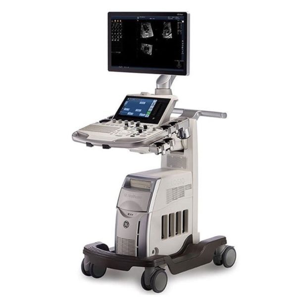
The GE Logiq S7 with XDclear Ultrasound is a mid range multi-purpose ultrasound machine. The GE Logiq S7 with XDClear has been fully upgraded, 23″ large LCD monitor and 10.1″ LCD touch screen. It becomes very reliable and trustworthy while reaching the revision 3.0, and GE incorporated single crystal transducers, so called XDClear transducers such as C1-6-D and C3-10-D. With one of the market most demanding technologies, XDClear, the user can enjoy premium image resolution, penetration, and wide bandwidth from two XDClear options. The GE Logiq S7 with XDClear built with the same system platform of GE Logiq S8 with XDClear, S-agile architecture, but just some of the features and transducer are not included or more economic options and transducers are integrated. Although the GE Logiq S7 with XDClear is classified as a mid range system, it offers a great benefit to users with advanced automated features such as Scan Assistant, OB Measure Assistant, and Breast Measure Assistant. Also, Automated Function Imaging(AFI), XDClear probes, large 23″ LCD monitor and 10.1″ touch screen are very unique features in mid-range.GE Logiq S7 With XDClear Dimensions and Weight:• Height: 1,210mm (Minimum), 1,760mm (Max.)
• Width: 530mm (Caster), 520mm (Keyboard), 565mm (Monitor)
• Depth: 865mm (Max.), 795mm (Caster)
• Weight: 90 kg (198 lb.) GE Logiq S7 With XDClear Specifications:• Digital beamformer
• Displayed Imaging Depth: 0 – 33 cm
• Minimum Depth of Field: 0 – 2 cm (Zoom) (probe dependent)
• Maximum Depth of Field: 0 – 33 cm (probe dependent)
• Continuous dynamic receive focus/continuous dynamic receive aperture GE Logiq S7 With XDClear Electrical power:• Voltage:100-120 Vac or 220-240 Vac
• Frequency: 50/60 Hz
• Power: Consumption maximum of 900 VA with peripherals GE Logiq S7 XDClear Features:• High resolution 23″ wide LCD monitor
• High resolution 10.1″ wide LCD touch screen
• 4 active probe ports and 1 parking probe port
• Integrated HDD
• Integrated DVD Multi Drive
• Integrated speakers
• Locking mechanism that provides rolling lock and caster swivel lock
• Integrated cable management
• Front and rear handles
• Easily removable air filters
• Automatic Optimization
• CrossXBeam
• Speckle Reduction Imaging (SRI-HD)
• Fine Angle Steer
• Coded Harmonic Imaging
• Virtual Convex
• Advanced 3D
• Patient information database
• Image Archive on integrated CD/DVD and hard disk drive
• Raw Data Analysis
• Real-time automatic Doppler calcs
• OB Calcs
• Fetal Trending
• Multi gestational Calcs
• Hip Dysplasia Calcs
• Gynecological Calcs
• Vascular Calcs
• Urological Calcs
• Renal Calcs
• Cardiac Calcs
• inSite ExC Capability
• On-board electronic documentation
• MPEGVue
• Key Macro
• Network Storage
• Quick Save
• Quick Patient Entry
• Email2MMS Accessories GE Logiq S7 with XDclear Ultrasound GE Logiq S7 With XDClear Peripheral Options:• Sony Digital UP-D711 Termal Printer
• Sony Fixture Kit for Digital UP-D711 Thermal Printer
• Sony Digital UP-D25 Color Thermal Printer
• Sony Digital UP-D897 BW Thermal Printer
• Mitsubishi P93W/E Thermal Printer
• Mitsubishi P95DW Thermal Printer
• Printer installation kit
• External USB printer connection
• Printing using Bluetooth connectivity (Inkjet printer option)
• Wireless LAN
• HDMI output available for compatible devices
• Footswitch with programmable functionality
• Universal Video Converter
• Power Assistant
• Battery Pack (for Power Assistant)
• Isolation Transformer
• Isolated 1Gb Ethernet connection
• Isolated USB connector
• EMI filter (PWR supply noise filter)
• RS-DLP Probe Adapter for TEE Probe
• Side Cabinet (Japan only)
• Drawer
• Small Probe Holder (Probe Holder Adapter for Small Probe)
• Multi Purpose Holder (Vertical Endocavitary Probe Holder)
• Optional Probe Holder (Side Probe Holder)
• Probe cable hanger
• ECG + AHA/IEC Cables
• USB 3 Pedal Foot Switch GE Logiq S7 With XDClear Supplies:Aquasonic ultrasound gel
Sono ultrasound wipes
Sony UPP-110HG thermal printing paper
Sony UPC-21L color thermal printing pack
Mitsubishi KP95HG thermal roll paper (new)
Mitsubishi KP65HM-CE High density thermal paper GE Logiq S7 With XDClear ports:• User accessible USB x 5 ports
• HDMI connector
• Ethernet 1000/100/10BaseT
• Audio Line Out (1.5mm pin jack) GE Logiq S7 With XDClear image storage:• On-board database of patient information from past exams
• Storage Formats:
• DICOM – compressed/ uncompressed, single/ multiframe, with/ without Raw Data
• Export JPEG, JPEG2000, WMV (MPEG 4), and AVI formats
• Storage Devices: – USB Flash Device: 64MB to 64GB (for exporting individual images/clips) – CD-R storage: 700MB – DVD storage: R (4.7GB) – Hard Drive Image Storage: ~300GB – Compare previous exam images with current exam images – Reload of archived data sets – Network Storage support for Import, Export, DICOM Read, SaveAs, SaveAs GE Logiq S7 with XDClear Options:• Auto IMT
• Auto EF
• Automated Volume Calculation (VOCAL II)
• Automated Function Imaging (AFI)
• Breast Measure Assistant
• OB Measure Assistant
• B-Flow/B-Flow Color
• B Steer+
• Breast Productivity Package
• Coded Contrast Imaging
• HRES Contrast
• Compare Assistant
• CW Doppler
• DICOM 3.0 connectivity
• Digital Video Recording Software (Software DVR)
• Elastography
• Elastography Quantification
• Flow Quantification
• LOGIQView
• Real Time 4D
• Report Writer
• Scan Assistant
• Stress Echo
• Thyroid Productivity Package
• Tissue Velocity Imaging (TVI)
• Tomographic Ultrasound Imaging (TUI)
• Volume Contrast Imaging (VCI) Static
• OmniView
• STIC
• Advanced Probes (XDclear)
• Cabinet: High/Mid/Low GE Logiq S7 with XDClear Applications:• Fetal/Obstetrics
• Abdominal (includes renal, GYN/Pelvic)
• Pediatric
• Small Organ (breast, testes, thyroid)
• Neonatal Cephalic
• Adult Cephalic
• Cardiac (adult and pediatric)
• Peripheral Vascular
• Musculo-skeletal Conventional and Superficial
• Urology (including prostate)
• Transrectal
• Transvaginal
• Transesophageal | Medison Accuvix XG Ultrasound System
Samsung Accuvix XG Ultrasound Machine2D Image Features
• Dynamic MR™ / Dynamic MR Plus is designed to enrich gray-scale resolution, as it enhances detection
and contrast resolution while also decreasing speckle echoes. This is particularly useful when evaluating
superficial structures, including thyroid, vessels, pelvic and abdominal anatomy.
• SRF (Speckle Reduction Filter™) enhances image quality by reducing or eliminating the appearance of
speckle echoes from ultrasound images. The degree of speckle reduction implemented is user-selectable.
• Wide Dynamic Range determines the number of gray shades utilized to map the gray-scale image.
It enables display of more details of bright areas and dark areas.
• SCI (Spatial Compounding Image) controls the ultrasound beam electronically by steering, and compounds
many scan lines.
• FSI (Full Spectrum Imaging) incorporates the penetration capabilities associated with lower frequencies,
yet maintains the fine pixel uniformity associated with higher frequencies, to deliver consistently high quality
images even in challenging diagnostic areas.3D Image Features Medison Accuvix XG Ultrasound System
• HDVI gives outstanding image quality and naturally clearer contrast, with excellent tissue differentiation,
edge depiction and speckle reduction, allowing consistent diagnoses with great confidence.
• 3D XI™, comprised of a suite of three innovative imaging applications – Multi-Slice View, Oblique View™ and
Volume CT – offers complete and precise control over 3D/4D volume data manipulation for maximum
diagnostic accuracy.
• 3D MXI is an innovative, cutting-edge 3D image processing technology. Comprising a comprehensive suite of
imaging tools – including Multi Volume Slice, Mirror View, Multi-OVIX, and 3D OH – 3D MXI lets you view,
examine and diagnose 3D volume data with supreme ease, speed and accuracy. • Fully adjustable system
The control panel can be adjusted to the user’s preferred height, for a better working environment and reduced
risk of back pain.
• Wide LED touch-screen
The Accuvix XG’s new LED touch-screen makes it easy to organize and operate.
• 19-inch HD LCD Monitor and articulating monitor arm
A 19-inch LCD monitor enables images to be displayed clearly even with a larger monitor, and the articulating
monitor arm enables easy mobility for a more comfortable and convenient working environment.
• Portability
The Accuvix XG is a lightweight system with 4 swivel wheels that allow easy steering, and a locking function. • Customizable measurement menus
Customizable measurement menus allow access to frequently-used functions, and enable a quicker and
more intuitive workflow.
• User keys and user knob
Accuvix XG offers user keys and a user knob that can map frequently-used functions, enabling the function to be
activated quickly and easily.
• Customized Annotation Menu and body marker
Users can preset up to 360 words of annotations, and body markers for each application, that reduce the time
needed for each examination.
• QuickScan
QuickScan maximizes workflow efficiency by automatically optimizing key imaging parameters with just the
push of a button. • HDVI
HD Volume Imaging(HDVI) gives outstanding image quality and naturally clearer contrast, with excellent tissue
differentiation, edge depiction and speckle reduction, allowing consistent diagnoses with great confidence.
It shows clearer images of subtle lesions, using multi slice image as well as C-plane.
• ElastoScan
ElastoScan™ translates stiff & soft tissue information into gray scale image. This technology defines the alteration
of the stiffness in tissue state more accurately, making it easier for the user to utilize the information in the image. | Sonosite Titan Ultrasound Machine
The SonoSite Titan Portable Ultrasound is SonoSite’s second generation portable ultrasound system. This system improved upon the technology of the SonoSite 180 with a larger screen and keyboard packed into a laptop style system. This system is well suited for Vascular access, Musculoskeletal, Anesthesiology, Internal Medecine, Emergency rooms and Family Physicians. Veterinary configurations are available. Veterinary configurations are available.8.4″ LCD monitor, 2 probe ports, Vascular, OB/GYN, Cardiac reports, Soft keys, Split screen, Zoom, Storage via 64 or 256 MB flash cards, 220 image CINE loop, USB & Ethernet image transfer, Volume calculations via diameters, THI [Tissue Harmonic Imaging], SonoCalc [IMT analysis, PC-based], DICOM, S-video (in/out), RGB, Composite video.
Dimensions: 3″ (H) x 10.9″ (W) x 11.9″ (D) Weight: 7.7 lbs Manufacturer
Sonosite Application
Cardiology, Endocrinology, Family Practice, Gastroenterology, Internal Medicine, Nephrology, Neurology, OB-GYN, Orthopedics, Otorhinolaryngology, Pain Management, Surgery, Urology, Vascular Probes - C8 – prostate – 8-5 MHz 8-mm broadband intracavitary array – 8.5 cm – Available – MicroMaxx/ TITAN
- C11 – abdominal/ pediatric cardiology/ neonatal/ vascular – 8-5 MHz 11-mm broadband curved array – 10 cm – N/A – TITAN
- C15 – adult cardiology/ abdominal – 4-2 MHz 15-mm broadband tightly curved array – 25 cm – N/A – TITAN
- C60 – abdominal/ obstetrics/ gynecology – 5-2 MHz 60-mm broadband curved array – 22 cm – Available – TITAN
- ICTe – obstetrics/ gynecology – 8-5 MHz 11-mm broadband tightly curved array – 10 cm – Available – MicroMaxx/ TITAN
- L25 – nerve/ musculoskeletal/ vascular and superficial – 10-5 MHz 25-mm broadband linear array – 6 cm – Transverse Guide Available – TITAN
- L38 – small parts/ breast/ vascular/ nerve/ musculoskeletal/ superficial – 10-5 MHz 38-mm broadband linear array – 6.5 cm – Available – TITAN
Options - Extended cardiac package
- Pulsed wave (PW) doppler
- Continuous wave (CW) doppler
- Tissue Harmonic Imaging
Accessories - Cart
- Black and White Video Printer
- DVD Recorder
The SonoSite Titan is the earliest version of the PoC dedicated portable ultrasound machine from Sonosite. Though its form factor is compact, it provides high resolution imaging, rapid responsive time, and supports a broad range of applications including cardiac. | Samsung Medison SonoAce R3 Ultrasound Machine
SonoAce R3 is a portable color ultrasound system with comprehensive diagnostic capabilities. Full Spectrum Imaging™ (FSI™) combines with Speckle Reduction Filter™ (SRF™) to deliver superior quality images, even in the challenging circumstances. The compact and user-friendly design features of the SonoAce R3 ensure portability and manoeuvrability in a lightweight unit. Sporting a 15-inch LCD monitor with Full-Featured Workflow, the SonoAceR3 offers your organisation flexibility, without compromising on precision and efficiency.Slim and Compact DesignThe compact design of SonoAce R3 offers Various-Featured Workflow with a thumbnail menu and shortcut, together with functional key groupings for increased efficiency. The user-friendly design combines a wide-view 15-inch LCD monitor with backlit control keys, customisable measurement package and body markers. The SonoAce R3 features 2 probe ports, 3 USB ports, rapid boot up capability and DICOM 3 compatible image filing. This slim, lightweight unit also has a convenient carrying handle for portability and flexibility.Samsung Medison SonoAce R3 Ultrasound MachineFull Spectrum Imaging™ (FSI™) produces the penetration capabilities associated with the lower frequencies, while still maintaining the fine pixel uniformity associated with the higher frequencies. This combination consistently delivers superior quality images, even in the challenging diagnostic circumstances. The resulting clarity delivers improvements in accuracy and efficiency for greater confidence in your diagnostic procedures. Harmonic Imaging is a technique that records higher frequency information, resulting in greater resolution and fewer artifacts.Synthetic Aperture
The physical radio channel may present some limits to imaging which can be overcome by using Synthetic Aperture Control software. This method uses information from diverse sources to form a more better picture of the area under examination. It creates one single scan line by performing the TX and RX twice to create enhanced penetration and resolution. Synthetic Aperture Control technology delivers a comprehensive picture and to aid accurate diagnosis.QuickScan
QuickScan has been designed to increase workflow efficiency by automatically optimising key imaging parameters at the touch of a button. The resulting increase in efficiency allows your organisation to offer patients an improved service with faster and more accurate diagnoses. This reliable and safe system helps your clinic or hospital to deliver a superior patient experience.Features
Full Spectrum ImagingTM
Quick ScanTM
Color and Power Doppler
Harmonic imaging
Freehand 3D
3D Multi Planar Imaging
Sonoview Image Management
PW Doppler
15″ LCD monitor
Two probe ports
Lightweight | GE Voluson E8 Ultrasound Machine15’’ LCD monitor [Rev BT06], 10.4” touchscreen, 3 Active Probe Ports, Floating Keyboard: Interactive back-lighting, Beta-View CrossXBeam, Virtual Convex, Extended View, ATO [Automatic Tissue Optimization], Coded Harmonic Imaging, High Resolution Zoom, Inversion Real-time automatic Doppler, OB/GYN; Vasc. Cardiac; Abd; Small-Parts; Urology; Ped; Ortho; Neuro reporting, Multigestational Calculations, HPRF [High Pulse Repetition Frequency], SonoVCAD [Matrix array volume technology], TruScan [On-board archive], VCI [Volume Contrast Imaging], SonoAVCfollicle [Sonography-based Automated Volume Count follicle], SonoVCADheart [Sonography-based Volume Computer Aided Display], TUI [Tomographic Ultrasound Imaging], CE [Coded Excitation], XTD, SRI III [Speckle reduction imaging], CRI [Compound Resolution Imaging], DICOM, Ethernet port, Video Out, 6 USB ports, Integrated HDD (160 GB) and DVD/CD-RW.
3D/4D, Endocrinology, Family Practice, Gastroenterology, Internal Medicine, Nephrology, Neurology, OB-GYN, Orthopedics, Otorhinolaryngology, Pain Management, Urology, Vascular Probes GE Voluson E8 Ultrasound Machine - 11L-D Linear Array
- 3S-D Cardiac Sector
- 4C-D Convex Array
- 9L-D Linear Array
- AB2-7 Convex
- IC5-9-D Intercavity
- M6C-D Convex
- P2D CWD Non-Imaging
- P6D CWD Non-Imaging
- PA6-8-D Cardiac
- RAB2-5-D 3D/ 4D Convex
- RAB4-8-D 3D/4D Convex
- RIC5-9-D 3D/4D Intracavity
- RIC6-12-D 3D/4D Intracavity
- RNA5-9-D 3D/4D Convex
- RRE6-10-D Endocavitary Micro-Convex
- RSP6-16-D 3D/D Linear
- SP10-16-D Linear
- 19″ monitor
- 4D Real Time
- CFM Doppler Mode
- Coded Contrast Imaging
- CrossBeam
- DICOM 4D
- HD flow
- Power Doppler Mode
- SonoVCAD
- STIC (Spacial-Temporal Image Correlation)
- T.U.I (Tomographic Ulrasound Imaging)
- VOCAL II VCI (Volume Contrast Imaging)
- Black and white printer
- Color printer
- DVD-Recorder
As your patient volume increases and your clinical cases become more complex, you will require an ultrasound system that enables you to deliver truly exceptional care – confidently and efficiently – every time. The Voluson E8 ultrasound system is designed to keep pace with busy practices – handling routine to complex women’s health exams with ease and precision. The premium image quality of the Voluson E8 combined with access to advanced capabilities, delivers the level of performance required by you and your patients. | GE Logiq 5 Ultrasound MachineThe LOGIQ 5 is a mid premium multipurpose color imaging system with high level image quality, computational power and workflow flexibility.Product Features: - Exclusive Raw Data Digital acquisition and storage
- Color Doppler, Spectral Dopler, B, M, Triplex, 3-D
- 150+ Frames per Second TrueDigital imaging
- All digital video clip and still image capture (4,000)
- On-board patient, image and reporting archive
- CD-ROM disk burner included, plus PCMCIA and USB
- Raw Data manipulation on image recall for Post Exam:
- Anatomic M-mode creation and adjustment
- Gain, magnification, and colorization control
- Playback speed, sweep speed, angle, baseline, and more…
- 3-D Digital processing and manipulation o Clip and still measure-annotate-enhance-store new
- DICOM 3.0 capable, plus VetPACS all Digital interfacing
- Angio
- ATO
- TMI
- AMM
- Virtual Convex
- PW/CW Doppler
- ECG/AUX
This unit comes with 2 probes:- Abdominal 3.5C
- Transvaginal E8C
GE Logiq 5 Applications:- Abdominal
- Vascular
- Obstetrics
- Gynecology
- Neonatal
- Urology
- Transcranial
- Cardiology
- Small Parts
GE Logiq P5 BT11 to BT06 Revisions
GE first launched the Logiq P5 in 2006. That first version was designated as BT06. “BT” is an abbreviation of “Break Through” and the number designates the year in which this version was launched. So the GE Logiq P5 premium BT08 was launched in 2008 and was in production till the next version in 2009, the Logiq P5 premium BT09. BT08 and BT09 added CrossXBeam and SRI as standard features and gave access to the 11L linear and E8CS endocavitary probesIn 2011, the Logiq P5 premium BT11 version was released and this is the latest version and is still in production in 2016. BT11 added support for the newer 3Sp adult cardiac sector probe, the 5Sp pediatric cardiac probe, and the 4D8c 4D microconvex probe. New options for Elastography, TVI, Stress Echo, Auto IMT, and SRI-HD were added. The Logiq P5 has always been a reliable ultrasound system, but the BT11 revision is especially stable because of it’s long 5 year history without the need for any further revisions! All this while the Logiq P5 premium became the #1 best selling GE ultrasound unit in production. |




Reviews
There are no reviews yet.