| Description | Applications:
• Fetal/Obstetrics
• Abdominal (includes renal, GYN/Pelvic)
• Pediatric
• Small Organ (breast, testes, thyroid)
• Neonatal Cephalic
• Adult Cephalic
• Cardiac (adult and pediatric)
• Peripheral Vascular
• Musculo-skeletal Conventional and Superficial
• Urology (including prostate)
• Transrectal
• Transvaginal
• Transesophageal | Highly Precise, Non-Surgical, Medical Procedure System for Professionals. Doublo HIFU’s Speed is Innovative Performance! Treatment time is dramatically reduced with Hironic’s High-capacity Technology and with Doublo HIFU System you can treat twice the amount of patients than beforeNew Standard of
MFU System
1.No. 1 MFU proven by the patents2.MFU proven by cumulative sales and clinical cases3.Fastest treatment by Two-way round shooting and Auto Shot mode | 15″ LCD monitor, 2 Hot-swappable Probe ports, Keyboard with programmable keys, HPRF (High Pulse Repetition Frequency), MBP (Multi-beam Processing), Trapezoidal Imaging, Panoramic Imaging, Advanced THI (Tissue Harmonic Imaging), TDI (Tissue Doppler Imaging) ,MicroScan (Noise & Artefact Reduction), Multi Parameter Compound Imaging, M Tuning (One Touch Image Optimization), Steerable M mode, uScan, Bi-Plane Transrectal Imaging, Multi-Plane Adult and Paediatric TEE/TOE, Endocavitary Imaging with Extended Field-of-View, Stress Echo, anatomic M mode, PDF report, Automatic flow volume analysis, IMT, HQNF (High Q Noise Filter), 2 USB ports, Video in/out ports, Li-ion battery. | Function • Gravity Sensor: You can operate the ultrasound system either in transversal layout or vertical layout.• Grid for estimation: With grid on the image area, you can easily estimate the size of the target substance like follicle fertilized egg etc. | 3D-4D ultrasound is a technique used during pregnancy that displays three-dimensional images and movement of a fetus (fetal face and other body parts). Other applications include multiplanar imaging of the pelvic organs. This developing technology is also being used in some diagnostic medical procedures such as biopsies and enhanced visualization of cardiac structures. National Ultrasound offers a variety of 3D-4D Ultrasound machines such as GE Voluson i, GE Voluson e, GE Voluson E8, Voluson 730 Expert, Medison Sonoace 8000, Sonoace 9900 or Toshiba with the Xario and Apilo. Our team of expert sales managers will help you find the right 3D-4D ultrasound machine to meet your diagnostic needs while staying within your budget. | Acuson Cypress cardiovascular system PLUS delivers advanced ergonomics and superior performance in a highly portable package. TheCypress System PLUSis a highly miniaturized, all digital, phased array echocardiography system that provides complete studies and outstanding images – even on patients who are difficult to image. |
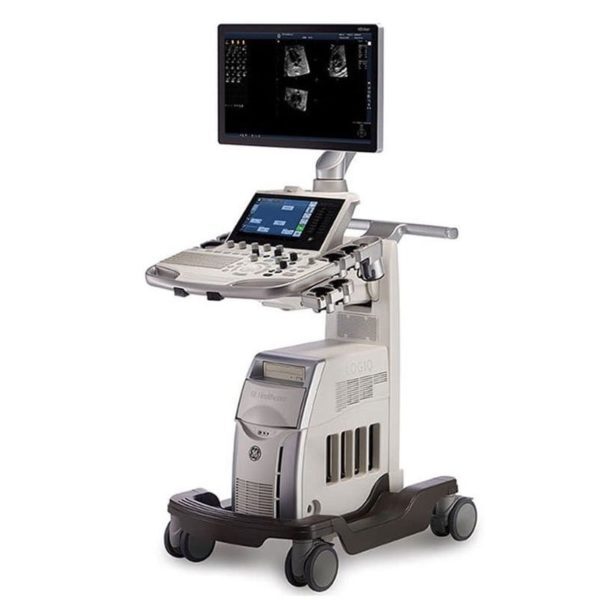
| Content | GE LOGIQ S7 WITH XDCLEAR ULTRASOUND
The GE Logiq S7 with XDclear Ultrasound is a mid range multi-purpose ultrasound machine. The GE Logiq S7 with XDClear has been fully upgraded, 23″ large LCD monitor and 10.1″ LCD touch screen. It becomes very reliable and trustworthy while reaching the revision 3.0, and GE incorporated single crystal transducers, so called XDClear transducers such as C1-6-D and C3-10-D. With one of the market most demanding technologies, XDClear, the user can enjoy premium image resolution, penetration, and wide bandwidth from two XDClear options. The GE Logiq S7 with XDClear built with the same system platform of GE Logiq S8 with XDClear, S-agile architecture, but just some of the features and transducer are not included or more economic options and transducers are integrated. Although the GE Logiq S7 with XDClear is classified as a mid range system, it offers a great benefit to users with advanced automated features such as Scan Assistant, OB Measure Assistant, and Breast Measure Assistant. Also, Automated Function Imaging(AFI), XDClear probes, large 23″ LCD monitor and 10.1″ touch screen are very unique features in mid-range.GE Logiq S7 With XDClear Dimensions and Weight:• Height: 1,210mm (Minimum), 1,760mm (Max.)
• Width: 530mm (Caster), 520mm (Keyboard), 565mm (Monitor)
• Depth: 865mm (Max.), 795mm (Caster)
• Weight: 90 kg (198 lb.) GE Logiq S7 With XDClear Specifications:• Digital beamformer
• Displayed Imaging Depth: 0 – 33 cm
• Minimum Depth of Field: 0 – 2 cm (Zoom) (probe dependent)
• Maximum Depth of Field: 0 – 33 cm (probe dependent)
• Continuous dynamic receive focus/continuous dynamic receive aperture GE Logiq S7 With XDClear Electrical power:• Voltage:100-120 Vac or 220-240 Vac
• Frequency: 50/60 Hz
• Power: Consumption maximum of 900 VA with peripherals GE Logiq S7 XDClear Features:• High resolution 23″ wide LCD monitor
• High resolution 10.1″ wide LCD touch screen
• 4 active probe ports and 1 parking probe port
• Integrated HDD
• Integrated DVD Multi Drive
• Integrated speakers
• Locking mechanism that provides rolling lock and caster swivel lock
• Integrated cable management
• Front and rear handles
• Easily removable air filters
• Automatic Optimization
• CrossXBeam
• Speckle Reduction Imaging (SRI-HD)
• Fine Angle Steer
• Coded Harmonic Imaging
• Virtual Convex
• Advanced 3D
• Patient information database
• Image Archive on integrated CD/DVD and hard disk drive
• Raw Data Analysis
• Real-time automatic Doppler calcs
• OB Calcs
• Fetal Trending
• Multi gestational Calcs
• Hip Dysplasia Calcs
• Gynecological Calcs
• Vascular Calcs
• Urological Calcs
• Renal Calcs
• Cardiac Calcs
• inSite ExC Capability
• On-board electronic documentation
• MPEGVue
• Key Macro
• Network Storage
• Quick Save
• Quick Patient Entry
• Email2MMS Accessories GE Logiq S7 with XDclear Ultrasound GE Logiq S7 With XDClear Peripheral Options:• Sony Digital UP-D711 Termal Printer
• Sony Fixture Kit for Digital UP-D711 Thermal Printer
• Sony Digital UP-D25 Color Thermal Printer
• Sony Digital UP-D897 BW Thermal Printer
• Mitsubishi P93W/E Thermal Printer
• Mitsubishi P95DW Thermal Printer
• Printer installation kit
• External USB printer connection
• Printing using Bluetooth connectivity (Inkjet printer option)
• Wireless LAN
• HDMI output available for compatible devices
• Footswitch with programmable functionality
• Universal Video Converter
• Power Assistant
• Battery Pack (for Power Assistant)
• Isolation Transformer
• Isolated 1Gb Ethernet connection
• Isolated USB connector
• EMI filter (PWR supply noise filter)
• RS-DLP Probe Adapter for TEE Probe
• Side Cabinet (Japan only)
• Drawer
• Small Probe Holder (Probe Holder Adapter for Small Probe)
• Multi Purpose Holder (Vertical Endocavitary Probe Holder)
• Optional Probe Holder (Side Probe Holder)
• Probe cable hanger
• ECG + AHA/IEC Cables
• USB 3 Pedal Foot Switch GE Logiq S7 With XDClear Supplies:Aquasonic ultrasound gel
Sono ultrasound wipes
Sony UPP-110HG thermal printing paper
Sony UPC-21L color thermal printing pack
Mitsubishi KP95HG thermal roll paper (new)
Mitsubishi KP65HM-CE High density thermal paper GE Logiq S7 With XDClear ports:• User accessible USB x 5 ports
• HDMI connector
• Ethernet 1000/100/10BaseT
• Audio Line Out (1.5mm pin jack) GE Logiq S7 With XDClear image storage:• On-board database of patient information from past exams
• Storage Formats:
• DICOM – compressed/ uncompressed, single/ multiframe, with/ without Raw Data
• Export JPEG, JPEG2000, WMV (MPEG 4), and AVI formats
• Storage Devices: – USB Flash Device: 64MB to 64GB (for exporting individual images/clips) – CD-R storage: 700MB – DVD storage: R (4.7GB) – Hard Drive Image Storage: ~300GB – Compare previous exam images with current exam images – Reload of archived data sets – Network Storage support for Import, Export, DICOM Read, SaveAs, SaveAs GE Logiq S7 with XDClear Options:• Auto IMT
• Auto EF
• Automated Volume Calculation (VOCAL II)
• Automated Function Imaging (AFI)
• Breast Measure Assistant
• OB Measure Assistant
• B-Flow/B-Flow Color
• B Steer+
• Breast Productivity Package
• Coded Contrast Imaging
• HRES Contrast
• Compare Assistant
• CW Doppler
• DICOM 3.0 connectivity
• Digital Video Recording Software (Software DVR)
• Elastography
• Elastography Quantification
• Flow Quantification
• LOGIQView
• Real Time 4D
• Report Writer
• Scan Assistant
• Stress Echo
• Thyroid Productivity Package
• Tissue Velocity Imaging (TVI)
• Tomographic Ultrasound Imaging (TUI)
• Volume Contrast Imaging (VCI) Static
• OmniView
• STIC
• Advanced Probes (XDclear)
• Cabinet: High/Mid/Low GE Logiq S7 with XDClear Applications:• Fetal/Obstetrics
• Abdominal (includes renal, GYN/Pelvic)
• Pediatric
• Small Organ (breast, testes, thyroid)
• Neonatal Cephalic
• Adult Cephalic
• Cardiac (adult and pediatric)
• Peripheral Vascular
• Musculo-skeletal Conventional and Superficial
• Urology (including prostate)
• Transrectal
• Transvaginal
• Transesophageal | DOUBLO GOLD HIFU SYSTEMWhy choose Doublo HIFU System?
HIFU (High-Intensity Focused Ultrasound System) is a highly precise medical procedure using high-intensity focused ultrasound to heat and destroy the pathogenic tissue rapidly. The earliest widespread use of HIFU was as a treatment for prostate cancer.HIFU hybrid system for Face & Body
Doublo Gold is the ultimate HIFU system for precise Face Lifting & Body Contouring. Offering the fastest treatment by Two-way round shooting and Auto Shot mode.Non Invasive – No Pain – No Bleeding – No Downtime
Faster shot speed : Efficacy improved by repetitious shooting with 300 shots in 8 minutes. Entertain more patients with a faster treatment procedure.Multiple treatment areas
By treating the upper dermis with wound healing effect, facial pores and skin tone is improved as the fibroblast is rebuilt and collagen is boosted.Dermal tissue elasticity and collagen increase as fibroblast is rebuilt by wound healing effect from damaging sub-cutaneous tissue and derma layer with thermal coagulation.
Skin tightening to reduce wrinkle and increase lifting effect is done by creating thermal coagulation at subcutaneous fat and SMASDOUBLO GOLD HIFU SYSTEM Highly Precise, Non-Surgical, Medical Procedure System for Professionals. Doublo HIFU’s Speed is Innovative Performance! Treatment time is dramatically reduced with Hironic’s High-capacity Technology and with Doublo HIFU System you can treat twice the amount of patients than beforeDOUBLO GOLD HIFU SYSTEM Accessories: Power cord for cart
Clear plastic tray insert
Large washer and small lock/spring washer
2x black attachements
2x treatment parameters guide
Simple user manual
Full user manual + CDHighly Precise, Non-Surgical, Medical Procedure System for Professionals. Doublo HIFU’s Speed is Innovative Performance! Treatment time is dramatically reduced with Hironic’s High-capacity Technology and with Doublo HIFU System you can treat twice the amount of patients than beforeNew Standard of
MFU System
1.No. 1 MFU proven by the patents2.MFU proven by cumulative sales and clinical cases3.Fastest treatment by Two-way round shooting and Auto Shot mode | SonoScape S8 Portable UltrasoundSystem Description: The SonoScape S8 portable ultrasound is an award winning portable ultrasound system with outstanding image quality in all modalities. SonoScape has designed a fully featured, shared service ultrasound system producing image quality one would expect from a much more expensive console system. Frost & Sullivan “Product Quality Leadership” Award:
“The S8 is versatile in performance,” notes Frost & Sullivan Research Analyst Shriram Shanmugham. “It uses a 15 MHz linear transducer, 512 super high density transducers, and is the only echocardiography system that also offers 4D imaging capability. None of the company’s competitors offers such a combination of 4D and high-end echocardiography technology affording portability.” “The high frame rate and high definition image quality provide users better diagnostic experience than ever” Application:- Shared Service!
- Ob/Gyn
- Anesthesia
- Urology
- Vascular / Vascular Access
- MSK
- Breast Imaging
- Abdominal
- Small Parts
- Cardiac
Features:- 15” LCD Monitor
- 2 Hot Swap Probe Ports
- High Pulse Repetition Frequency [HPRF]
- Multi Beam Processing [MBP]
- Trapezoidal Imaging
- Panoramic
- Imaging
- Advanced Tissue Harmonic Imaging
- M-Tuning [One touch image optimization]
- uScan
- Multi Plane Adult & Pediatric TEE
- Intimae Media Thickness [IMT]
- High Q Nosie Filter [HQNF]
- B-Mode
- M-Mode
- Color Doppler
- Power Doppler
- Spectral Steered Doppler
- Real time Triplex: B, Flow & PW Doppler / B, Flow & Color M
- ECG
- 4D
- Tissue Doppler Imaging
- B-Color
- Anatomic M-Mode
- DICOM 3.0
- 2 USB Ports
- Video In/Out
| | Options:- Mobile Trolley
- DVD/CD-RW
- Printer
- Stress Echo
Transducers:- C344, 5-2MHz/R40mm, Abdominal Convex Probe
- C362, 5-2MHz/R60mm, Abdominal Convex probe
- C542, 7-4MHz/R40mm, Abdominal & Pediatric Probe
- C311, 4-2MHz/R15mm, Micro-convex Cardiac Probe
- C611, 8-4MHz/R11mm, Micro-convex Pediatric Cardiac Probe
- L741, 10-5MHz/46mm, Vascular Small part Linear Probe
- L742, 15-5MHz/38mm, Vascular Small part linear probe
- L743, 15-5MHz/46mm, Vascular Small part linear probe
- L541, 7-4MHz/38mm, Vascular Linear probe
- 10L1, 12-6MHz/36mm, Vascular Small Parts Probe
- 10I2, 15-5MHz/25mm, Linear Surgical Probe
- 6V1, 8-4MHz/R11mm, Endovaginal Probe (135 degree)
- 6V3, 9-5MHz/R10mm, Endovaginal probe (200 degree)
- EC9-5, 9-5MHz/R8mm, micro-convex Endocavity probe (150 degree)
- 2P1, 4-2MHz, Phased Array Cardiac Probe
- 5P1, 7-4MHz, Phased Array Pediatric Cardiac Probe
- MPTEE, 7-4MHz, Multi-plane Phased Array TEE Probe
- MPTEE Mini, 7-4MHz Pediatric Multi-plane Phased Array TEE Probe
- VC6-2, 6-2MHz/R40mm, Curved volumetric probe for Live 3D
Biopsy Guides:- C344, C362, L741, L742, L743, 10LI, L541, 6V1, 6V3, EC9-5, 2P1, 7U2
|
| SIUI CTS-800 Veterinary Ultrasound Machine with Linear Rectal Probe
This small and convenient “Palm-Sized” portable veterinary ultrasound scanner fits in the palm of your hand, and is mostly used for farm animals. Its long battery life and waterproof features makes it the perfect ultrasound scanner for veterinarians on the field.The screen of the SIUI CTS-800V will rotate horizontally or vertically, according to how you are holding it.Gravity Sensor: You can operate the ultrasound system either in transversal layout or vertical layout. It can automatically adapt to two different layouts when you rotate it.Grid for estimation: With grid on the image area you can easily estimate the size of the target substance like follicle fertilized egg etc.SPECIFICATIONS SIUI CTS-800 Veterinary Ultrasound Machine with Linear Rectal ProbeMonitor: 7-inch WVGA LCD monitorWeight: 0.8kg Environmental rating: IP54 (main unit) IP67 (probe head) Battery: 3 hours operation time Display mode: B 2B ZOOM B B/M M Depth: Max to 250mm B gain: 0-100 Image storage: 256 frames Cine loop: 256 frames Probe: L50/7.5 MHz linear rectal probe (standard) L65/5.0 MHz linear rectal probe (optional) R20/5.0 MHz micro convex probe (optional)
– Main unit with 7-inch LCD monitor.
– 1 Probe connector.
– 1 Li-ion battery. 3 hours operation time.
– 1 Adaptor.
– 1 Car Charger.
– Belt and sun shield.
– 1 USB port (Mini).
– Carrying case.PROBES– Rectal Linear L7FVC (6.5/7.1/7.5/8.3/9.0MHz).
– Rectal Linear L5FVC (4.0/4.7/5.0/5.5/6.2MHz).
– Backfat Linear L1FC (150mm, 2.5/3.0/3.5/4.0/5.0MHz).
– Convex C3FC (2.5/3.0/3.5/4.0/5.0MHz).
– Microconvex C5FC (3.5/4.0/5.0/6.5/7.1MHz).CTS-800 is a palm-sized veterinary ultrasound system. Features Weight: 0.8kg Environmental rating: IP54 (main unit) IP67 (probe head) Battery: 3 hours operation time Display mode: B 2B ZOOM B B/M M Depth: Max to 250mm B gain: 0-100 Image storage: 256 frames Cine loop: 256 frames Probe: L50/7.5 MHz linear rectal probe (standard) L65/5.0 MHz linear rectal probe (optional) R20/5.0 MHz micro convex probe (optional) | GE Voluson i Portable 3D/4D UltrasoundFeatures* Lightweight and portable
* Battery operation
* Real-Time 4D
* 3D Multiplanar Display
* 3D Power Doppler
* Automatic Optimization (AO)
* Speckle Reduction Imaging (SRI) and CrossXBeam deliver advanced computational power, allowing for simultaneous processing of CrossXBeam CRI and SRI together – enabling added speckle reduction, contrast resolution and image clarity.
* HD-Flow uses a bi-directional Doppler feature to achieve a more sensitive vascular study and reduce overwriting.
* Volume Contrast Imaging (VCI) allows for image quality in either a single plane or all three planes of a volume acquisition
* Spatio-Temporal Image Correlation (STIC) captures a full virtual fetal heart cycle in real time, and the volume can be saved for offline analysis.
* Tomographic Ultrasound Imaging (TUI) makes analysis and documentation of dynamic studies easier with a simultaneous view of multiple parallel slices of a volume data set.
* Virtual Organ Computer-aided AnaLysis (VOCAL) is a semi-automated volume measurement tool that utilizes computer technology to provide accurate volume calculations.Comes with:GE Voluson i Portable 3D/4D Ultrasound* 1x Voluson i Portable 3D/4D Ultrasound
* 1x RAB2-5RS (2-5MHz) 3/4D Convex Abdominal Probe
* 1x E8C-RS (4-10MHz) Wideband Microconvex Endocavity (Vaginal) Probe
* 1x RIC5-9-RS (5-9MHz) 2D/3D RealTime 4D Endocavitary ProbeThe GE Voluson i ultrasound machine is primarily used for OB-GYN applications, but is versatile and powerful enough to be used for a variety of other applications as well. The Voluson i ultrasound machine carries the power of the GE Voluson line with access to premium 2D and 4D imaging in a portable form factor. The GE Voluson i provides the extraordinary image quality needed to confidently scan for women’s health. The GE Voluson i portable ultrasound machine offers features such as speckle reduction imaging and CrossXBeamCRI, which both lend to the incredible image clarity and quality of this system. The Voluson I portable ultrasound system is known for ease of use, allowing the operator to scan patients faster and more efficiently with better accuracy. | Acuson Cypress PLUS UltrasoundAcuson Cypress cardiovascular system PLUS delivers advanced ergonomics and superior performance in a highly portable package. TheCypress System PLUSis a highly miniaturized, all digital, phased array echocardiography system that provides complete studies and outstanding images – even on patients who are difficult to image.The Cypress PLUS transitions easily from your small private office or hospital echo lab to the operating theatre or your patient’s bedside. It’s ultrasound where you need it.The Cypress PLUS delivers high performance in a complete range of cardiovascular capabilities.Designed with advanced ergonomics in mind, the Cypress system PLUS streamlines your workflow and increases your productivity.If you already own a Cypress system, keeping technologically current is simple with the Cypress Upgrade program.The Cypress system is the smallest, lightest, high performance, phased array echo system ever built. Weighing in at just 19 pounds, with digital storage capacity for over 100 studies and network capabilities to send studies from remote locations, clinicians can perform complete echocardiography studies from almost anywhere. TheCypress system was designed for portability, durability and reliability, and to withstand the rigors of constant transportation. Internal system interconnects are minimized. Key components are shock mounted and the entire system sits inside a robust chassis. From hospital to office or from airplane to mountain village, the Cypress system will perform.Full Range of Capabilities
The Cypress system is a complete, full-featured echo cardiography system that provides complete studies and outstanding image quality even on the most difficult to image patients. It offers a full range of features including:Acuson Cypress PLUS Ultrasound– 2D with Harmonics
– Patient Report with audio capture
– M-Mode
– Embedded DICOM conformance
– Color PW and CW Doppler
– High frame rate color flow imaging
– Stress echo
– Network output
– Vascular imaging
– Comprehensive calculations package
– Contrast agent imaging
– USB peripheral support (PC printer connectivity)Physical Characteristics
• Single unit houses ultrasound system with control panel, flat panel display and MO drive
• Weight: 8.6 Kg (19 pounds)
• Height: 35.6 cm (14 in)
• Width: 40.6 cm (16 in)
• Depth: 20.3 cm (8 in) – with keyboard folded up 34.3 cm (13.5 in) – with keyboard down
• Heat dissipation: 490 BTU/hour (nominal)User Interface
• Fold-up control panel for portability
• Direct access to system functions through dedicated keyboard controls and multifunctional soft keys
• Easy-access main controls, including multifunctional trackball, enter and select keys, for minimum hand movement while controlling the system
• Adjustable trackball speed
• Functional grouping of keys, rotational knobs, switches and sliding TGC controls for direct access to critical functions
• Tactile feedback on control activation
• Backlighting of control panel labels
• Mode-dependent soft keys
• User-interface tools
• Pre-defined and user-entered labeling textMonitor
• 10.4 inch/26.4 cm flat panel active matrix display screen
• Protective anti-glare, anti-reflective lensHard Drive
• Internal hard drive for image and data storage
• Internal storage of approximately 50 patient studies (25 – 30 loops per study)Transducer Ports
• One transducer connector (type IEC601BF)Operating/Display Modes
• 2-D, fundamental and harmonic imaging
• Color Doppler Velocity (CDV)
• Color Doppler Energy (CDE)
• M-mode, vertical split screen display with 2-D
• Pulsed Wave (PW) Spectral Doppler
• Continuous Wave (CW) Spectral Doppler
• Duplex Doppler (combined 2-D and Spectral Doppler display)2D Calipers – Generic Measurements and Calculations
• Measurements and calculations in 2D, M-mode, color Doppler, PW and CW spectral Doppler modes
• Basic measurements and report calculations
• Comprehensive patient calculations report
• On-screen “mini-reports”
• Complete or configured patient report
• Cardiac package
• Carotid package
• Arterial package
• Venous package
• Abdominal package
• User configurable Sites
• Selectable menu position
• Four cursor types
• Angle correction
• Distance
• Area
• Slope
• Time
• Velocity
• Velocity/pressure
• Mean velocity/pressure gradient
• Velocity Time Integral
• Pressure halftime
• Continuity Equation
• Ejection Fraction
• Left Ventricular Volumes, including End-Systolic and End-Diastolic volumes
• Cardiac output
• Cardiac index
• Fractional area changeCine Review
• 2D Cine memory up to 15 seconds of history
• Color Doppler cine memory up to 15 seconds of history
• M-mode strip cine along with PW and CW Doppler mode
• strip cine memory determined by sweep speed (selectable by the user) 10, 20, 30 or 40 secondsTransducer Technology
• 3V2c: 2-4 MHz wideband phased array
• 7V3c: 3-7 MHz wideband phased array
• 7L3: 5-7 MHz wideband linear array (Vascular)
• 4C1: 2-4 MHz wideband curved linear array (Abdominal)
• V5Ms: 3-6 MHz wideband phased array (micro-pinless (MP) connector) |


Reviews
There are no reviews yet.