PERKASA MEDICAL

Free shipping worldwide. Orders over $200
Availability: In Stock

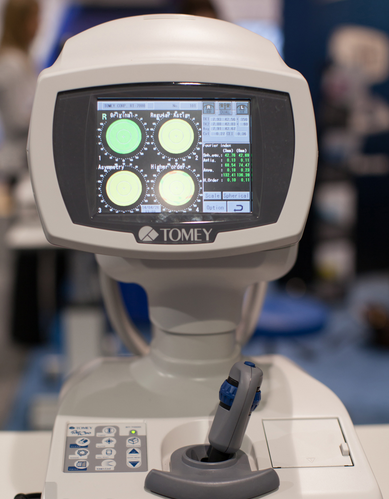
Tomey RT-7000 Autorefractor Keratometer Topographer For Sale

$11,350.00

The Tomey Refractometer Keratometer and Topographer only in 1 unit senior citizens and children can also get the inspection with out any trouble because they dont have to move the seat for three measurements.

Other people want this. 15 people have this in their carts right now.

Description

Tomey RT-7000 Autorefractor Keratometer Topographer

-Three Functions in One Instrument

-Easy & Speedy Touch Screen Alignment

-6.4 inches color TFT LCD

-Dual  CCD technology for Refractometry

-IOL/ Cataract Mode

-New incites for Keratometry KAI, KRI

-Auto Alignment / Auto Shot of Topography

Corneal Topography

– Auto Alignment / Auto Shot

Any one can easily capture measurement with Auto Alignment and Auto Shot. The Measurement variation is significantly reduced no matter what the skill level is of the operator.

CL Fitting Simulation

-A pseudo-fluorescent pattern of the contact lens selected from the built-in data base can be confirmed and the fitting can be stimulated before the patient wears  the trial lens.

Corneal Eccentricity Index

-The index of CEI (Corneal Eccentricity Index) indicates the ratio of flattering in percentage from the central cornea to the periphery.

Three Function in One Instrument

– Refractometer Keratometer and Topographer only in 1 unit senior citizens and children can also get the inspection with out any trouble because they don’t have to move the seat for three measurements. Switch from Ref-Kerato Mode to Corneal Topography Mode with only ONE TOUCH. The light come appears with only one button from the measuring head and the Ref-Keratometer is transformed to the topographer.

Refractometry

– Dual CCD technology for Refractometry

Two CCD cameras are used to capture images for observation and for measurement while providing highly accurate measurement data.

Keratometory

– New Indices for Keratometry

The Corneal Irregular astigmatism display function is installed to expand the possibility of Keratometer. This is the new function to measure the level of corneal irregular astigmatism which was difficult in the past.

SPECIFICATIONS

MeasurmentRanges

Refraction Measuremnt

Sphere  -25.00D to ±22.00DIVD=12.0mm

Cylinder  0D to ± 10.00DIVD=12.0mm

Axis  0 to 180

Corneal Curvature Measurement  5.00mm to 11.00mm

Measurement Zone 3mm R:8.00mm

Corneal Mapping

Display Range  9 to 100 D

-Normal Mode

1.0 to 8.0mm (R:8.00mm)

-Special Mode

0.9 to 7.0 mm (R:8.00mm)

Measurement Time

REF 0.2 Seconds

KRT 0.1 Seconds

Minimum Pupil Diameter  2.2mm

Minimum Measurment Step


Refraction Measurement

Sphere  0.01D

Cylinder  0.01D

Axis  1

Corneal Curvature Measurement  0.01mm

PD (Pupil Distance)  50mm to 86mm

Recording  Built-In Thermal Printer

Vertex Distance  0mm, 12.0mm, 13.5mm, 14.0mm, 15.5mm, 16.0mm

Exterior Output  (AN port / 4USB ports)

Dimensions Tomey RT-7000 Autorefractor Keratometer Topographer

High  466mmm or 18 Inches

Width  307mm or 12 Inches

Depth  490mm or 19 Inches

Weight  Approx 20,000Kg / 44.4 Pounds

Display  6.4 Inches color liquid crystal

Power Supply

Voltage  AC 100 to 240V

Frequency  50/60 Hz

Consumption Power  120VA to 150VA

Temperature  +10C to + 40C

Display  30% to 75%

Reviews

There are no reviews yet.

Be the first to review “Tomey RT-7000 Autorefractor Keratometer Topographer For Sale”

Your email address will not be published. Required fields are marked *

Quick Comparison

SettingsTomey RT-7000 Autorefractor Keratometer Topographer For Sale removeZeiss Humphrey Hark 599 Autorefractor / Keratometer For Sale removeTomey RC-5000 Auto Refractor Keratometer For Sale removeNidek RS-330 Retina Scan Duo 2 with FAF removeCarl Zeiss Cirrus HD-OCT 5000 Smart Advanced Topography HD removeNidek CEM-530 Specular Microscope For Sale remove
NameTomey RT-7000 Autorefractor Keratometer Topographer For Sale removeZeiss Humphrey Hark 599 Autorefractor / Keratometer For Sale removeTomey RC-5000 Auto Refractor Keratometer For Sale removeNidek RS-330 Retina Scan Duo 2 with FAF removeCarl Zeiss Cirrus HD-OCT 5000 Smart Advanced Topography HD removeNidek CEM-530 Specular Microscope For Sale remove
Image
SKU
Rating
Price $11,350.00 $2,500.00 $4,800.00 $3,150.00 $6,950.00 $3,250.00
Stock
Availability
Add to cart

Add to cart

Add to cart

Add to cart

Add to cart

Add to cart

Add to cart

DescriptionThe Tomey Refractometer Keratometer and Topographer only in 1 unit senior citizens and children can also get the inspection with out any trouble because they dont have to move the seat for three measurements.

The HUMPHREY 599 is the best combination refractor-keratometer to offer subjective refinement of sphere, cylinder, and axis as well as isolated acuity lines, children’s targets, near testing, and low contrast sensitivity testing.

The Tomey RC-5000 has highly accurate measurement combined with short examination time and ease of use, makes working with the Tomey RC-5000 Autorefractor Keratometer both professional and quick. The advanced touch-screen display allows you to perform both refraction and keratometry automatically with one simple movement, capture one eye on the display and the Tomey RC-5000 will do both eyes automatically. With the new features, the Tomey RC-5000 Autorefractor Keratometer will surely make your office more efficient.
The Nidek RS-330 OCT is a combination of a High-Definition OCT and fundus camera. Optional delivery with FAF (Auto-fluorescence function)
CIRRUS HD-OCT 5000 is configured with the highest resolution visualization capabilities and sophisticated clinical applications such as Advanced RPE analysis to track retinal pigment epithelial integrity and Ganglion Cell Analysis to assess glaucomatous loss in the macula that may not be evident in the peripapillary region. The new FastTrac  retinal tracking reduces eye motion artifacts without sacrificing patient throughput with a proprietary scan acquisition strategy, high speed 20 Hz LSO camera, and single-pass alignment scanning. This feature enables the highest resolution B-Scans to be captured in identical locations from visit to visit providing precise assessment of change in targeted pathologies.

Combination of auto and manual analyses

All three manual analysis methods can be performed on the same image and also on auto-analyzed images. The versatility of combining automated and manual analyses allows analysis of the range of pathology in a comprehensive practice.
The CEM-530 with new advanced software and enhanced image capture system allows rapid image acquisition. 
Content

Tomey RT-7000 Autorefractor Keratometer Topographer

-Three Functions in One Instrument-Easy & Speedy Touch Screen Alignment-6.4 inches color TFT LCD-Dual  CCD technology for Refractometry-IOL/ Cataract Mode-New incites for Keratometry KAI, KRI-Auto Alignment / Auto Shot of TopographyCorneal Topography– Auto Alignment / Auto ShotAny one can easily capture measurement with Auto Alignment and Auto Shot. The Measurement variation is significantly reduced no matter what the skill level is of the operator.CL Fitting Simulation-A pseudo-fluorescent pattern of the contact lens selected from the built-in data base can be confirmed and the fitting can be stimulated before the patient wears  the trial lens.Corneal Eccentricity Index-The index of CEI (Corneal Eccentricity Index) indicates the ratio of flattering in percentage from the central cornea to the periphery.Three Function in One Instrument– Refractometer Keratometer and Topographer only in 1 unit senior citizens and children can also get the inspection with out any trouble because they don’t have to move the seat for three measurements. Switch from Ref-Kerato Mode to Corneal Topography Mode with only ONE TOUCH. The light come appears with only one button from the measuring head and the Ref-Keratometer is transformed to the topographer.Refractometry– Dual CCD technology for RefractometryTwo CCD cameras are used to capture images for observation and for measurement while providing highly accurate measurement data.Keratometory– New Indices for KeratometryThe Corneal Irregular astigmatism display function is installed to expand the possibility of Keratometer. This is the new function to measure the level of corneal irregular astigmatism which was difficult in the past.SPECIFICATIONSMeasurmentRangesRefraction MeasuremntSphere  -25.00D to ±22.00DIVD=12.0mmCylinder  0D to ± 10.00DIVD=12.0mmAxis  0 to 180Corneal Curvature Measurement  5.00mm to 11.00mmMeasurement Zone 3mm R:8.00mmCorneal MappingDisplay Range  9 to 100 D-Normal Mode1.0 to 8.0mm (R:8.00mm)-Special Mode0.9 to 7.0 mm (R:8.00mm)Measurement TimeREF 0.2 SecondsKRT 0.1 SecondsMinimum Pupil Diameter  2.2mmMinimum Measurment Step Refraction MeasurementSphere  0.01DCylinder  0.01DAxis  1Corneal Curvature Measurement  0.01mmPD (Pupil Distance)  50mm to 86mmRecording  Built-In Thermal PrinterVertex Distance  0mm, 12.0mm, 13.5mm, 14.0mm, 15.5mm, 16.0mmExterior Output  (AN port / 4USB ports)Dimensions Tomey RT-7000 Autorefractor Keratometer TopographerHigh  466mmm or 18 InchesWidth  307mm or 12 InchesDepth  490mm or 19 InchesWeight  Approx 20,000Kg / 44.4 PoundsDisplay  6.4 Inches color liquid crystalPower SupplyVoltage  AC 100 to 240VFrequency  50/60 HzConsumption Power  120VA to 150VATemperature  +10C to + 40CDisplay  30% to 75%

Zeiss Humphrey Hark 599 Autorefractor / Keratometer

The HUMPHREY 599 allows for immediate patient feedback on the prescription and can be linked to the Humphrey Lens Analyzer to allow patients to compare their current RX to the new refraction results. Precise keratometer readings can be instantly obtained for all clinical needs.Fast, accurate and objective measurements with complete printout Automatic cycle for objective measurements Complete automatic alignment and tracking Acuity line isolation Cylinder and axis refinement Duochrome target Jackson Cross Cylinder Test Near vision testing Glare testing Spherical equivalents PD displayed on printout Children’s acuity targets Recall for old and new Rx comparisons Communicom Interface Keratometry Reading including average and delta K’s 
  • Fast, accurate and objective measurements with complete printout
  • Automatic cycle for objective measurements
  • Complete automatic alignment and tracking
  • Acuity line isolation
  • Cylinder and axis refinement
  • Duochrome target
  • Jackson Cross Cylinder Test
  • Near vision testing
  • Spherical equivalents
  • PD displayed on printout
  • Children’s acuity targets
  • Recall for old and new Rx comparisons
  • Communicom Interface
  • Keratometry Reading including average and delta K’s
Features:Fast, accurate and objective measurements with complete printout Automatic cycle for objective measurements Complete automatic alignment and tracking Acuity line isolation Cylinder and axis refinement Duochrome target Jackson Cross Cylinder Test Near vision testing Glare testing Spherical equivalents PD displayed on printout Children’s acuity targets Recall for old and new Rx comparisons Communicom Interface Keratometry Reading including average and delta K’s in the Humphrey HARK 599About us:Zeiss Humphrey Hark 599 Autorefractor / Keratometer ALT Medical Device is your complete eye care equipment solution. Is comprehensive company offering only high end, quality equipment. As a company, ALT Medical Device combines outstanding business practices with an unparalleled approach to customer service. We have 100% customer satisfaction by delivering quality equipment and full customer service with every transaction.

Tomey RC-5000 Auto Refractor Keratometer

Tomey RC-5000 RefractometryNormal Mode:-Fogging is applied automatically for each measurement for more Accurate ResultsQuick Mode:-Fogging is applied as needed for consecutive measurements.IOL/CAT Mode:-This mode is used to measure cataract or pseudophakic eyes.Keratometry:-Short consecutive measurements and time provide reliable data. -Measurements at 6.00mm (R=8.0mm) as well as at 3.00mm give you enough information for contact lens fitting. -base curves of contact lenses are stored, categorized and suggested manufacturers are automatically printed.Diameter Measurements of Cornea and Pupil:-Measurement can be done easily by moving the two cursors on the display to the boundary of cornea or pupil. This is useful for deciding the diameter of a contact lens and for other contact lens fitting practice.Power Joystick:-Move the joystick ring up/down and the instrument head moves up/down quickly. -Move the joystick hand rest to the right/left or backward/forward and the instrument head moves to the right/left or backward/forward quickly. -Rotate the joystick ring and the instrument head moves up/down slowly. -Tilt the joystick and the instrument head moves in each direction slowly.Power Chinrest:-The chin rest can be moved by pushing the buttons on the front panel. No need to reach out to the chin rest for manual adjustments.Autorefractor Keratometer Features:Refractometory : Tomey RC-5000 Auto Refractor Keratometer-Sphere 25.00 to +22.00D (VD =12.00mm) Cylinder -10.00 to +10.00D (VD = 12.00mm) -Axis 0 to 180 Degree -Minimum Pupil Dia. 2.2mm -Measurements Time 0.2 Sec.Keratometry :-Range 5.00mm to 11.00mm -Corneal Refraction 30.68D to 67.50D (n = 1.3375) -Corneal Astigmatism 0D to 10D (n = 1.3375) -Axis 0 to 180 Degree -Measured Area 3.0mm / 6.0 mm -Measured Time 0.1 Sec PD -Measurement Yes (50 to 86mm) -Diameter Measuremnt of Cornea & Pupil 1.0 to 14.0mm -Chinrest : Power Chin Rest -Printer : High Speed Thermal Line Printer -OutputPort RS-232C -Power Requirements: AC 100V to 240V -50/60Hz 130VA

Nidek RS-330 Retina Scan Duo 2 with FAF

The Retina Scan Duo 2 is a combined OCT and fundus camera system that incorporates new features enhancing screening and clinical efficiency in addition to user-friendly features that were incorporated from the previous model.FEATURES : Nidek RS-330 Retina Scan Duo 2 with FAF
  • Fundus image acquisition with macula and disc capture in one image on OCT
  • Combined diagnosis of macular and disc pathologies
  • Wide area scan (12 x 9 mm) Wide area normative database (macula: 9 x 9 mm, disc: 6 x 6 mm): A 12 x 9 mm wide area image can be acquired. The retina map captures both the macula and disc in a single shot. The normative database provides a wide area color-coded map comparing the patient’s macular thickness to a population of normal eyes.
  • Denoising technique with deep learning: A new image enhancement technique using deep learning automatically displays a denoised image once B-scan acquisition is complete. With deep learning of a large data set of images averaged from 120 images, this denoising technique provides high definition images comparable to a multiple-image-averaging technique. The denoising function generates high definition images from a single frame while decreasing image acquisition time and increasing patient comfort.
  • Quick acquisition of high definition B-scan images from a single-frame image
  • Fundus autofluorescence (FAF): The FAF function is an advanced screening feature that allows non-invasive evaluation of the RPE without contrast dye. *Available for the FAF model
Nidek RS-330 Retina Scan Duo 2– Easy operation with 3-D auto tracking, auto shot, and user friendly interface – High definition images – Wide area scan (12 x 9 mm) / Wide area normative database (9 x 9 mm) – Multiple OCT scan patterns – Value added features – Various reports
Nidek High speed image capture OCT images are captured at scan speeds of 70,000 A-scans/s which is 32% faster than acquisition with the Retina Scan Duo™ using Regular OCT sensitivity*Wide area scan (12 x 9 mm) Wide area normative database (macula: 9 x 9 mm, disc: 6 x 6 mm) A 12 x 9 mm wide area image can be acquired. The retina map captures both the macula and disc in a single shot. The normative database provides a wide area color-coded map comparing the patient’s macular thickness to a population of normal eyes.Denoising using deep learning A new image enhancement technique using deep learning automatically displays a denoised image once B-scan acquisition is complete. With deep learning of a large data set of images averaged from 120 images, this denoising technique provides high definition images comparable to a multiple-image-averaging technique. The denoising function generates high definition images from a single frame while decreasing image acquisition time and increasing patient comfort
Carl Zeiss Cirrus HD-OCT 5000 Smart Advanced Topography HD

Advancing Smart OCT

for Anterior Segment, Glaucoma and Retina
  • Anterior Segment Premier Module
  • PanoMap Wide-Field Display
  • Smart HD Scans
  • En face report
Measurement centering with Foveafinder

Visualization at the speed of CIRRUS

Analyzing a single pathology from multiple views provides comprehensive insight and analysis of the clinical situation. How this helps you:
  • Spot small areas of pathology. Tightly spaced B-scans, (either 30 or 47 μm apart), in the cube ensure that small areas of pathology are imaged. For reference, a human hair is about 40-120 μm in diameter.
  • Visualize the fovea. Scans that are spaced further apart than in the CIRRUS cube may miss the central fovea.
  • Fuel for analysis. Millions of data points from the cube are fed into the Zeiss proprietary algorithms for accurate segmentation, reproducible measurements and registration for change analysis.
  • Take the pressure off the operator. As long as the scan is placed in the vicinity of the fovea or optic nerve, the software automatically centers the measurements after the capture.
  • See the tissue from different perspectives. View the cube data from all angles, with 3D rendering, OCT fundus images and Advanced Visualization™.
  • Future ready. Previously captured CIRRUS cubes can be analyzed using new analyses.
 
Measurement centering with Foveafinder

Tracking at the speed of CIRRUS

FastTrac™ reduces eye motion artifacts without sacrificing patient throughput with a proprietary scan acquisition strategy, high speed 20 Hz LSO camera, and single-pass alignment scanning With FastTrac, scan at the highest resolution at the same location at each visit.
Watch video
Measurement centering with Foveafinder

Assessment at the speed of CIRRUS

Measurement centering with FoveaFinder and AutoCenter
On the macula, the unique FoveaFinder technology ensures the ETDRS and ganglion cell plus inner plexiform layer measurement frameworks are centered on the fovea.AutoCenter™ function automatically centers the 3.4 mm diameter peripapillary RNFL calculation circle around the disc for precise placement and repeatable registration. The placement of the circle is not operator dependent. Accuracy, registration and reproducibility are assured.
Measurement centering with Foveafinder

Manage at the speed of CIRRUS Carl Zeiss Cirrus HD-OCT 5000 Smart Advanced Topography HD

Guided Progression Analysis (GPA™) for Assessing RNFL and Optic Disc Change
GPA™ compares RNFL thickness and optic disc measurements over time and determines if statistically significant change has occurred.Focal Progression Analysis – Up to 6 progression maps are compared to two baselines. Areas of statistically significant change are color-coded yellow when first noted and then red when the change is sustained over consecutive visits. Trend Analysis with Rate of Change – RNFL thickness values are plotted over time. Trends are calculated and confidence intervals are shaded when rate of change is statistically significant.
 

Nidek CEM-530 Specular Microscope

The analysis results with graphic and color-coded cell images helps the clinician to rapidly and effectively evaluate the endothelial cell layer.In addition to conventional central and peripheral specular microscopy, the CEM-530 includes a unique function to capture paracentral images.
  • Paracentral specular microscopy
  • Faster measurements and two-second auto analysis
  • Comprehensive analysis
  • Advanced manual analysis functions
  • Easy operation
  • Additional features with CEM Viewer for NAVIS-EX
Combination of auto and manual analyses
All three manual analysis methods can be performed on the same image and also on auto-analyzed images. The versatility of combining automated and manual analyses allows analysis of the range of pathology in a comprehensive practice.Features
  • Multi area specular microscopy
  • Enhanced usability and quick analysis
  • Advanced manual analysis functions
  • Combination of auto and manual analyses
  • Additional features with CEM Viewer for NAVIS-EX
Multi area specular microscopy
In addition to conventional central and peripheral specular microscopy, the CEM-530 includes a NIDEK original function that captures paracentral images. The combination of central, paracentral, and peripheral imaging provides a broader, overall view that can be used for detailed morphological and quantitative evaluation of the endothelial layer and individual cells.


Enhanced usability and quick analysis

The 3D auto tracking and auto shot functions result in a user friendly and patient friendly experience. Data analysis within 2 seconds allows efficient patient flow.

Detail analysis


Advanced manual analysis functions
Center point

Select the approximate center of a cell. The cells are detected based on the surrounding points. This method is effective for areas where groups of cells are clumped together.


Corner point

Trace the outlines of the cells to be analyzed by selecting the corners of each cell. This method is suitable for detailed identification of the size and dimension of isolated cells.


Pattern select

Select a hexagonal reference pattern that is similar to the cell size and drag it onto the cell to be analyzed. This method is effective for rough identification of the size and dimension of the cells.


 Combination of auto and manual analyses 
All three manual analysis methods can be performed on the same image and on auto-analyzed images. The versatility of combining automated and manual analysis on the same image allows for better clinical interpretation of the diverse range of pathology in a comprehensive practice.


Additional features with CEM Viewer for NAVIS-EX* (optional)
CEM Viewer is software used for viewing and working with CEM-530 data via NAVIS-EX. This function enhances the capability of the CEM-530 with additional features and increases the efficiency of any clinic. *NAVIS-EX is optional software and is required for use of the CEM Viewer.
Data management and endothelial cell count

Unlimited NAVIS-EX database is available for review on the CEM Viewer. The basic functions of the CEM-530 such as endothelial cell count are available on the CEM Viewer.


Progression follow-up and comparison

Multiple examination data sets are displayed in chronological order for follow-up. Additionally, two data sets are displayed for comparison. Endothelial changes can be monitored over time with this function.


Paracentral display with peripheral
The images and analyses of the paracentral and peripheral areas are displayed providing a comprehensive image of endothelial cells.

WeightN/AN/AN/AN/AN/AN/A
DimensionsN/AN/AN/AN/AN/AN/A
Additional information
You're viewing: Tomey RT-7000 Autorefractor Keratometer Topographer For Sale $11,350.00
Add to cart
Select the fields to be shown. Others will be hidden. Drag and drop to rearrange the order.
  • Image
  • SKU
  • Rating
  • Price
  • Stock
  • Availability
  • Add to cart
  • Description
  • Content
  • Weight
  • Dimensions
  • Additional information
  • Attributes
  • Custom attributes
  • Custom fields
Click outside to hide the comparison bar
Compare
Shopping cart close