| Description | All necessary adjustments and operations are realized automatically.
By touching the mode and measurement button, semi-automatic measurement also achieved.
This function supports easy workflow for blood vessels and blood flow volume measurements. | The more patients you see, the more you need Voluson E8.
The Voluson E8 ultrasound system is designed to keep pace with busy practices that conduct a wide range of Women’s Health exams, from routine scanning to complex assessments.
The imaging excellence of Radiance System Architecture – your diagnostic confidence will be enhanced by the extraordinary image clarity and speed of Radiant System Architecture. | M-Turbo is a compact ultrasound imaging system from SonoSite, featuring the image quality and functionality needed to meet your diverse needs. The standard M-Turbo is also still available exclusively from BCF. | MedCorp is a trusted source for new and refurbished ultrasound systems with a combined 50 years experience in the industry and a detailed process for ensuring that each system they sell is as good as the day it was manufactured. | RealTime 4D imaging – continuous, 3D scanning with simultaneous visualization of the A, B and C planes.
The system allows you to explore images in any plane to reveal the smallest details with stunning clarity and apply sophisticated analytical tools to answer virtually any of your clinical questions.
• 3D Static/4D Real-time Imaging (15 FPS) State-of-the-art user interface with on screen menu’s Automatic Tissue Optimization (ATO) 15″ high resolution non-interlaced flat CRT 4 active probe ports Image Archive on CD, MOD, and Hard Drive (40 GB) B-Mode, M-Mode Tissue Harmonic Imaging Digital Beamformer with 512 system processing channel technology CINE memory: up to 256 MB (up to 3000 2D Images) Power Doppler Imaging (PDI) Tissue Doppler Imaging | The SonoSite Titan is the earliest version of the PoC dedicated portable ultrasound machine from Sonosite. Though its form factor is compact, it provides high resolution imaging, rapid responsive time, and supports a broad range of applications including cardiac. |
| Content | Konica Sonimage HS2 Ultrasound
Konica Sonimage HS2 Ultrasound is a Point-of-Care portable ultrasound system designed to support a wide range of applications and patient types. Konica Minolta’s advanced technology features provide high resolution image quality and efficient workflow for daily clinical practice.Konica Sonimage HS2 Ultrasound Unique Nanofabrication Technology, combines both material and machining expertise, to produce broad frequency linear probe “L18-4”. It offers both high sensitivity as well as greater penetration.T2HI (Traid Tissue Harmonic Imaging)
Dual Sonic Technology
By combining T2HI and Dual Sonic, high quality THI signal is formed around the center of ultrasound beam in receiving area.
As a result, it suppresses the acoustic noise and ensure the optimum image from deep to superficial structure.iXRET
The SONIMAGE HS2 achieves higher resolution and faster frame rates by utilizing Konica Minolta’s unique technologySNV (Simple Needle Visualization)
The SONIMAGE HS2 automatically detects needle insertion.
This funciton can be used both ‘In plane’ and ‘Out of planeEfficient Workflow for Daily Clinical Practice The SONIMAGE HS2 delivers intuitive workflow by customizing 8 physical buttons and touch panel. MPA (Multi Parameter Adjuster) MPA enables to change multiple image parameters like frequency change and turning trapezoid on in conjunction with depth change. Vascular NAVI
All necessary adjustments and operations are realized automatically.
By touching the mode and measurement button, semi-automatic measurement also achieved.
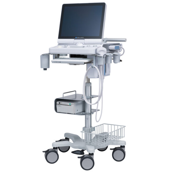
This function supports easy workflow for blood vessels and blood flow volume measurements. The SONIMAGE HS2 offers the unique capability to write text and draw lines and figures using fingers on the screen. This is an excellent tool for training and communication with patients. | GE Voluson E8 Ultrasound Machine15’’ LCD monitor [Rev BT06], 10.4” touchscreen, 3 Active Probe Ports, Floating Keyboard: Interactive back-lighting, Beta-View CrossXBeam, Virtual Convex, Extended View, ATO [Automatic Tissue Optimization], Coded Harmonic Imaging, High Resolution Zoom, Inversion Real-time automatic Doppler, OB/GYN; Vasc. Cardiac; Abd; Small-Parts; Urology; Ped; Ortho; Neuro reporting, Multigestational Calculations, HPRF [High Pulse Repetition Frequency], SonoVCAD [Matrix array volume technology], TruScan [On-board archive], VCI [Volume Contrast Imaging], SonoAVCfollicle [Sonography-based Automated Volume Count follicle], SonoVCADheart [Sonography-based Volume Computer Aided Display], TUI [Tomographic Ultrasound Imaging], CE [Coded Excitation], XTD, SRI III [Speckle reduction imaging], CRI [Compound Resolution Imaging], DICOM, Ethernet port, Video Out, 6 USB ports, Integrated HDD (160 GB) and DVD/CD-RW.
3D/4D, Endocrinology, Family Practice, Gastroenterology, Internal Medicine, Nephrology, Neurology, OB-GYN, Orthopedics, Otorhinolaryngology, Pain Management, Urology, Vascular Probes GE Voluson E8 Ultrasound Machine - 11L-D Linear Array
- 3S-D Cardiac Sector
- 4C-D Convex Array
- 9L-D Linear Array
- AB2-7 Convex
- IC5-9-D Intercavity
- M6C-D Convex
- P2D CWD Non-Imaging
- P6D CWD Non-Imaging
- PA6-8-D Cardiac
- RAB2-5-D 3D/ 4D Convex
- RAB4-8-D 3D/4D Convex
- RIC5-9-D 3D/4D Intracavity
- RIC6-12-D 3D/4D Intracavity
- RNA5-9-D 3D/4D Convex
- RRE6-10-D Endocavitary Micro-Convex
- RSP6-16-D 3D/D Linear
- SP10-16-D Linear
- 19″ monitor
- 4D Real Time
- CFM Doppler Mode
- Coded Contrast Imaging
- CrossBeam
- DICOM 4D
- HD flow
- Power Doppler Mode
- SonoVCAD
- STIC (Spacial-Temporal Image Correlation)
- T.U.I (Tomographic Ulrasound Imaging)
- VOCAL II VCI (Volume Contrast Imaging)
- Black and white printer
- Color printer
- DVD-Recorder
As your patient volume increases and your clinical cases become more complex, you will require an ultrasound system that enables you to deliver truly exceptional care – confidently and efficiently – every time. The Voluson E8 ultrasound system is designed to keep pace with busy practices – handling routine to complex women’s health exams with ease and precision. The premium image quality of the Voluson E8 combined with access to advanced capabilities, delivers the level of performance required by you and your patients. | Sonosite M-Turbo Ultrasound Machine
M-Turbo is a compact ultrasound imaging system from SonoSite, featuring the image quality and functionality needed to meet your diverse needs.Ideal for the general practitioner looking to do more advanced cardiac and vascular exams.Highlights of the new Sonosite M-Turbo :- Now available the M-Turbo with probe.
- This special version of the M-Turbo comes with a one year warranty, however the probes still have the standard 5 year Sonosite warranty.
- With the M-Turbo you can still add Colour, Pulse Wave and Continuous Wave Doppler.
- The system is now available with DICOM networking.
- Speak to your local Account Manager now to arrange a demonstration.
The standard M-Turbo is also still available exclusively from BCF.Highlights of the Sonosite M-Turbo:- More processing power than MicroMaxx – the next step up in image quality
- 5 year manufacturer’s warranty
- Mains or battery powered so can be used anywhere, anytime
- Able to upgrade from standard black and white system to high quality Colour/Doppler in future
- Large range of probes for all applications
. - Quick start up in just 12 seconds.
More… Sonosite M-Turbo Ultrasound MachinePortable, tough and easy to use. So tough it was originally designed for the American military and comes with a full manufacturer’s 5 year warranty.Despite its size, the M-Turbo produces outstanding cardiac images. It has very high colour Doppler sensitivity.Fully upgradeable to Colour, Pulsed Wave (PW) and Continuous Wave (CW) Doppler and capability to upgrade to DICOM or Wireless DICOM. You can connect it to a PACS system to allow viewing and secure storage of images.Stores still images and movie clips which can be exported via USB in PC friendly formats (jpeg, mpeg and avi).Optional enhanced needle visualisation software (unique to Sonosite) easily highlights needles within the patient allowing for accurate acquisition of a FNA or Tru-Cut biopsy sample.M-Turbo is a compact ultrasound imaging system from SonoSite, featuring the image quality and functionality needed to meet your diverse needs. The standard M-Turbo is also still available exclusively from BCF. | SonoSite Nanomaxx Ultrasound MachineThe SonoSite NanoMaxx ultrasound system combines premium functionality and affordability with the versatility and portability that makes SonoSite a top competitor in the portable ultrasound market. The SonoSite NanoMaxx comes in a sleek, hand-held package that never compromises performance, with applications ranging from emergency medicine and anesthesia to vascular.MedCorp offers the refurbished SonoSite NanoMaxx for purchase. Used SonoSite NanoMaxx from MedCorp offer everything you’d expect from a newer machine, including PC/MAC export configurations, 2D, Color and Color Power Doppler modes, and 2GB of internal memory that can hold 50 images each for 40 patients, all in an 8” touch-screen interface. However, because this is a refurbished SonoSite NanoMaxx, the price is lower than the new models. - 8″ Touch-screen interface
- 2D, Color and Color Power Doppler modes
- 2GB internal memory that can hold 50 images each for 40 patients
- 2 hours of battery power
- PC/MAC export configuration
- Four preset pictograph screen positions
- Drop resistant magnesium case
System Description:SonoSite Nanomaxx Ultrasound Machine
The NanoMaxx ultrasound system offers excellent performance at an affordable price. It is easy to use and offers top of the line diagnostic imaging, color power Doppler and color-flow velocity. The NanoMaxx can aid users in making clinical decisions or guide interventional procedures. The unit uses proprietary technology which allows the optimization of many system settings by simply pressing a button. Application:
Abdominal, Cardiology, Gynecology, Nerve, Obstetrics, CIMT, Musculoskeletal, Small Parts, Superficial, Vascular, Venous | GE Voluson 730 Pro Ultrasound MachineThe Voluson 730 Pro is built on the same platform as GE’s leading Real-Time 4D system, the Voluson 730 Expert. With an easy to use interface and exceptional 2D image quality, in addition to GE’s exclusive 4D imaging technology, the Voluson 730 Pro is ideal for any clinical practice with a variety of patient workloads. Recognized world-wide for its 4D imaging capabilities, the Voluson 730 Pro also features exceptional 2D image quality.RealTime 4D imaging – continuous, 3D scanning with simultaneous visualization of the A, B and C planes.
The system allows you to explore images in any plane to reveal the smallest details with stunning clarity and apply sophisticated analytical tools to answer virtually any of your clinical questions.Performance:GE Voluson 730 Pro Ultrasound Machine
State-of-the-art user interface with high resolution 10.4 inch LCD touch panel
Automatic Tissue Optimization
Tissue Doppler
Coded Harmonic Imaging
Coded Excitation (CE)
HD-Flow
CrossXBeamCRI (Compound Resolution Imaging)
Static 3D Mode
B Mode
Focus & Frequency Composite (FFC)
High Resolution Zoom
Pan Zoom
Steering
Virtual Convex
Beta-View
Patient information database
Image Archive on MOD and hard drive
3D/4D data compression (lossy/lossless)
Inversion
Real-time automatic Doppler calcsCapabilities:
OB/GYN, Vascular, Cardio, Abdominal, Small-Parts, Urology, Pediatrics, Ortho, Neurology, Multigestational Calculations, B-Flow, VCI (Volume Contrast Imaging)Available Probes:
AB2-7 Wide Band Convex Probe
AC2-5 Wide Band Convex Probe
4C-A Wide Band Convex Probe
M7C-H Wide Band Convex Probe
IC5-9H Wide Band Convex Probe
PA2-5P Wide Band Phased Array Probe
PA6-8 Wide Band Phased Array Probe
SP10-16 Wide Band Linear Probe
SP4-10 Wide Band Linear Probe
SP6-12 Wide Band Linear Probe
M12L-H Wide Band Linear Probe (1.25D Array)
RAB2-5L Wide Band Convex Volume Probe
RAB4-8L Wide Band Convex Volume Probe
RIC5-9H Wide Band Convex Volume Probe
RIC5-9W Wide Band Convex Volume Probe
RRE6-10 Wide Band Convex Volume Probe
RSP6-16 Wide Band Linear Volume Probe
RNA5-9 Wide Band Convex Volume Probe
SCW 2.0; non imaging suprasternal CW-Pencil Probe
PCW 4.0; non imaging peripheral CW-Pencil Probe | Sonosite Titan Ultrasound Machine
The SonoSite Titan Portable Ultrasound is SonoSite’s second generation portable ultrasound system. This system improved upon the technology of the SonoSite 180 with a larger screen and keyboard packed into a laptop style system. This system is well suited for Vascular access, Musculoskeletal, Anesthesiology, Internal Medecine, Emergency rooms and Family Physicians. Veterinary configurations are available. Veterinary configurations are available.8.4″ LCD monitor, 2 probe ports, Vascular, OB/GYN, Cardiac reports, Soft keys, Split screen, Zoom, Storage via 64 or 256 MB flash cards, 220 image CINE loop, USB & Ethernet image transfer, Volume calculations via diameters, THI [Tissue Harmonic Imaging], SonoCalc [IMT analysis, PC-based], DICOM, S-video (in/out), RGB, Composite video.
Dimensions: 3″ (H) x 10.9″ (W) x 11.9″ (D) Weight: 7.7 lbs Manufacturer
Sonosite Application
Cardiology, Endocrinology, Family Practice, Gastroenterology, Internal Medicine, Nephrology, Neurology, OB-GYN, Orthopedics, Otorhinolaryngology, Pain Management, Surgery, Urology, Vascular Probes - C8 – prostate – 8-5 MHz 8-mm broadband intracavitary array – 8.5 cm – Available – MicroMaxx/ TITAN
- C11 – abdominal/ pediatric cardiology/ neonatal/ vascular – 8-5 MHz 11-mm broadband curved array – 10 cm – N/A – TITAN
- C15 – adult cardiology/ abdominal – 4-2 MHz 15-mm broadband tightly curved array – 25 cm – N/A – TITAN
- C60 – abdominal/ obstetrics/ gynecology – 5-2 MHz 60-mm broadband curved array – 22 cm – Available – TITAN
- ICTe – obstetrics/ gynecology – 8-5 MHz 11-mm broadband tightly curved array – 10 cm – Available – MicroMaxx/ TITAN
- L25 – nerve/ musculoskeletal/ vascular and superficial – 10-5 MHz 25-mm broadband linear array – 6 cm – Transverse Guide Available – TITAN
- L38 – small parts/ breast/ vascular/ nerve/ musculoskeletal/ superficial – 10-5 MHz 38-mm broadband linear array – 6.5 cm – Available – TITAN
Options - Extended cardiac package
- Pulsed wave (PW) doppler
- Continuous wave (CW) doppler
- Tissue Harmonic Imaging
Accessories - Cart
- Black and White Video Printer
- DVD Recorder
The SonoSite Titan is the earliest version of the PoC dedicated portable ultrasound machine from Sonosite. Though its form factor is compact, it provides high resolution imaging, rapid responsive time, and supports a broad range of applications including cardiac. |


Reviews
There are no reviews yet.