| Description | All necessary adjustments and operations are realized automatically.
By touching the mode and measurement button, semi-automatic measurement also achieved.
This function supports easy workflow for blood vessels and blood flow volume measurements. | The SonoSite Titan is the earliest version of the PoC dedicated portable ultrasound machine from Sonosite. Though its form factor is compact, it provides high resolution imaging, rapid responsive time, and supports a broad range of applications including cardiac. | Acuson sequoia 512 ultrasound click images to enlarge item description acuson sequoia 512 ultrasoundsold as picturedincludes: 2 probes, 4c1 convex & 6l4 linearsony up 895 md printerunit will be cleaned, function checked and certified for electrical safety. Checked, cleaned, certified for electrical safetyguaranteed working when arrives on the guarantee working we need to be notified in 48 hrs. And the seals must be intact. | The GE Vivid q portable ultrasound machine is a more powerful version of the Vivid i that adds further quantitative features including ICE, Auto EF, AFI, TVI,TT, TSI, Blood Flow and Blood Flow imaging. The Vivid q is the pinnacle of GE’s portable cardiovascular ultrasounds. | RealTime 4D imaging – continuous, 3D scanning with simultaneous visualization of the A, B and C planes.
The system allows you to explore images in any plane to reveal the smallest details with stunning clarity and apply sophisticated analytical tools to answer virtually any of your clinical questions.
• 3D Static/4D Real-time Imaging (15 FPS) State-of-the-art user interface with on screen menu’s Automatic Tissue Optimization (ATO) 15″ high resolution non-interlaced flat CRT 4 active probe ports Image Archive on CD, MOD, and Hard Drive (40 GB) B-Mode, M-Mode Tissue Harmonic Imaging Digital Beamformer with 512 system processing channel technology CINE memory: up to 256 MB (up to 3000 2D Images) Power Doppler Imaging (PDI) Tissue Doppler Imaging | The Samsung Medison SonoAce R3 portable ultrasound provides the image quality and features of a premium system at a fraction of the cost. The Samsung Medison SonoAce R3 portable ultrasound is brought to you by Samsung / Medison, a company whose name has been trusted in ultrasound since 1985. The Samsung Medison SonoAce R3 portable ultrasound is intuitive and easy to navigate with sophisticated software to increase workflow. |
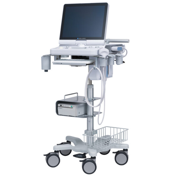
| Content | Konica Sonimage HS2 Ultrasound
Konica Sonimage HS2 Ultrasound is a Point-of-Care portable ultrasound system designed to support a wide range of applications and patient types. Konica Minolta’s advanced technology features provide high resolution image quality and efficient workflow for daily clinical practice.Konica Sonimage HS2 Ultrasound Unique Nanofabrication Technology, combines both material and machining expertise, to produce broad frequency linear probe “L18-4”. It offers both high sensitivity as well as greater penetration.T2HI (Traid Tissue Harmonic Imaging)
Dual Sonic Technology
By combining T2HI and Dual Sonic, high quality THI signal is formed around the center of ultrasound beam in receiving area.
As a result, it suppresses the acoustic noise and ensure the optimum image from deep to superficial structure.iXRET
The SONIMAGE HS2 achieves higher resolution and faster frame rates by utilizing Konica Minolta’s unique technologySNV (Simple Needle Visualization)
The SONIMAGE HS2 automatically detects needle insertion.
This funciton can be used both ‘In plane’ and ‘Out of planeEfficient Workflow for Daily Clinical Practice The SONIMAGE HS2 delivers intuitive workflow by customizing 8 physical buttons and touch panel. MPA (Multi Parameter Adjuster) MPA enables to change multiple image parameters like frequency change and turning trapezoid on in conjunction with depth change. Vascular NAVI
All necessary adjustments and operations are realized automatically.
By touching the mode and measurement button, semi-automatic measurement also achieved.
This function supports easy workflow for blood vessels and blood flow volume measurements. The SONIMAGE HS2 offers the unique capability to write text and draw lines and figures using fingers on the screen. This is an excellent tool for training and communication with patients. | Sonosite Titan Ultrasound Machine
The SonoSite Titan Portable Ultrasound is SonoSite’s second generation portable ultrasound system. This system improved upon the technology of the SonoSite 180 with a larger screen and keyboard packed into a laptop style system. This system is well suited for Vascular access, Musculoskeletal, Anesthesiology, Internal Medecine, Emergency rooms and Family Physicians. Veterinary configurations are available. Veterinary configurations are available.8.4″ LCD monitor, 2 probe ports, Vascular, OB/GYN, Cardiac reports, Soft keys, Split screen, Zoom, Storage via 64 or 256 MB flash cards, 220 image CINE loop, USB & Ethernet image transfer, Volume calculations via diameters, THI [Tissue Harmonic Imaging], SonoCalc [IMT analysis, PC-based], DICOM, S-video (in/out), RGB, Composite video.
Dimensions: 3″ (H) x 10.9″ (W) x 11.9″ (D) Weight: 7.7 lbs Manufacturer
Sonosite Application
Cardiology, Endocrinology, Family Practice, Gastroenterology, Internal Medicine, Nephrology, Neurology, OB-GYN, Orthopedics, Otorhinolaryngology, Pain Management, Surgery, Urology, Vascular Probes - C8 – prostate – 8-5 MHz 8-mm broadband intracavitary array – 8.5 cm – Available – MicroMaxx/ TITAN
- C11 – abdominal/ pediatric cardiology/ neonatal/ vascular – 8-5 MHz 11-mm broadband curved array – 10 cm – N/A – TITAN
- C15 – adult cardiology/ abdominal – 4-2 MHz 15-mm broadband tightly curved array – 25 cm – N/A – TITAN
- C60 – abdominal/ obstetrics/ gynecology – 5-2 MHz 60-mm broadband curved array – 22 cm – Available – TITAN
- ICTe – obstetrics/ gynecology – 8-5 MHz 11-mm broadband tightly curved array – 10 cm – Available – MicroMaxx/ TITAN
- L25 – nerve/ musculoskeletal/ vascular and superficial – 10-5 MHz 25-mm broadband linear array – 6 cm – Transverse Guide Available – TITAN
- L38 – small parts/ breast/ vascular/ nerve/ musculoskeletal/ superficial – 10-5 MHz 38-mm broadband linear array – 6.5 cm – Available – TITAN
Options - Extended cardiac package
- Pulsed wave (PW) doppler
- Continuous wave (CW) doppler
- Tissue Harmonic Imaging
Accessories - Cart
- Black and White Video Printer
- DVD Recorder
The SonoSite Titan is the earliest version of the PoC dedicated portable ultrasound machine from Sonosite. Though its form factor is compact, it provides high resolution imaging, rapid responsive time, and supports a broad range of applications including cardiac. | Siemens / Acuson Sequoia 512 Ultrasound Machine
The ACUSON Sequoia 512 ultrasound is an ultra-premium system that utilizes revolutionary technology, dedicated to the advancement of diagnostic imaging. It represents some of the most advanced technology in ultrasound imaging. The Acuson Sequoia 512 has unparalleled imaging performance, greater image content, enhanced detail, superior contrast resolution & penetration, and excellent color Doppler sensitivity.One of the best selling ultrasounds in the United States, the Sequois 512 provides superior imaging with Native Patient Specific Imaging that detects, measures and adapts (in real time) to each patient’s individual acoustics.Performance:Reduce image optimization time by up to 80% and allow for an easier, faster and more confident diagnosis:- 2D
- SST Color Doppler
- Color Doppler Energy
- Color Doppler Velocity
- Solo Spectral Doppler
- M-mode and Color Doppler M-mode
- MultiHertz Multiple Frequency Imaging
- DELTA Differential Echo Amplification
- Home Base user interface
- One-Touch Image Control with exam and Image Presets
- Digital acquistion, storage, review and transfer of static images, dynamic clips, and cine
- Digital storage on an integrated hard disk
- Digital transfer via MO Disk
- Integrated Compression engine
- Embedded DICOM conformance
- Patient- oriented file structure
- Native Tissue Harmonic Imaging
- Tissue Equalization™ Technology (Optional)
- Signature II™ (Optional)
- Advanced Imaging package (Optional)
- Auto Doppler (Optional)
- Freestyle Compounding (Optional)
- FreeStyle Extended Imaging (Optional)
- 3D surface Rendering (Optional)
- Cadence™ ADI (Optional)
- Cadence CPS for 512 (Optional)
- Axius™ Edge Assisted EF (Optional)
- Doppler Tissue Imaging (Optional)
- DICOM Print And Store (Optional)
Capabilities:Siemens / Acuson Sequoia 512 Ultrasound MachineGeneral Radiology, Abdominal, Intraoperative Monitoring, OB/GYN, Neonatal, Vascular, Cardiac, Small PartsAvailable Transducers:4V1, 4V2, 8V5, 15L8W, 6L3, 8L5, 6C2, 5C2, 4C1, 3V2C, 5V2C,7V3C, EVC8-4V, EC-10C5, V5M TEE, V7B neo TEE, AcuNav Transducer, 2.0 Non Imaging CW Doppler probe.Acuson sequoia 512 ultrasound click images to enlarge item description acuson sequoia 512 ultrasoundsold as picturedincludes: 2 probes, 4c1 convex & 6l4 linearsony up 895 md printerunit will be cleaned, function checked and certified for electrical safety. Checked, cleaned, certified for electrical safetyguaranteed working when arrives on the guarantee working we need to be notified in 48 hrs. And the seals must be intact. | GE Vivid Q Ultrasound Machine
The powerful GE Vivid Q ultrasound machine is the ultimate lightweight, compact high-performance cardiovascular system for clinicians needing a machine that combines user efficiency and reliable performance. This unique system performs consistently thanks to its TruScan architecture and can support up to 17 probes. The GE Vivid Q is recognized for its premium image quality, advanced image management, and high-performance quantification tools.This advanced GE ultrasound delivers improved patient care and enhanced user productivity with the new M4s-Matrix array technology transducer, which provides greatly enhanced image quality in electronic beamforming, raw data image acquisition and archiving, optimal onboard and post processing technology.This ultrasound system provides high clinical values for quantification and review of stored images, making it one of today’s most sought-after models. Some of its powerful features include compatibility with EchoPac clinical workstation and DICOM networking, speckle tracking strain, integrated stress echo, quantification tools, and reporting capabilities. Also of note is one of the Vivid Q’s best new features, which is its TEE transducer capability.A one-touch button optimization provides premium image quality for workflow efficiency, and maintains AFI- automatic function imaging with complete wallFeatures- Advanced QScan Imaging (incl TSI)
- AdvStress
- ATO
- Auto EF
- Automated Function Imaging
- Blood Flow Imaging
- CardiacApp
- Cardio Lab
- Color Doppler
- CW Doppler
- DICOM Networking
- DICOM: Print
- DICOM: Worklist
- DVD/RW
- ECG
- ECG Cable
- EchoDicom
- EchoPAC
- Evue
- ICE Imaging
- Intima Media Thickness (IMT)
- LVO Contrast
- M Mode
- M4S
- MPEGvue
- PW Doppler
- Small Depth
- TEE
- THI
- Tissue Harmonics Imaging (THI/NTHI)
- Tissue Velocity Imagine & Tissue Tracking
- TSI
- VascAbdContrast
- VascularApp
- VirtualPrinter
- Wide Aperture
GE Vivid Q FeaturesThe GE Vivid Qis a Portable Laptop-style Ultrasound that can be used to help diagnose cardiovascular anatomy. Features and tools based on the previous Vivid 7 and the Vivid E9 have been added to the new lightweight Vivid Q, mainly the M4S-RS transducer that allows for 3D image focusing and enables a higher level of image quality. The Vivid Q ultrasound is versatile and can additionally be used for Cardiac, vascular, Abdominal, Pediatric, Obstetrics, and Emergency medicine. The Vivid Q Ultrasound comes with a 15-inch color LCD monitor that can display a variety of imaging modes including; ICE, Auto EF, AFI, TVI, TT, TSI, and Blood Flow. Additional the Ultrasound system is comparable with 17 different transducers and probes to fit every imaging need. - Auto EF - Automated ejection fraction measurement program designed specifically for the GE Vivid q.
- Tissue Synchronization Imaging (TSI).
- Intracardiac Echo (ICE).
- 16 different compatible probes.
- Coded Octave imaging - enhances image color flow and Doppler imaging.
| GE Voluson 730 Pro Ultrasound MachineThe Voluson 730 Pro is built on the same platform as GE’s leading Real-Time 4D system, the Voluson 730 Expert. With an easy to use interface and exceptional 2D image quality, in addition to GE’s exclusive 4D imaging technology, the Voluson 730 Pro is ideal for any clinical practice with a variety of patient workloads. Recognized world-wide for its 4D imaging capabilities, the Voluson 730 Pro also features exceptional 2D image quality.RealTime 4D imaging – continuous, 3D scanning with simultaneous visualization of the A, B and C planes.
The system allows you to explore images in any plane to reveal the smallest details with stunning clarity and apply sophisticated analytical tools to answer virtually any of your clinical questions.Performance:GE Voluson 730 Pro Ultrasound Machine
State-of-the-art user interface with high resolution 10.4 inch LCD touch panel
Automatic Tissue Optimization
Tissue Doppler
Coded Harmonic Imaging
Coded Excitation (CE)
HD-Flow
CrossXBeamCRI (Compound Resolution Imaging)
Static 3D Mode
B Mode
Focus & Frequency Composite (FFC)
High Resolution Zoom
Pan Zoom
Steering
Virtual Convex
Beta-View
Patient information database
Image Archive on MOD and hard drive
3D/4D data compression (lossy/lossless)
Inversion
Real-time automatic Doppler calcsCapabilities:
OB/GYN, Vascular, Cardio, Abdominal, Small-Parts, Urology, Pediatrics, Ortho, Neurology, Multigestational Calculations, B-Flow, VCI (Volume Contrast Imaging)Available Probes:
AB2-7 Wide Band Convex Probe
AC2-5 Wide Band Convex Probe
4C-A Wide Band Convex Probe
M7C-H Wide Band Convex Probe
IC5-9H Wide Band Convex Probe
PA2-5P Wide Band Phased Array Probe
PA6-8 Wide Band Phased Array Probe
SP10-16 Wide Band Linear Probe
SP4-10 Wide Band Linear Probe
SP6-12 Wide Band Linear Probe
M12L-H Wide Band Linear Probe (1.25D Array)
RAB2-5L Wide Band Convex Volume Probe
RAB4-8L Wide Band Convex Volume Probe
RIC5-9H Wide Band Convex Volume Probe
RIC5-9W Wide Band Convex Volume Probe
RRE6-10 Wide Band Convex Volume Probe
RSP6-16 Wide Band Linear Volume Probe
RNA5-9 Wide Band Convex Volume Probe
SCW 2.0; non imaging suprasternal CW-Pencil Probe
PCW 4.0; non imaging peripheral CW-Pencil Probe | Samsung Medison SonoAce R3 Ultrasound Machine
SonoAce R3 is a portable color ultrasound system with comprehensive diagnostic capabilities. Full Spectrum Imaging™ (FSI™) combines with Speckle Reduction Filter™ (SRF™) to deliver superior quality images, even in the challenging circumstances. The compact and user-friendly design features of the SonoAce R3 ensure portability and manoeuvrability in a lightweight unit. Sporting a 15-inch LCD monitor with Full-Featured Workflow, the SonoAceR3 offers your organisation flexibility, without compromising on precision and efficiency.Slim and Compact DesignThe compact design of SonoAce R3 offers Various-Featured Workflow with a thumbnail menu and shortcut, together with functional key groupings for increased efficiency. The user-friendly design combines a wide-view 15-inch LCD monitor with backlit control keys, customisable measurement package and body markers. The SonoAce R3 features 2 probe ports, 3 USB ports, rapid boot up capability and DICOM 3 compatible image filing. This slim, lightweight unit also has a convenient carrying handle for portability and flexibility.Samsung Medison SonoAce R3 Ultrasound MachineFull Spectrum Imaging™ (FSI™) produces the penetration capabilities associated with the lower frequencies, while still maintaining the fine pixel uniformity associated with the higher frequencies. This combination consistently delivers superior quality images, even in the challenging diagnostic circumstances. The resulting clarity delivers improvements in accuracy and efficiency for greater confidence in your diagnostic procedures. Harmonic Imaging is a technique that records higher frequency information, resulting in greater resolution and fewer artifacts.Synthetic Aperture
The physical radio channel may present some limits to imaging which can be overcome by using Synthetic Aperture Control software. This method uses information from diverse sources to form a more better picture of the area under examination. It creates one single scan line by performing the TX and RX twice to create enhanced penetration and resolution. Synthetic Aperture Control technology delivers a comprehensive picture and to aid accurate diagnosis.QuickScan
QuickScan has been designed to increase workflow efficiency by automatically optimising key imaging parameters at the touch of a button. The resulting increase in efficiency allows your organisation to offer patients an improved service with faster and more accurate diagnoses. This reliable and safe system helps your clinic or hospital to deliver a superior patient experience.Features
Full Spectrum ImagingTM
Quick ScanTM
Color and Power Doppler
Harmonic imaging
Freehand 3D
3D Multi Planar Imaging
Sonoview Image Management
PW Doppler
15″ LCD monitor
Two probe ports
Lightweight |


Reviews
There are no reviews yet.