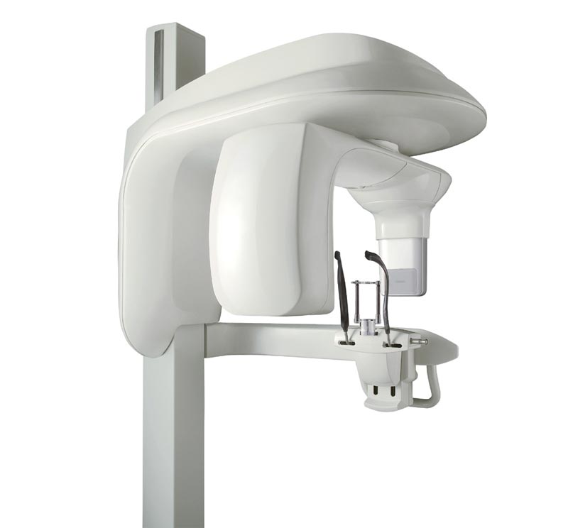
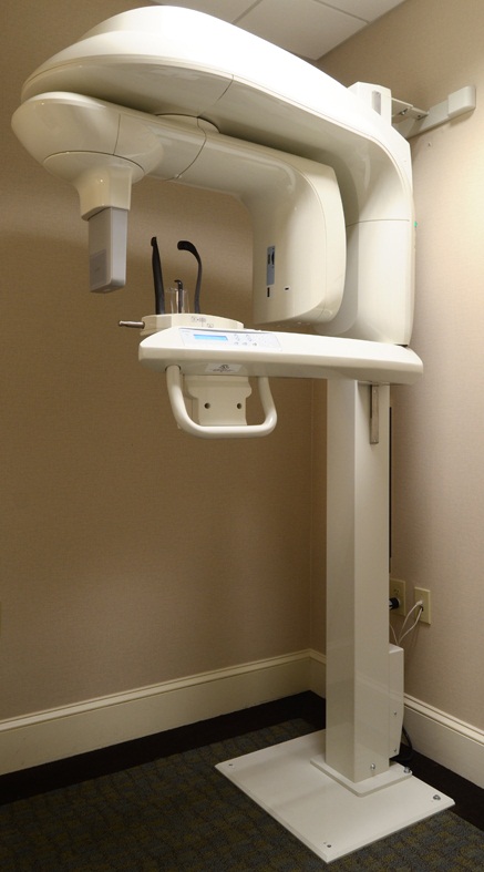
| Description | As one of the most user-friendly Cone Beam Computed Tomography (CBCT) systems on the market, the 9000 3D Extraoral Imaging System offers several versatile solutions for your extraoral imaging needs. Combining focused-field 3D technology with dedicated panoramic imaging, this affordable 2-in-1 unit delivers the best of both worlds, offering the highest resolution and the lowest radiation dose. | TRIOS Color automatically measures the teeth shades as you scan. The dentist has the option to evaluate and highlight relevant areas for the lab. The complete shade information sis sent to the lab together with the impression. | Mr Blue 2.0 is on the cutting edge of innovation – serving as a genuine ECP partner since its launch. Now featuring a whole range of new functions and even greater flexibility, Mr Blue is the perfect solution for demanding workshop. | The image can then be displayed on a monitor, helping you to educate your patients The HRK can provide peripheral keratometry measurement data which is very useful when fitting contact lenses. The eye can be displayed in full colour mode on a tilting touch screen which enables you to see the patient’s contact lens fitting status, previously only possible using a slit lamp. As the HRK has the world’s first contact lens fitting function in an auto keratometer, you can now see the flourescein liquid with blue illumination along with automatic calculation and recommendation for contact lens fitting. | With the intraoral scanner dwio the canadian company Dental Wings presented at the IDS a really revolutionary approach in the field of intraoral scanner. The digitally interested users will have noticed the similarity of the dwio with the Steinbichler intraoral scanner which was launched at the IDS 2 years before. In fact, the dwio intraoral scanner represents a further development. | CIRRUS HD-OCT 5000 is configured with the highest resolution visualization capabilities and sophisticated clinical applications such as Advanced RPE analysis to track retinal pigment epithelial integrity and Ganglion Cell Analysis to assess glaucomatous loss in the macula that may not be evident in the peripapillary region. The new FastTrac retinal tracking reduces eye motion artifacts without sacrificing patient throughput with a proprietary scan acquisition strategy, high speed 20 Hz LSO camera, and single-pass alignment scanning. This feature enables the highest resolution B-Scans to be captured in identical locations from visit to visit providing precise assessment of change in targeted pathologies. |
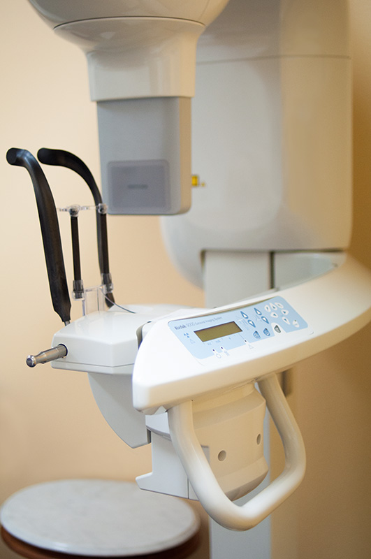
| Content | Kodak 9000 3D Extraoral Imaging System
Add cephalometric imaging abilities to your 9000 3D System for even more abilities. Through our revolutionary one-shot technology, cephalometric image acquisition takes less than a second—thereby reducing exposure time and the risk of retakes due to patient movement. With the 9000C 3D System, you can perform a wider range of diagnoses and treatments right in your office, rather than referring patients to imaging centers. Reduce multiple visits, prevent rescheduling issues, and enhance patient care with three versatile imaging abilities right at your fingertips: panoramic, cephalometric, and 3D imaging.Superb image quality, all three modalitiesVariable focal trough adapts to patient’s jaw morphology – generating high-quality, true panoramic images Best-in-class cephalometric image quality with reduced risk of motion blur Ultra-high resolution, low-dose, focused-field CBCT images provide superior visualization of fine bony anatomy, such as radicular structures
Optimized for radiology workflowsDR technology reduces exam time and eliminates steps, such as film processing and cassette handling One-shot cephalometric technology captures images in less than a second, reducing exam time and risk of retakes Dedicated sensors for each modality save technologist time, reduce risk of breakage DICOM conformance ensures easy integration with PACS, RIS, and printing systems
Simple by design Kodak 9000 3D Extraoral Imaging SystemStreamlined user interface, preset programs and face-to-face positioning makes exams quick and simple Accommodates patients of all shapes and sizes and wheelchair accessible Intuitive, easy-to-use imaging software with improved features for reviewing, reporting and sharing images Includes a comprehensive implant planning model to help referring dentists identify exact measurements and create simulations of proposed treatments
A trusted company, proven productsProven product reliability – even in demanding, high-volume environments Broad range of Carestream equipment and professional services such equipment service agreements, site planning and DICOM integration services. - One-stop shopping for digital imaging and IT solutions from a company with over a century of medical and dental imaging expertise
| Panoramic Modality | Cephalometric Modality | Cone Beam CT Modality | Technology CCD – Optical | CCD – Optical Fiber Sensor | One-Shot with CCD Sensor with Optical Fiber | Cone Beam with CMOS Sensor with Optical Fiber | Gray Scale | 14 bit | 14 bit | 15 bit | 3D Field-of-View | n/a | n/a | • Single Volume: 5 cm x 3.75 cm • Stitched Volumes (up to 3): 9 cm x 7 cm x 3.75 cm | Exam Options | • Panoramic • Child Mode • Segmented Panoramic • Maxillary Sinus • LA TMJ x 2 • LA TMJ x 4 | • Cephalometric • Lateral • Oblique • Frontal (AP/PA) • Submento-Vertex • Carpus • Multiple Cephalometric Field Choices: 30 x 30, 24 x 30, 24 x 24, 18 x 24, 18 x 18 | • CBCT • Resolution Options: 76, 100, or 200 micrometers • Location of Exam: User-defined (mandible, maxilla, TMJ) | Input Voltage | 230/240 V – 60 Hz | 230/240 V – 60 Hz | 230/240 V – 60 Hz | Tube Voltage | 60-90 kV (max) | 60-90 kV (max) | 60-90 kV (max) | Tube Current | 2-15 mA (max | 2-15 mA (max | 2-15 mA (max | Frequency | 140 kHZ (max) | 140 kHZ (max) | 140 kHZ (max) | Total Filtration | > 3.5 mm eq. AL | > 3.5 mm eq. AL | > 3.5 mm eq. AL | Weight | 353 lb (160 kg) | 438 lb (199 kg) | 353 lb (160 kg) |
| 3Shape Trios 3 Pod ColorNew ! 3Shape TRIOS Color POD Digital impressions3Shape TRIOS Pod is a new configuration solution and an alternative to the TRIOS cart. It enables scanning with the TRIOS handheld scanner and software using selected laptop PCs. The solution offers a high degree of mobility and flexibility for dentists working in multiple locations or for clinics with limited space. The TRIOS Pod lets users control scanning from an iPad or mirror the 3D view on other displays in the clinic such as monitors integrated in the chair.Connect to your laptop or PC
Use the Pod with your laptop or the PC in your treatment room. Simply connect to the USB port and start scanning.FEATURES : 3Shape Trios 3 Pod Color
– RealColor scans with many clinical advantages: With TRIOS color you can scan in natural colors to clearly distinguish between teeth, gingiva, and restorative materials. Easily identify true preparation margins and improve scanning experience.
– Powder-free for optimal accuracy and comfort: Applying powder is technique demanding, can ruin scan accuracy, is uncomfortable for patients, and prolongs chair time.
– Autoclaveable tip with anti-mist heater: Achieve optimal hygiene and meet clinical requirements. The integrated antimist heater automatically ensures an optimal temperature for undistorted and crystal clear scanning.
– Extreme Mobility: The light and handy Pod solution is easy to share among treatment rooms or different locations
– Use your iPad with scanning: Control TRIOS from your iPad or mirror the live 3D view directly on other displays in the clinic such as monitors integrated in the chair.
– Connect to multiple laptops or PCs: Use with laptops, PCs in your treatment rooms, or with the PCs integrated in your chair units. Simply connect to the USB port and start scanning. Note: Use with approved PCs.
– Small footprint: The compact TRIOS Pod can be placed anywhere in the treatment room or even in rooms where space is very limited.Included : Mac laptop | Essilor Mr Blue 2.0 Lens Edgers and TracerComplete Control of Lens Mounting
- Optimal monitoring of all trajectories, irrespective of finishing materials or shapes
- Absolute axis control thanks to hybrid edging technology: grinding + milling
- Ideally suited for quality finishing even on highest base curves
- Fast and accurate drilling with data acquired in a single operation
- Automatic centering and blocking for simple operation
Unrivalled Ergonomics- Completely integrated functions without external peripheral devices
- Vertical architecture with <Heads-up displays> for optimum comfort
- Storage features ensure all accessories are within reach.
- Intelligent interface adapts to the way you work.
Highest Quality Guaranteed Essilor Mr Blue 2.0 Lens Edgers and TracerPerfect Centering- Binocular 3-D tracing of full-rim frames up to base 9*
- Automatic adjustment of centering data based on the frames’ 30D parameters*
Aesthetic and Robust Mountings- FIT-4-Frame technology with dedicated small wheel*
- Drilling and grooving tools can be tilted up to 30o, making it safe to mount lenses of up to base 10. *Essilor patents and exclusives
Safe Process- Roughing cycle automatically adapts to the curves of lenses up to base 10, avoiding all risk of breakage
- Bevel placement and shelf depth optimized for safe mounting without undue stress of lens
Aesthetically Pleasing and Easy-to-Mount Bevels- The bevel heights are adjustable to perfectly adjust to the thinnest grooves
- The shelf bevel heights are optimized to follow the frame’s shape perfectly
- The step bevel is particularly suited for sport eyeglasses with deep, rectangular or uneven groove profile
Mr Blue 2.0 is on the cutting edge of innovation – serving as a genuine ECP partner since its launch. Now featuring a whole range of new functions and even greater flexibility, Mr Blue is the perfect solution for demanding workshop. | Huvitz HRK-8000A Auto Refractor KeratometerHuvitz Auto Ref – Keratometer HRK-8000A- Touch and tilting 7″ LCD Screen that allows for free and smooth tilting for viewing at any angle.
- Optimized Optical System
- Micro Lens Array
- Aberration Measurement which also can be displayed in a graphical Zernike Refraction Map
- PSF & Image Simulation
- Colour View Mode
- Peripheral Keratometry Measurement
- Ultra High Precision KER Data
- Contact Lens Fitting Assistance Guide
- Contact Lens Prescription Guide
- Auto Tracking
- Animated Guide
- Faster Measurement Speed
- Quiet and speedy auto cutting printer
- More data on aberration measurement
With its extreme precision and accuracy, the HRK8000A uses the most advanced wavefront technology to measure the light reflected from the retina and helps you to diagnose any refractive error. With a micro lens array developed by Huvitz, the HRK creates a number of focal points, the pattern of which can give a valuable insight into the patients ocular system. The HRK not only displays the conventional data such as spherical, cylinder and axis, the high order aberration data is displayed in a graphical Zernike refractive map, giving you a deeper understanding of your patient’s eyes.Optimized Optical System Huvitz HRK-8000A Auto Refractor Keratometer
A wavefront is a physical representation of the optical quality of a light beam. Using various methods, aberrometers map how light rays travel through the eye. The HR-8000A is engineered according to the Hartmann-Shack wavefront measurement technique, which is widely preferred for its speed and repeatability.Touch & Tilting Color Display Screen
A bright, high contrast 7″ VGA color TFT LCD screen provides high resolution video images. A smooth, free tilting function also offers you a comfortable and clear view at any angle.The image can then be displayed on a monitor, helping you to educate your patients The HRK can provide peripheral keratometry measurement data which is very useful when fitting contact lenses. The eye can be displayed in full colour mode on a tilting touch screen which enables you to see the patient’s contact lens fitting status, previously only possible using a slit lamp. As the HRK has the world’s first contact lens fitting function in an auto keratometer, you can now see the flourescein liquid with blue illumination along with automatic calculation and recommendation for contact lens fitting. | Dwio Intraoral Scanner Dental WingsIntraoral scanner
Based on a novel and incredibly compact 3D capture technique called Multiscan Imaging, the Dental Wings intraoral scanner allows dentists to take digital impressions in a natural, fluid manner. Its remarkably small handpiece, gesture control, and easy maintenance make it a compelling digital impression option for clinicians.Design approach: the patient first
The intraoral scanner was designed to address the needs of dental professionals looking for an intuitive technology for capturing digital impressions. With the overall objective to help dentists focus on the patient rather than the technology, the Dental Wings intraoral scanner offers:– A remarkably small handpiece very similar in size, shape, and weight to existing dental handpieces. Its familiar shape allows to assume a natural position relative to the patient.
– Visual and auditive feedback — a luminescent ring and audible signals indicate when data is being successfully captured, allowing the user to focus in the mouth rather than on the screen.
– Easy maintenance of the lightweight all-metal handpiece that has no moving parts, no complex illumination or cooling system.
– Motion control technology for a no-touch operation of the system while the user has gloves on.Scanning technology Dwio Intraoral Scanner Dental Wings
Five miniaturized 3D scanners in the handpiece tip directly view the teeth and soft tissue from multiple orientations simultaneously, capturing even the most difficult to see areas of preparations with minimal effort by the user.Open workflows
The intraoral scanner is a dedicated “scan only” chairside system with multiple options for the design and fabrication steps. Captured data is seamlessly transmitted to in-office or remote systems via DWOS Connect for prosthesis design and production. Users of Dental Wings’ simple to use yet powerful DWOS CAD suite of design applications, as well as other open dental CAD software, are able to import the scan data directly. | Carl Zeiss Cirrus HD-OCT 5000 Smart Advanced Topography HDAdvancing Smart OCTfor Anterior Segment, Glaucoma and Retina- Anterior Segment Premier Module
- PanoMap Wide-Field Display
- Smart HD Scans
- En face report
Visualization at the speed of CIRRUSAnalyzing a single pathology from multiple views provides comprehensive insight and analysis of the clinical situation. How this helps you: - Spot small areas of pathology. Tightly spaced B-scans, (either 30 or 47 μm apart), in the cube ensure that small areas of pathology are imaged. For reference, a human hair is about 40-120 μm in diameter.
- Visualize the fovea. Scans that are spaced further apart than in the CIRRUS cube may miss the central fovea.
- Fuel for analysis. Millions of data points from the cube are fed into the Zeiss proprietary algorithms for accurate segmentation, reproducible measurements and registration for change analysis.
- Take the pressure off the operator. As long as the scan is placed in the vicinity of the fovea or optic nerve, the software automatically centers the measurements after the capture.
- See the tissue from different perspectives. View the cube data from all angles, with 3D rendering, OCT fundus images and Advanced Visualization™.
- Future ready. Previously captured CIRRUS cubes can be analyzed using new analyses.
Tracking at the speed of CIRRUSFastTrac™ reduces eye motion artifacts without sacrificing patient throughput with a proprietary scan acquisition strategy, high speed 20 Hz LSO camera, and single-pass alignment scanning
With FastTrac, scan at the highest resolution at the same location at each visit. Assessment at the speed of CIRRUSMeasurement centering with FoveaFinder and AutoCenterOn the macula, the unique FoveaFinder technology ensures the ETDRS and ganglion cell plus inner plexiform layer measurement frameworks are centered on the fovea.AutoCenter™ function automatically centers the 3.4 mm diameter peripapillary RNFL calculation circle around the disc for precise placement and repeatable registration. The placement of the circle is not operator dependent. Accuracy, registration and reproducibility are assured. Manage at the speed of CIRRUS Carl Zeiss Cirrus HD-OCT 5000 Smart Advanced Topography HDGuided Progression Analysis (GPA™) for Assessing RNFL and Optic Disc ChangeGPA™ compares RNFL thickness and optic disc measurements over time and determines if statistically significant change has occurred.Focal Progression Analysis – Up to 6 progression maps are compared to two baselines. Areas of statistically significant change are color-coded yellow when first noted and then red when the change is sustained over consecutive visits.
Trend Analysis with Rate of Change – RNFL thickness values are plotted over time. Trends are calculated and confidence intervals are shaded when rate of change is statistically significant. |



Reviews
There are no reviews yet.