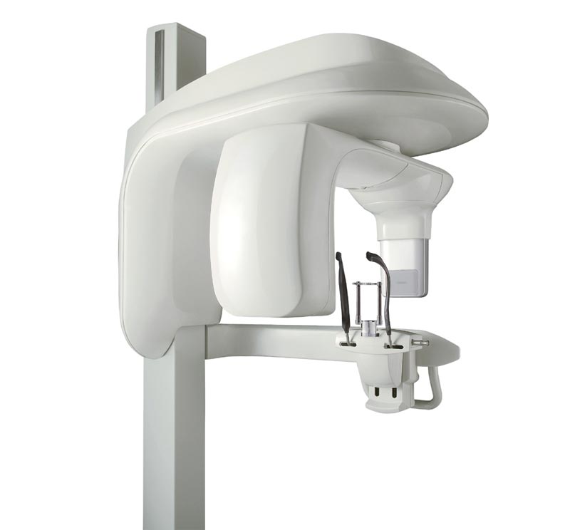
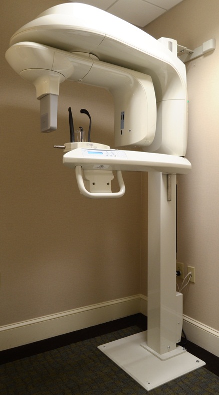
| Description | As one of the most user-friendly Cone Beam Computed Tomography (CBCT) systems on the market, the 9000 3D Extraoral Imaging System offers several versatile solutions for your extraoral imaging needs. Combining focused-field 3D technology with dedicated panoramic imaging, this affordable 2-in-1 unit delivers the best of both worlds, offering the highest resolution and the lowest radiation dose. | CIRRUS HD-OCT 5000 is configured with the highest resolution visualization capabilities and sophisticated clinical applications such as Advanced RPE analysis to track retinal pigment epithelial integrity and Ganglion Cell Analysis to assess glaucomatous loss in the macula that may not be evident in the peripapillary region. The new FastTrac retinal tracking reduces eye motion artifacts without sacrificing patient throughput with a proprietary scan acquisition strategy, high speed 20 Hz LSO camera, and single-pass alignment scanning. This feature enables the highest resolution B-Scans to be captured in identical locations from visit to visit providing precise assessment of change in targeted pathologies. | workflow, because some letters inspire confidence.With Neksia, Essilor combines years of experience in high-performance and precision edging into one of the most user-friendly and productive systems in the world. | The E4 boasts double the speed, double the accuracy, and double as many cameras as its predecessor the E3, delivering improved efficiency and consistency with every scan. From small labs seeking the best-of-the-best, to high-volume, full-service labs striving for a leaner workflow, our E4 lab scanner is the right choice for you. | InMode Aesthetics InmodeRF BodyTite with BodyTite and FaceTite ApplicatorsBenefits
Controls heating to ensure that large volume contraction is consistent, avoiding overtreated or undertreated area
When treating different areas of the body, the operator is always receiving real time temperature monitoring, even in deep fat areas where laser temperature readers cannot reach
Allows focal treatment of all body areas with large volume
Treats deep fat, up to 50 mm (2 inches) | The 3Shape D710 is optimized for scanning both the impression and gypsum models and enables high throughput for both advanced and standard indications. These innovative scanners are so easy to use that staff training is barely required. Item fixation is fast, and the scan starts with a click on the program. |
| Content | Kodak 9000 3D Extraoral Imaging System
Add cephalometric imaging abilities to your 9000 3D System for even more abilities. Through our revolutionary one-shot technology, cephalometric image acquisition takes less than a second—thereby reducing exposure time and the risk of retakes due to patient movement. With the 9000C 3D System, you can perform a wider range of diagnoses and treatments right in your office, rather than referring patients to imaging centers. Reduce multiple visits, prevent rescheduling issues, and enhance patient care with three versatile imaging abilities right at your fingertips: panoramic, cephalometric, and 3D imaging.Superb image quality, all three modalitiesVariable focal trough adapts to patient’s jaw morphology – generating high-quality, true panoramic images Best-in-class cephalometric image quality with reduced risk of motion blur Ultra-high resolution, low-dose, focused-field CBCT images provide superior visualization of fine bony anatomy, such as radicular structures
Optimized for radiology workflowsDR technology reduces exam time and eliminates steps, such as film processing and cassette handling One-shot cephalometric technology captures images in less than a second, reducing exam time and risk of retakes Dedicated sensors for each modality save technologist time, reduce risk of breakage DICOM conformance ensures easy integration with PACS, RIS, and printing systems
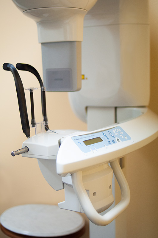
Simple by design Kodak 9000 3D Extraoral Imaging SystemStreamlined user interface, preset programs and face-to-face positioning makes exams quick and simple Accommodates patients of all shapes and sizes and wheelchair accessible Intuitive, easy-to-use imaging software with improved features for reviewing, reporting and sharing images Includes a comprehensive implant planning model to help referring dentists identify exact measurements and create simulations of proposed treatments
A trusted company, proven productsProven product reliability – even in demanding, high-volume environments Broad range of Carestream equipment and professional services such equipment service agreements, site planning and DICOM integration services. - One-stop shopping for digital imaging and IT solutions from a company with over a century of medical and dental imaging expertise
| Panoramic Modality | Cephalometric Modality | Cone Beam CT Modality | Technology CCD – Optical | CCD – Optical Fiber Sensor | One-Shot with CCD Sensor with Optical Fiber | Cone Beam with CMOS Sensor with Optical Fiber | Gray Scale | 14 bit | 14 bit | 15 bit | 3D Field-of-View | n/a | n/a | • Single Volume: 5 cm x 3.75 cm • Stitched Volumes (up to 3): 9 cm x 7 cm x 3.75 cm | Exam Options | • Panoramic • Child Mode • Segmented Panoramic • Maxillary Sinus • LA TMJ x 2 • LA TMJ x 4 | • Cephalometric • Lateral • Oblique • Frontal (AP/PA) • Submento-Vertex • Carpus • Multiple Cephalometric Field Choices: 30 x 30, 24 x 30, 24 x 24, 18 x 24, 18 x 18 | • CBCT • Resolution Options: 76, 100, or 200 micrometers • Location of Exam: User-defined (mandible, maxilla, TMJ) | Input Voltage | 230/240 V – 60 Hz | 230/240 V – 60 Hz | 230/240 V – 60 Hz | Tube Voltage | 60-90 kV (max) | 60-90 kV (max) | 60-90 kV (max) | Tube Current | 2-15 mA (max | 2-15 mA (max | 2-15 mA (max | Frequency | 140 kHZ (max) | 140 kHZ (max) | 140 kHZ (max) | Total Filtration | > 3.5 mm eq. AL | > 3.5 mm eq. AL | > 3.5 mm eq. AL | Weight | 353 lb (160 kg) | 438 lb (199 kg) | 353 lb (160 kg) |
| Carl Zeiss Cirrus HD-OCT 5000 Smart Advanced Topography HDAdvancing Smart OCTfor Anterior Segment, Glaucoma and Retina- Anterior Segment Premier Module
- PanoMap Wide-Field Display
- Smart HD Scans
- En face report
Visualization at the speed of CIRRUSAnalyzing a single pathology from multiple views provides comprehensive insight and analysis of the clinical situation. How this helps you: - Spot small areas of pathology. Tightly spaced B-scans, (either 30 or 47 μm apart), in the cube ensure that small areas of pathology are imaged. For reference, a human hair is about 40-120 μm in diameter.
- Visualize the fovea. Scans that are spaced further apart than in the CIRRUS cube may miss the central fovea.
- Fuel for analysis. Millions of data points from the cube are fed into the Zeiss proprietary algorithms for accurate segmentation, reproducible measurements and registration for change analysis.
- Take the pressure off the operator. As long as the scan is placed in the vicinity of the fovea or optic nerve, the software automatically centers the measurements after the capture.
- See the tissue from different perspectives. View the cube data from all angles, with 3D rendering, OCT fundus images and Advanced Visualization™.
- Future ready. Previously captured CIRRUS cubes can be analyzed using new analyses.
Tracking at the speed of CIRRUSFastTrac™ reduces eye motion artifacts without sacrificing patient throughput with a proprietary scan acquisition strategy, high speed 20 Hz LSO camera, and single-pass alignment scanning
With FastTrac, scan at the highest resolution at the same location at each visit. Assessment at the speed of CIRRUSMeasurement centering with FoveaFinder and AutoCenterOn the macula, the unique FoveaFinder technology ensures the ETDRS and ganglion cell plus inner plexiform layer measurement frameworks are centered on the fovea.AutoCenter™ function automatically centers the 3.4 mm diameter peripapillary RNFL calculation circle around the disc for precise placement and repeatable registration. The placement of the circle is not operator dependent. Accuracy, registration and reproducibility are assured. Manage at the speed of CIRRUS Carl Zeiss Cirrus HD-OCT 5000 Smart Advanced Topography HDGuided Progression Analysis (GPA™) for Assessing RNFL and Optic Disc ChangeGPA™ compares RNFL thickness and optic disc measurements over time and determines if statistically significant change has occurred.Focal Progression Analysis – Up to 6 progression maps are compared to two baselines. Areas of statistically significant change are color-coded yellow when first noted and then red when the change is sustained over consecutive visits.
Trend Analysis with Rate of Change – RNFL thickness values are plotted over time. Trends are calculated and confidence intervals are shaded when rate of change is statistically significant. | Essilor Neksia Lens Edging SystemWork More Easily- A touch of efficiency – The 8.4-inch color touch-screen display makes navigation a breeze. And you can select the icon-based functions quickly and easily with just the touch of a finger.
- Fast-access settings – With all job settings located conveniently on the centering part of the system, you can define your finishing settings quickly and launch edging jobs in just a few clicks. If necessary, you can still adjust settings after the feeler measurement.
- A user-friendly 1,000-position job database – Save precious time by easily accessing or storing complete jobs in the database via the touch screen or optional barcode reader.
Experience Workflow Efficiency- Fast, easy and extended frame tracking – Equipped with auto frame-type selection, simple clamping and rapid tracing cycles, Neksia helps you trace any type of frame effortlessly, including children’s frames, small B-size and large A-size frames
- Automatic drilling-hole recognition – Achieve even greater productivity thanks to optional features dedicated to automatic detection of drilling holes and flat bevel shapes.
- Convenient centering and blocking – Neksia arms you with valuable features that make centering and blocking straight-forward and precise.
- Handle lenses easily with the wide-open centering area
- Increase precision with intelligent targeting and parallax-free working
- Benefit from the intuitive on-screen interface
Achieve High Performance Essilor Neksia Lens Edging System- Improve axis accuracy – By combining new edging cycle algorithms and cutting-force regulation, you are guaranteed optimal results whatever the edging situation.
- Optimized edging speed based on the lens material, shape and lens thickness guarantees fast cycle times, lens positioning and alignment.
- The New Edging Assisted System cycle avoids axis deviation on new- generation hydrophobic lenses, rectangular shapes or fragile lenses.
- Best automatic cycles – For 90% of your jobs, all it takes are a few clicks to achieve a perfectly calculated bevel, groove or drilling cycle. The lens is well positioned for an aesthetic and perfectly fitted result, no matter the material or frame type.
- Consistent intelligent chamfering – With Neksia’s patented flexible chamfering wheel, the pressure applied to the wheel adapts seamlessly to the lens edge depending on the shape and curve of the lens.
| 3Shape E4 Dental Lab ScannerThe E4 is a dental 3D scanner produced by 3Shape. 3Shape is a 3D scanner manufacturer based in Denmark.E4 dental scanner main features- Full arch scan in 9 seconds
- 4-micron accuracy
- Four 5-MP cameras
- Color acquisition
3SHAPE E4
Ultimate productivity with our fastest scanner ever
For those seeking the speed and accuracy to drive lab productivity and growth, look no further. The E4 boasts double the speed, double the accuracy, and double as many cameras as its predecessor the E3, delivering improved efficiency and consistency with every scan. From small labs seeking the best-of-the-best, to high-volume, full-service labs striving for a leaner workflow, our E4 lab scanner is the right choice for you.
4 micron
Scan with an accuracy of 4 microns (ISO 12836) for great case results. Combine this precision with increased speed and you can take on more implant cases, for example.
4 cameras
Four cameras save you time by scanning dies in the model instead of individually. You can even scan impressions instead of producing a gypsum model or combine impression and die at the same time.
5MP cameras
5MP cameras are super high definition cameras. They record 5 megapixel images with enhanced camera sensors, meaning you get superior image clarity and can do high-precision scanning.
HARDWARE SPECIFICS
The E4 is a marriage of iconic 3Shape design and superior technical specifications.
Cameras
Four 5 MP cameras enables efficient die scanning and impression scanning, improving your workflows by saving time
Accuracy
4 microns (ISO 12836), giving you the accuracy and precision you need for implant cases
Full arch scan speed 9 seconds (versus 22 seconds for the 3Shape E3)
Full arch impressions scan speed 45 seconds
Texture Color
Scanning strategy Die-in-model | INMODE Bodytite Laser – Radio Frequency (RF)BodyTite
Cannula Length:17 cm
Cannula Diameter:2.2 mm
Depth of Treatment:Up to 50 mm (2)
Internal Temperature:Operator adjustable between 50°C – 70°C
External Temperature:Operator adjustable up to 42°C
Output Temperature:1 MHz+
FaceTite
Cannula Length:10 cm
Cannula Diameter:1.3 mm
Depth of Treatment:Up to 25 mm (1″)
Internal Temperature:Operator adjustable between 50°C 70°C
External Temperature:Operator adjustable up to 42°C
Output Frequency:1 MHzBodyTite WorkstationMinimally Invasive Radio-Frequency Assisted Lipolysis (RFAL) Surgical Results Without the Scars.BodyTite is InModes latest minimally-invasive body contouring procedure that provides superior internal and external lipolysis, delivering results only achieved through more extensive excisional surgical procedures. BodyTite is powered by RFAL (Radio-Frequency Assisted Lipolysis) resulting in three-dimensional remodeling.BodyTite provides doctors with a tool to perform arm lifts without excisions, breast lifts without the lollipop scar and contour areas of non-adherence with results previously only achieved through more extensive surgical procedures.Benefits
Controls heating to ensure that large volume contraction is consistent, avoiding overtreated or undertreated area
When treating different areas of the body, the operator is always receiving real time temperature monitoring, even in deep fat areas where laser temperature readers cannot reach
Allows focal treatment of all body areas with large volume
Treats deep fat, up to 50 mm (2 inches)PATIENT BENEFITS
Patients today want to avoid surgery when possible. With BodyTite and FaceTite, your patients can reduce fat without leaving behind saggy, wrinkly skin. They are left with surgical-like results with no scars, less downtime and long-lasting, beautiful results.PRACTICE BENEFITS
Adopting RFAL with three-dimensional skin contraction can help innovate your practice. Leveraging BodyTite and FaceTite for your practice allows you to add a suite of minimally invasive procedures that contours and lifts abdominal, breast, face, neck, arm, thigh and knee tissue.FaceTite
HOW DOES IT WORK
FaceTite is a minimally-invasive contouring solution for the face and small areas of the body. Physicians can achieve results similar to a facelift or brachioplasty without excisional surgery. This is a safe, effective and exciting treatment that offers patients significant aesthetic changes.FaceTite is powered by RFAL (Radio-Frequency Assisted Lipolysis) and provides superior internal and external lipolysis, resulting in three-dimensional remodeling of the fibrous septa. Built-in safeguards including real time measurements of skin temperature, impedance monitoring, power cut-off and audible feedback help ensure patient safetyBenefits INMODE Bodytite Laser – Radio Frequency (RF)
Uniform thermal field provides an even distribution of heat.
Dual temperature technologies provide greater safety and control compared to other technologies.
100% energy concentration in the treatment zone.
Physician sets and controls the temperatures. Heat gradient enables optimal treatment results.AccuTite
ACCURACY IS EVERYTHING
PRECISION TARGETING AND PROVEN PERFORMANCE
ACCURACY IS EVERYTHING
The new AccuTite makes focal RF contraction without excisional surgery the new standard in cosmetic medicine. Small, but mighty, the AccuTite delivers precision heating in addition to the science and safety recognized with the Radio-Frequency Assisted Lipolysis (RFAL) family of technologies.
FOR FACE AND BODY
FACEBrow
Periorbital
Nasolabial Fold
Lower Face
Neck
BODYAxilla Bra Fat
Upper Arms
Hands
Abdomen Etching
Inner Thigh
Knees
Womens Health & WellnessSIMPLE, SAFE & FOCUSED
The AccuTite is the smallest minimally-invasive radio-frequency contraction device with the most number of clinical studies and peer-reviewed publications backing its safety and efficacy. Patients who need precision fat reduction or skin contraction now have a new solution that can be done under local anesthesia, in an office setting, in less than 20 minutes, without excisional surgery. | 3shape D710 Dental Scanner3Shape D710 Scanner series can scan gypsum and impression versions. The series scanners can integrate into the clinic with minimum staff training. The scanners provide greater practice productivity, a comprehensive list of signals, and the most extensive 3D scans.Where flexibility and productivity are the key standards 3Shape chain scanners are for medium to large labs.The 3Shape D710 is optimized for scanning both the impression and gypsum models and enables high throughput for both advanced and standard indications. These innovative scanners are so easy to use that staff training is barely required. Item fixation is fast, and the scan starts with a click on the program.3SHAPE D710 For Sale3Shape D700 Scanner is both a scanner and impression scanner in 1 unit. It utilizes three measurement movement and two cameras a laser and creates another pass to especially hone. Just one device can be scanned by the apparatus in under 30 seconds and a whole model in about a minute with a degree of accuracy of 20 microns.Features:
-3-axis motion system
-Red-laser Tech
-Gypsum Version scanning
-Impression scanning Alternative
-Multi-die scanningBenefits:
-Easy operation, extreme Precision, and Rapid Scan
-Enables impressions, deep inlays, and Complete undercuts to scan Efficiently
-Supply labs Using ultra-efficient batches and multi-case processingHigh-speed scanning
The D710 is optimized for scanning gypsum and impression versions and enables high throughput for both advanced and standard indications3Shape – leading impression scanning technologies
3Shape’s patented adaptive scanning technology, combined with the scanner’s 3-axis motion and 2 cameras with reduced angle, facilitate the unique impression scanning capability.Adaptive scanning intelligently detects incomplete areas and automatically builds
adaptive scan sequences for capturing full object geometry. |



Reviews
There are no reviews yet.