| Description | The ZEISS Cirrus HD-OCT 4000 enable examination of the posterior and anterior of the eye at an extremely fine spatial scale, without surgical biopsy or even any contact with the eye. The Cirrus HD-OCT builds on and refines the retinal imaging technology first introduced with the ZEISS Stratus OCT™. HD-OCT stands for “high-definition optical coherence tomography.”Employing the advanced imaging technology of spectral domain optical coherence tomography, Cirrus HD-OCT acquires OCT data about 70 times faster (27,000 vs. 400 A-scans per second) and with better resolution (5 μm vs. ~10 μm axial resolution in tissue), compared to first-generation OCT technology. Cirrus acquires whole cubes of OCT image data, composed of hundreds of line scans, in about the same time as Stratus acquires a six-line scan. You can view these data cubes in three planes, or through three dimensions, giving you access to an extensive amount of retinal image data in one scan. | The iCare IC200 tonometer is designed for professional use in the surgical operation room, the emergency room and also the clinic. It uses the same patented rebound measuring principle found in other iCare tonometers. There is no need for drops, air or specialised skills for its use. The IC200 gives you positional freedom with a new premium design and user interface. It allows measuring whether the patient is sitting, standing, half-sitting or in the supine or lateral recumbent position. | Features- NEW TruFit 3D Virtualization allows you to simulate the lens inside the frame before edging, allowing you to minimize retouches and maximize your profit.
- One of the fastest lens edgers on the market.
- Process nearly any job type to avoid having to send out costly jobs to labs.
- Superior quality of finish and polish provides a premium look and feel that you can be proud of.
- Smart Design shape customization.
Wavefront Power-mapping technology for lens analysis and verification.
- Shelf Bevel to easily process high base-curve wrap frames.
- TruScan High base curve frame tracing
| Laser-Like Precision
- Tactile control with minimal tissue drag
- Surface only thermal effect
- Predictable, char-free layer-by-layer dissection with optimal visualization of tissue planes
- Safe to use near nerves, vessels, and all delicate organs, including the heart | The simplicity is another advantage
Energy-saving and auto-sensing feature will surprise you
White & Black colors are available
1. Compact and sleek design with luxurious feel
Simple, and it makes your work environment look great
2. Adjustable LED intensity
(1) Lifetime durability LED lamp
(2) Easy to mark and block even with dark-tinted lenses by brightness control function
3. Auto power-saving mode
(1) Automatic power off after confirming the marking points
(2) Automatic power off and sleep mode after preset time
Adopting high efficiency LED light source which can easily recognize dark-tinted lenses, and adjust brightness.
• Compact design, intuitive graphic, auto power on/off function. | With California, Optos has incorporated new hardware and software technology enabling practitioners to see more, discover more and effectively treat more ocular pathology thus promoting patient health.In addition to the benefits found with all of the UWF devices from Optos such as 200 degrees or up to 82% of the retina captured in a single image, in multiple modalities, as well as eyecare professionals being able to see 50% more of the retina when compared to other conventional imaging devices, California rg, offers the following benefits:– Motorized head and chin rest to more easily assist those patients requiring additional assistance during imaging– Multiple imaging modalities including; 3-in-1 color depth imaging (color, red-free and choroidal in a single image) as well as
Autofluorescence |
| Content | ZEISS Cirrus HD OCT 4000 Quad Core OCT
The ZEISS Cirrus HD-OCT 4000 (Cirrus HD-OCT or Cirrus) enable examination of the posterior and anterior of the eye at an extremely fine spatial scale, without surgical biopsy or even any contact with the eye. The Cirrus HD-OCT builds on and refines the retinal imaging technology first introduced with the ZEISS Stratus OCT™. HD-OCT stands for “high-definition optical coherence tomography.”Employing the advanced imaging technology of spectral domain optical coherence tomography, Cirrus HD-OCT acquires OCT data about 70 times faster (27,000 vs. 400 A-scans per second) and with better resolution (5 μm vs. ~10 μm axial resolution in tissue), compared to first-generation OCT technology. Cirrus acquires whole cubes of OCT image data, composed of hundreds of line scans, in about the same time as Stratus acquires a six-line scan. You can view these data cubes in three planes, or through three dimensions, giving you access to an extensive amount of retinal image data in one scan.Intended UseThe Cirrus HD-OCT with Retinal Nerve Fiber Layer (RNFL), Macular, Optic Nerve Head, and Ganglion Cell Normative Databases is indicated for in-vivo viewing, axial cross-sectional, and three-dimensional imaging and measurement of anterior and posterior ocular structures.Indications for UseThe Cirrus HD-OCT is a non-contact, high resolution tomographic and biomicroscopic imaging device. It is indicated for in-vivo viewing, axial cross-sectional, and three-dimensional imaging and measurement of anterior and posterior ocular structures, including cornea, retina, retinal nerve fiber layer, ganglion cell plus inner plexiform layer, macula, and optic nerve head. The Cirrus normative databases are quantitative tools for the comparison of retinal nerve fiber layer thickness, macular thickness, ganglion cell plus inner plexiform layer thickness, and optic nerve head measurements to a database of normal subjects. The Cirrus HD-OCT is intended for use as a diagnostic device to aid in the detection and management of ocular diseases including, but not limited to, macular holes, cystoid macular edema, diabetic retinopathy, age-related macular degeneration, and glaucoma.Note: The Cirrus HD-OCT is not intended to be used as the sole diagnostic for disease.Essential Performance ZEISS Cirrus HD OCT 4000 Quad Core OCTThe Essential Performance of the instrument is to provide accurate measurements of anterior and posterior ocular structure.Patient PopulationThe Cirrus HD-OCT may be used on all adults in need of diagnostic evaluation of the eye. This includes (but is not limited to) patients with the following disabilities or challenges:• Wheelchair user
• Very low or not measurable visual acuity
• Fixation problems
• Postural problems
• Deafness
• Large body, but not those above 99th percentile based on anthropomorphic dataThere is a general requirement that the patient be able to sit upright and be able to place their face in the chin and forehead rest of the instrument (with or without supplemental human or mechanical support).Cirrus HD-OCT is designed for in-vivo viewing, axial cross-sectional, and three-dimensional imaging and measurement of anterior and posterior ocular structures. | Icare ic200 TonometerThe device is based on a rebound measuring principle that requires no drops, air or specialized skills for its use. The new premium design and user interface bring IOP measuring to a new level. The Icare ic200 tonometer is designed for professional use in the surgical operation room and emergency room as well as the clinic. The ic200 is fully portable, requires no anesthesia and its freedom of positioning allows measuring whether the patient is sitting, standing, half-sitting or in the supine or lateral recumbent position.A high-visibility indicator at the probe base confirms your positioning of the tonometer prior to measurement. A green light indicates measurement will be reliable, a red light indicates incorrect positioning. Features:
- Effortless probe loading with the same probe as for the Icare TA01i and ic100 tonometers
- The probe can’t accidentally drop from the device
- Easy menu in many languages
- Wireless (Bluetooth) printing and measurement transfer - Revolutionary design to allow for positional freedom when taking IOP measurements
- Innovative automatic measuring sequence allows for 6 automatic measurements or single measurements to be taken with the same button
- Intelligent positioning system for correct alignment of the tonometer
- Powered by AA batteries
Specifications:
- Dimensions : 43mm[W] x 104mm[H] x 214mm[L]
- Weight : 165 g (without batteries), 260 g (with 4 x AA batteries)
- Power supply : 4 x AA non-rechargeable batteries, 1.5V alkaline LR6
- Measurement range : 7 - 50 mmHg
- Accuracy : ±1.2 mmHg (≤20 mmHg) and
: ±2.2 mmHg (>20 mmHg)
- Repeatability (coefficient of variation) : <8% Package Includes:
- Icare ic200 tonometer
- 4 x AA batteries
- Aluminum case
- IOP pad
- Probe base cover
- Quick guide
- Screw driver
- Silicone grip
- Spare probe base
- Instruction manual
- Warranty card
- Wrist strap | Briot Couture Lens Finishing System
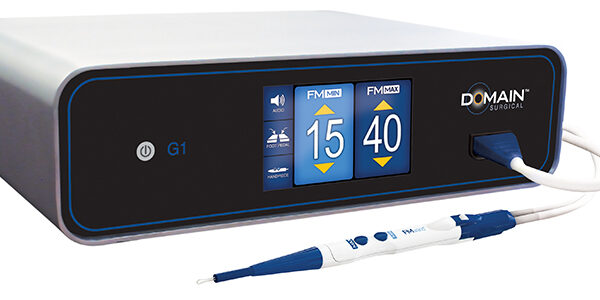
When your standards are high, this is the partner you deserve. A revolution in edging, Briot Couture gives you a flawless finish with every frame – whatever the style, shape or curve. 3D pre-visualization lets you see your results before you even start working. TrueFit technology calculates curvatures to ensure seamless lens-frame pairing. Additionally, new sensors coupled with advanced software maintain constant pressure on the lens, reducing cycle time with no risk of misalignment – especially on hydrophobic lenses.Wavefront, The Future Of Blocking Briot Couture Lens Finishing System Our exclusive wavefront lens analysis allows you to see progressive lens designs on the frame trace before you even cut the lens, giving you the opportunity to validate that the shape and lens design are compatible without wasting time or money redoing the job. Shape Creation Limited Only By Your Imagination With Smart Design Milling And Shelf Beveling Create complex shapes and a true shelf bevel for high base-curve wrap frames with the Smart Design Milling feature in the Briot Couture. Exclusive TrueScan High-Curve Tracing Featuring our patented TrueScan technology, the Attitude Tracer/Blocker has a specially designed angled probe that will gently measure the contour of even the thinnest, most delicate metal frames or high base-curve wrap frames effortlessly. Brushless Technology For Faster, Quieter Operation Edge lenses faster and more efficiently while also reducing noise with new brusheless motor technology featured on the Evolution. Now, you can plug this edger in anywhere that has a 120V line adding extra placement flexibility in your optical lab. Provide The Best Finish On High Base-Curve Frames With Briot’s Best-Fit Technology Our advanced drive technology in conjunction with a small 90mm diamond wheel allows flawless finishing on high base-curve lenses and complex frames with sharp corners. Damaged Demo Lens? No Problem With Shape Creator 2.0 Accurately reconstruct broken or defective demo lenses with minimal effort using the new Shape Creator 2.0 technology on the Couture Blocker. | Domain Surgical FMX and FMwand Ferromagnetic Surgical SystemThe FMX Generator intelligently delivers electrical energy to the surgical instruments. The advanced generator features a patented software control algorithm that continuously monitors and adjusts the delivery of energy, ensuring that the optimal amount of heat is delivered to the tissue at all times. The large touchscreen display provides the entire surgical team immediate feedback on power setting levels and changing settings is as easy as touching the display. The FMX Generator has earned the Type CF (“Cardiac Floating”) rating.Laser-Like Precision
- Tactile control with minimal tissue drag
- Surface only thermal effect
- Predictable, char-free layer-by-layer dissection with optimal visualization of tissue planes
- Safe to use near nerves, vessels, and all delicate organs, including the heartMinimal Thermal Injury
- As little as 1/10th the thermal injury of monopolar electrosurgery
- As few as 80 microns (0.08 mm) of thermal spread
- Clear margins for reliable pathology specimens
- Surgeons note less unintended damage to tissue, leading to reduced use of blood products during surgery, and less post-operative edema and drainageElectrical Silence
- No electrical current passes through tissue
- No grounding pad
- No spark, arcing, or stray currentPrepare the FMwand by following these simple steps:
- Plug the FMX Power Module into the front panel of the FMX Generator
- Snap the power module into the back of the FMwand Handpiece
- Set the power settings on the generator and the FMwand is readyFMX Generator Technical Specifications Domain Surgical FMX and FMwand Ferromagnetic
- Operating Environment Conditions
Temp: 10° to 30° C (50° to 86° F)
Humidity: 15% to 85% Non-condensing
- Storage Conditions
Temp: -20° to 65° C (-4° to 149° F)
Humidity: 15% to 85% Non-condensing
- Shipping Conditions
Temp: -20° to 65° C (-4° to 149° F)
Humidity: 15% to 85% Non-condensing
- Moisture Protection
Generator: IPX1
Foot Pedal: IPX7 minimum
- Generator Dimensions : 14″ x 4″ x 12″ 35.6 cm x 10.2 cm x 30.5 cm
- Generator Weight : Approximately 20 lbs. (9 kg)
- Power Requirements
Voltage: 110/220/230 Vac
Current: 4/2A rms max
Frequency: 50/60 Hz
- Power Output : 60W Max
- Fuses
For 110V: T 5A L 250V
For 220V/230V: T 2.5A L 250V
- Operating Frequency : 40.68 MHz
- Mode of Operation : Intermittent Operation 10 sec. ON, 30 sec. OFF
- Protection Class : Class I
- Output Type : Type CF | Huvitz HMB8000 Manual Blocker
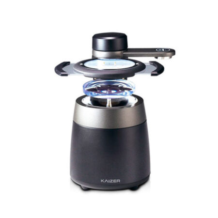
The HMB-8000 manual blocker is part of the Huvitz KAIZER series. Kaizer is the next generation to follow the Huvitz Excelon series and is designed with a wide array of features intended to simplify and perfect in-house lens finishing.The KAIZER series is truly a system like no other featuring top-of-the-line technology and unsurpassed performance. When you select the HMB-8000, you will enjoy a lifetime of LED durability. The integrated brightness control function makes it easy to mark and block any lens—even those with dark tint. The HMB-8000 blocking system is also easy to use and features intuitive graphics with outstanding functionality. With its sleek, compact design and luxurious feel, the HBM-8000 is an outstanding component of the KAIZER series.Features of the HMB-8000 Blocker
▪ Compatible with High-End Finishing Systems
▪ Adjustable LED Intensity
▪ Small Footprint
▪ Easy-to-Use
▪ Energy Efficient
▪ VersatilityKAIZER Compatible
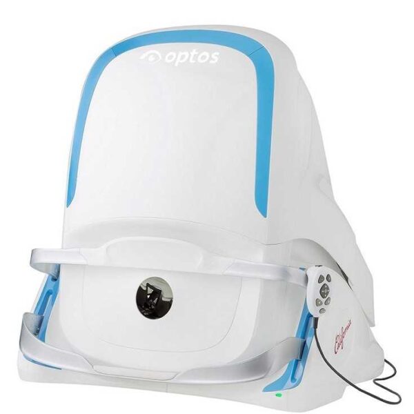
The HMB-8000 manual blocker is part of the Huvitz KAIZER series. Kaizer is the next generation to follow the Huvitz Excelon series and is designed with a wide array of features intended to simplify and perfect in-house lens finishing. The KAIZER series is truly a system like no other featuring top-of-the-line technology and unsurpassed performance.The Huvitz HAB-8000 is a one-stop solution, combining the functions of a tracer, lensmeter, and blocker in one powerful machine. Simply place the lens in position and the HAB-8000 will do the rest. The HAB-8000 is user-friendly enough for in-office use as well as robust enough for higher-volume settings. In fact, with the HAB-8000 you can process up to four jobs at the same time while connecting to up two separate edgers and a drill unit. The possibilities are endless. A 10.4-inch high-resolution touch screen monitor allows operators to control all of the system's functions. Featuring Automatic Lens Center Recognition, the HAB-8000 automatically detects the lens shape and knows what type of lens you’re using. The integrated high-performance lensmeter automatically recognizes the lens as it is set in position. Whether it’s a progressive, bi-focal, or other lens, there’s no need for marking with the HAB-8000. In addition, this high-end Huvitz model offers Digital Scan Hole Detection, Advanced 3D Tracing, Drag-and-Drop Hole Editing, Real-Time Data Transmission and so much more. Fully automated lens centering and blocking is now available at the push of a button. | Optos California RG Ultra-widefield Retinal Imaging SystemEffectively Cure more ocular conditions
Optos introduces its newest ultra-widefield (UWF™) imaging apparatus, California, which make with specific for vitreoretinal specialists and ophthalmologists.The compact desktop device was said to provide increased imaging functionality and has introduced a new imaging modality, indocyanine green angiography, used for imaging the choroidal vasculature, which Optos said was the key in the diagnosis, management, and treatment of certain conditions such as neovascular AMD and ailments with choroidal neovascular membranes.Why California?
With California, Optos has integrated innovative hardware and software technologies allowing professionals to see more, discover more and effectively cure more ocular pathology thus promoting patient wellness. We’re committed to further strengthening our clinical evidence while demonstrating the importance of imaging the entire retina.[su_quote cite=” Dr. David M. Brown” url=”https://houstonretina.com/David-M-Brown-MD”]”If you had another fluorescein camera, then California will fit in the same area if much smaller. It also does autofluorescence, which is helpful for evaluating geographic atrophy,”[/su_quote]New proprietary optical hardware optimizes and maintains the resolution of their optomap images throughout the scan of the retina leading to more clarity at the far periphery.Additionally, comparisons can make between different images or various dates by scrolling through all saved pictures.The Optos California directional retinal imaging apparatus makes it possible for an immediate 200-degree perspective of a patient’s eye at a single scan, supplying a high-resolution picture in under half a second. Additionally, our advanced 3D OCT scanning instrument, the Topcon Triton, has updated software making glaucoma detection and decision making much easier and stronger[su_highlight background=”#f5e791″]With optos California, an optometrist may generate over $9,000 – $15,000 a month. California Billed: $55 each picture for each single patients. Earning money every month with optos California[/su_highlight]
- Compact to reduce space requirements
- New design leads to ease of use and faster image capture
- Non-mydriatic high-resolution imaging through many cataracts and 2mm pupils saves time in busy practices
- Browser-based image review enables simple integration and easy access to your data from any connected pc or tablet in a HIPAA compliant environment
- Interweaved angiography enables parallel capture of fa and icg images without manually switching between imaging modalities
Optos California is available in three models:
California icg design for retinal specialists to maximize management of AMD, uveitic conditions along with other choroidal pathology; it offers the following imaging modalities and image viewing options.
California af was made to facilitate retinal tests and document findings.
California fa was made to help in the identification and management of diabetic eye disease and other cardiovascular ailments.optomap and optomap plus (red and green laser):- Color composite
- green laser View
- red laser View
- optomap af (green laser): autofluorescence
- optomap fa (blue laser): fluorescein angiography
- optomap icg (infra-red): indocyanine green angiography
|


Reviews
There are no reviews yet.