| Description | The Easyfield perimeter is operated via an external computer (notebook or PC). You can enjoy the full freedom of networking the examination data with the familiar user interface of the OCULUS programs. The use of translucent eye shields allows you to conduct measurements without the usual eye patch, thus saving you valuable time of preparing examinations. | Features- NEW TruFit 3D Virtualization allows you to simulate the lens inside the frame before edging, allowing you to minimize retouches and maximize your profit.
- One of the fastest lens edgers on the market.
- Process nearly any job type to avoid having to send out costly jobs to labs.
- Superior quality of finish and polish provides a premium look and feel that you can be proud of.
- Smart Design shape customization.
Wavefront Power-mapping technology for lens analysis and verification.
- Shelf Bevel to easily process high base-curve wrap frames.
- TruScan High base curve frame tracing
| The simplicity is another advantage
Energy-saving and auto-sensing feature will surprise you
White & Black colors are available
1. Compact and sleek design with luxurious feel
Simple, and it makes your work environment look great
2. Adjustable LED intensity
(1) Lifetime durability LED lamp
(2) Easy to mark and block even with dark-tinted lenses by brightness control function
3. Auto power-saving mode
(1) Automatic power off after confirming the marking points
(2) Automatic power off and sleep mode after preset time
Adopting high efficiency LED light source which can easily recognize dark-tinted lenses, and adjust brightness.
• Compact design, intuitive graphic, auto power on/off function. | The Canon RK-F2 Fully Automatic Refractor-Keratometer helps accelerate the exam process by automatically aligning and acquiring a reading for one eye and continuing to the opposite eye to perform the same function – all with one touch of a button. | Retinomax Series, the world’s leading handheld Ref and Refract Keratometer, delivers wide measurement range on par with table type Ref, now boasts greater mobility, stability and accuracy.Current informatical society, eye fatigue, one of the typical modern diseases cause by eye accommodation issue is now easily discover by ACOMOREF2 series during screening basis. Accommodative Micro Fluctuation result will show in graph for easy reading, plus opacity detection function makes ACOMOREF2 the world first All-in-one Refractometer. | The iCare IC200 tonometer is designed for professional use in the surgical operation room, the emergency room and also the clinic. It uses the same patented rebound measuring principle found in other iCare tonometers. There is no need for drops, air or specialised skills for its use. The IC200 gives you positional freedom with a new premium design and user interface. It allows measuring whether the patient is sitting, standing, half-sitting or in the supine or lateral recumbent position. |
| Content | Oculus Easyfield Visual Field Analyzer Perimeter
KEY FEATURES
– Full-fledged compact perimeter capable of performing standard automated perimetry of the central visual field up to 30° eccentricity– Designed for the combined use as visual field screener and perimeter, offering features usually available only in large units– Spherical bowl with 30 cm (11.81â) radius is enclosed into an ergonomically movable cone equipped with a distance adapted lens– Conforms to the Goldmann standard and fulfills the ISO-12866 norm for perimeters– Measurements of the Easyfield are carried out using an LED grid with 135 fixed test locations, including the common 30-2, 24-2 and 10-2 patterns– Novel SPARK test strategy leads to faster and more stable threshold tests providing improved diagnostic capabilities– Besides the standard field indices the Easyfield delivers evaluations of the innovative Glaucoma Staging Program (GSP) and the classifications provided by the Glaucoma Staging SystemADVANTAGES Oculus Easyfield Visual Field Analyzer Perimeter
– Fast: Shorter examination times even for threshold tests– Compact: No completely dark room required thanks to the closed construction– Lightweight: Minimal footprint and maximal transportability– Full Threshold: The classical 4-2 dB staircase strategy using two reversals in the patients answer to deliver a threshold value– Fast Threshold: Bracketing strategy using variable steps and taking advantage of already measured locations– CLIP: Strategy using stimuli with continuously increasing luminance– Threshold value is assigned the moment the stimulus is perceived– SPARK: Fast and averaged threshold strategy based on statistical correlations between threshold values measured in different locationsThe Oculus Easyfield is a compact visual field that is packed with options with a very small footprint. This system is ideal for tight spaces or a smaller practice. Easy to use and very user friendly. The Oculus Easyfield is an exciting compact perimeter that performs static perimetry up to 30°. It has been designed as a visual field screener and perimeter, offering features usually available only in large units. The Easyfield’s integrated bowl with a 11.81” (30 cm) radius and distance corrected lens is adapted to the Goldmann standard and fulfills the ISO-norm 12866 for perimeters. The Easyfield has a fixed point grid with 135 test locations including 30-2. | Briot Couture Lens Finishing System
When your standards are high, this is the partner you deserve. A revolution in edging, Briot Couture gives you a flawless finish with every frame – whatever the style, shape or curve. 3D pre-visualization lets you see your results before you even start working. TrueFit technology calculates curvatures to ensure seamless lens-frame pairing. Additionally, new sensors coupled with advanced software maintain constant pressure on the lens, reducing cycle time with no risk of misalignment – especially on hydrophobic lenses.Wavefront, The Future Of Blocking Briot Couture Lens Finishing System Our exclusive wavefront lens analysis allows you to see progressive lens designs on the frame trace before you even cut the lens, giving you the opportunity to validate that the shape and lens design are compatible without wasting time or money redoing the job. Shape Creation Limited Only By Your Imagination With Smart Design Milling And Shelf Beveling Create complex shapes and a true shelf bevel for high base-curve wrap frames with the Smart Design Milling feature in the Briot Couture. Exclusive TrueScan High-Curve Tracing Featuring our patented TrueScan technology, the Attitude Tracer/Blocker has a specially designed angled probe that will gently measure the contour of even the thinnest, most delicate metal frames or high base-curve wrap frames effortlessly. Brushless Technology For Faster, Quieter Operation Edge lenses faster and more efficiently while also reducing noise with new brusheless motor technology featured on the Evolution. Now, you can plug this edger in anywhere that has a 120V line adding extra placement flexibility in your optical lab. Provide The Best Finish On High Base-Curve Frames With Briot’s Best-Fit Technology Our advanced drive technology in conjunction with a small 90mm diamond wheel allows flawless finishing on high base-curve lenses and complex frames with sharp corners. Damaged Demo Lens? No Problem With Shape Creator 2.0 Accurately reconstruct broken or defective demo lenses with minimal effort using the new Shape Creator 2.0 technology on the Couture Blocker. | Huvitz HMB8000 Manual Blocker
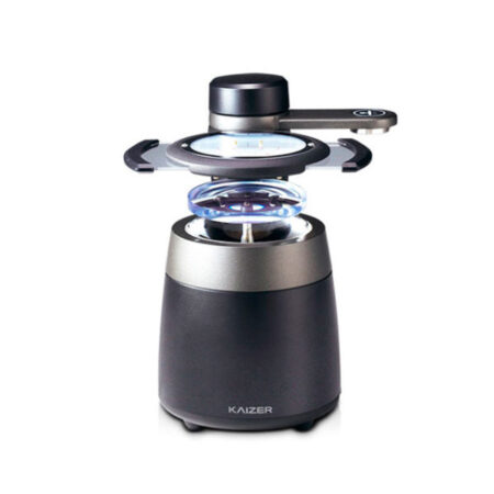
The HMB-8000 manual blocker is part of the Huvitz KAIZER series. Kaizer is the next generation to follow the Huvitz Excelon series and is designed with a wide array of features intended to simplify and perfect in-house lens finishing.The KAIZER series is truly a system like no other featuring top-of-the-line technology and unsurpassed performance. When you select the HMB-8000, you will enjoy a lifetime of LED durability. The integrated brightness control function makes it easy to mark and block any lens—even those with dark tint. The HMB-8000 blocking system is also easy to use and features intuitive graphics with outstanding functionality. With its sleek, compact design and luxurious feel, the HBM-8000 is an outstanding component of the KAIZER series.Features of the HMB-8000 Blocker
▪ Compatible with High-End Finishing Systems
▪ Adjustable LED Intensity
▪ Small Footprint
▪ Easy-to-Use
▪ Energy Efficient
▪ VersatilityKAIZER Compatible
The HMB-8000 manual blocker is part of the Huvitz KAIZER series. Kaizer is the next generation to follow the Huvitz Excelon series and is designed with a wide array of features intended to simplify and perfect in-house lens finishing. The KAIZER series is truly a system like no other featuring top-of-the-line technology and unsurpassed performance.The Huvitz HAB-8000 is a one-stop solution, combining the functions of a tracer, lensmeter, and blocker in one powerful machine. Simply place the lens in position and the HAB-8000 will do the rest. The HAB-8000 is user-friendly enough for in-office use as well as robust enough for higher-volume settings. In fact, with the HAB-8000 you can process up to four jobs at the same time while connecting to up two separate edgers and a drill unit. The possibilities are endless. A 10.4-inch high-resolution touch screen monitor allows operators to control all of the system's functions. Featuring Automatic Lens Center Recognition, the HAB-8000 automatically detects the lens shape and knows what type of lens you’re using. The integrated high-performance lensmeter automatically recognizes the lens as it is set in position. Whether it’s a progressive, bi-focal, or other lens, there’s no need for marking with the HAB-8000. In addition, this high-end Huvitz model offers Digital Scan Hole Detection, Advanced 3D Tracing, Drag-and-Drop Hole Editing, Real-Time Data Transmission and so much more. Fully automated lens centering and blocking is now available at the push of a button. | ZEISS Cirrus HD OCT 4000 Quad Core OCT- 3D Auto-Alignment
- Auto-Focus
- Auto-Shooting Retro-Illumination
- 10 Ref/Kera measurement data memory for each eye
- Small pupil size
- Corneal periphery measurement
- Wide diopter measurement range (-33D through +22D)
- Integrated thermal printer
- Motorized joystick control
- Motorized chin rest
SMALL PUPIL SIZE
At 2.0 mm, the small pupil mode helps contribute to an efficient screening and is useful when a patient’s pupils are difficult to dilate. The short reaching distance between the patient and operator allows closer personal interaction and easy access to the patient’s eyes.RETRO-ILLUMINATION
Using infrared retro-illumination, the RK-F2 Ref-Keratometer provides a detailed view of the eye which is helpful for identifying cataracts, vitreous opacity, scars, and other serious eye problems.CORNEAL PERIPHERY MEASUREMENT
The peripheral keratometry mode provides accurate measurements for examining oblique astigmatisms and helps determine the best fit for a contact lens. The RK-F2 Ref-Keratometer can be used to make a series of measurements at a 30 degree angle from the eye’s center along the attentive meridians. WIDE MEASUREMENT RANGE The dioptric measurement range of -30D to +22D allows examination of strong myopia or hyperopia. The astigmatic measurement range extends from 0 to 10D. The radius of curvature extends from 5 to 10 mm (33.75D to 67.5D) for keratometry.IMAGE ERROR DETECTION
Advanced software automatically confirms both the correct alignment and focus.COMPACT AND LIGHTWEIGHT
The RK-F2 Ref-Keratometer is compact and designed to be operated either sitting or standing, whichever is more comfortable for the operator. Weighing a little more than 33 pounds, the lightweight RK-F2 Ref-Keratometer maximizes small work areas and comfortably fits on a Canon instrument table (sold separately) with a Canon Full Auto Tonometer or Canon Non-Mydriatic Retinal Camera (both sold separately). Keeping the Canon machines next to one another can help expedite the exam process.MULTI-FUNCTIONAL COLOR LCD MONITOR
The 5.7 inch color LCD monitor tilts 40° making the RK-F2 Ref-Keratometer easy to use if the operator is sitting or standing. The clear LCD display also includes multi-functional screen buttons allowing the operator to switch menus as needed.MOTORIZED OMNI-DIRECTIONAL JOYSTICK
Aligning the patient’s eye is easy using the motorized omni-directional joystick with a fine focus dial. All positioning may be performed with one hand allowing the other hand to work with the patient, if needed. The top of the joystick button has the start button which can immediately be pressed once the patient is properly positioned.BUILT-IN PRINTER ZEISS Cirrus HD OCT 4000 Quad Core OCT
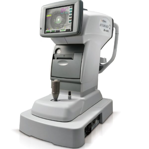
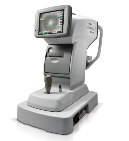
The front-loaded, high speed built-in printer includes an auto cutter making it simple to remove the print-out. The ECO setting reduces the printing density and thereby conserves the paper necessary for printing by up to 50% as compared to printing without using the ECO setting. | Righton BON ACOMOREF 2 K-Model Autoref-Keratometer3 in 1 Function
With combination of Refractometer (Keratometer), Accommodative Micro Fluctuation, Opaque Media (opacity) screening. For even more information view lightning link casino. Reduce of accommodation testing time
Accommodation measurement speed is remarkably improved that can be tested in 49 seconds per eye in SCR (screening) mode. Objectively find the cause of eye fatigue
Patient’s individual AMF (Accommodative Micro Fluctuation) value will be analyzed and from the data, High Frequency Components (1 – 2.3Hz) will be extracted and based on measurement data, chart location, accommodation reaction value and pupil size to formulate FK map (Frequency of Kinetic reaction). ADD Function Righton BON ACOMOREF 2 K-Model Autoref-Keratometer
The ADD function enable a patient’s near point power to be measured objectively by applying accommodative stimulation. And auto AMF can detects if the power gives the patient eye strain or not by color indication. Residual Astigmatism Calculation (Only for K-model)
Residual astigmatism calculation, necessary for CYL contact lenses and CYL IOL prescriptions, is automatically conducted and results printed out (K-Model). Astigmatism corresponding meridian target
Retinomax Series, the world’s leading handheld Ref and Refract Keratometer, delivers wide measurement range on par with table type Ref, now boasts greater mobility, stability and accuracy.Current informatical society, eye fatigue, one of the typical modern diseases cause by eye accommodation issue is now easily discover by ACOMOREF2 series during screening basis. Accommodative Micro Fluctuation result will show in graph for easy reading, plus opacity detection function makes ACOMOREF2 the world first All-in-one Refractometer. | Icare ic200 TonometerThe device is based on a rebound measuring principle that requires no drops, air or specialized skills for its use. The new premium design and user interface bring IOP measuring to a new level. The Icare ic200 tonometer is designed for professional use in the surgical operation room and emergency room as well as the clinic. The ic200 is fully portable, requires no anesthesia and its freedom of positioning allows measuring whether the patient is sitting, standing, half-sitting or in the supine or lateral recumbent position.A high-visibility indicator at the probe base confirms your positioning of the tonometer prior to measurement. A green light indicates measurement will be reliable, a red light indicates incorrect positioning. Features:
- Effortless probe loading with the same probe as for the Icare TA01i and ic100 tonometers
- The probe can’t accidentally drop from the device
- Easy menu in many languages
- Wireless (Bluetooth) printing and measurement transfer - Revolutionary design to allow for positional freedom when taking IOP measurements
- Innovative automatic measuring sequence allows for 6 automatic measurements or single measurements to be taken with the same button
- Intelligent positioning system for correct alignment of the tonometer
- Powered by AA batteries
Specifications:
- Dimensions : 43mm[W] x 104mm[H] x 214mm[L]
- Weight : 165 g (without batteries), 260 g (with 4 x AA batteries)
- Power supply : 4 x AA non-rechargeable batteries, 1.5V alkaline LR6
- Measurement range : 7 - 50 mmHg
- Accuracy : ±1.2 mmHg (≤20 mmHg) and
: ±2.2 mmHg (>20 mmHg)
- Repeatability (coefficient of variation) : <8% Package Includes:
- Icare ic200 tonometer
- 4 x AA batteries
- Aluminum case
- IOP pad
- Probe base cover
- Quick guide
- Screw driver
- Silicone grip
- Spare probe base
- Instruction manual
- Warranty card
- Wrist strap |


Reviews
There are no reviews yet.