| Description | The weight of each Platinum series is still high, above 320 lbs. The monitor size is 17-inches, but upgradable to 19-inches. Although the holds good quality and reputation in Radiology, they still need to be improved in cardiology and OB/GYN’s workflow, automation, and 4D quality, comparing GE or Samsung products to satisfy customers. | The Sequoia system’s advanced technologies are designed to make image acquisition easier, reduce exam time and increase overall efficiency. Along with many other included features, the Sequoia system offers Native, a patient specific imaging which adapts both phase and amplitude information of each unique patient’s properties which delivers a consistently better image across a broad range of patients. | Ultraformer III’s 7 clip-in transducer cartridges project uniform ultrasound beams directly to multiple layers underneath the skin to promote tighter collagen formation, tissue contraction and reduction in the volume of adipocytes that form bulging areas of tissue.Patients notice an immediate benefit in improved skin tone and texture, with ongoing improvements over 3 to 6 months
Traditionally difficult areas such as eye lifting are made easier with Ultraformer III’s specialised cartridges. It is possible for patients to experience up to 6mm of eyebrow lift, depending on their particular situation. | The more patients you see, the more you need Voluson E8.
The Voluson E8 ultrasound system is designed to keep pace with busy practices that conduct a wide range of Women’s Health exams, from routine scanning to complex assessments.
The imaging excellence of Radiance System Architecture – your diagnostic confidence will be enhanced by the extraordinary image clarity and speed of Radiant System Architecture. | The SonoSite Titan is the earliest version of the PoC dedicated portable ultrasound machine from Sonosite. Though its form factor is compact, it provides high resolution imaging, rapid responsive time, and supports a broad range of applications including cardiac. | Portable Color ultrasound System
This is a portable color ultrasound system that displays good image quality for diverse applications. This small and compact system has various diagnostic capabilities. |
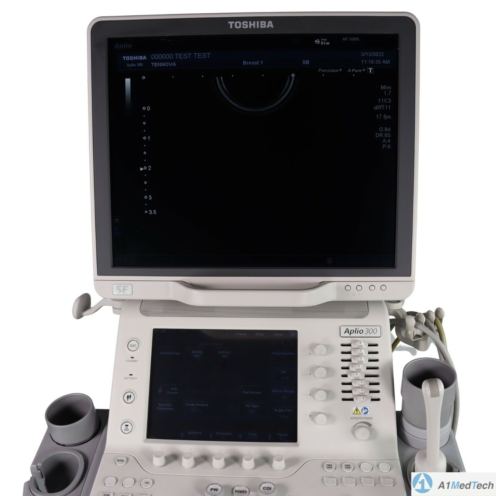
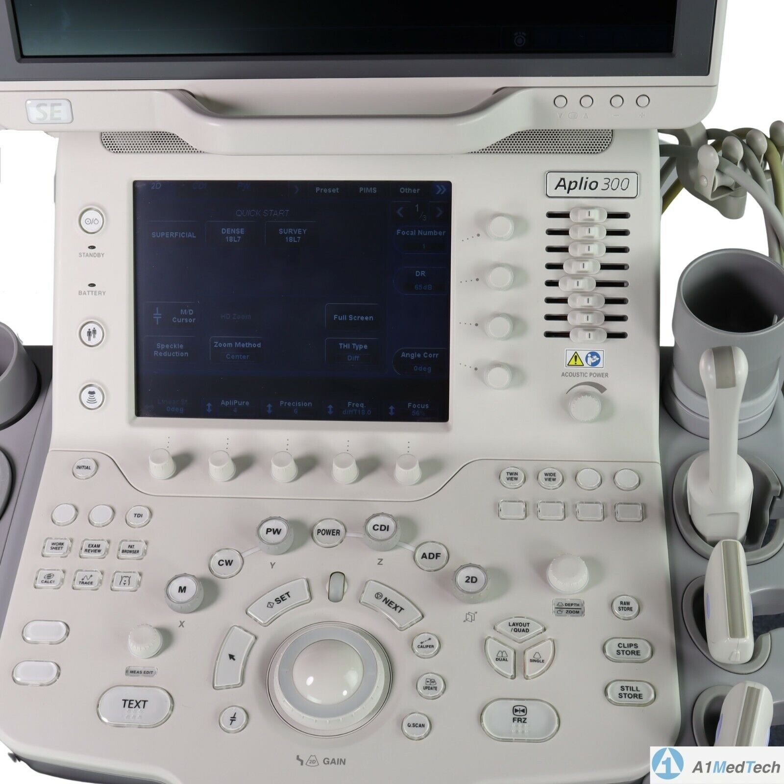
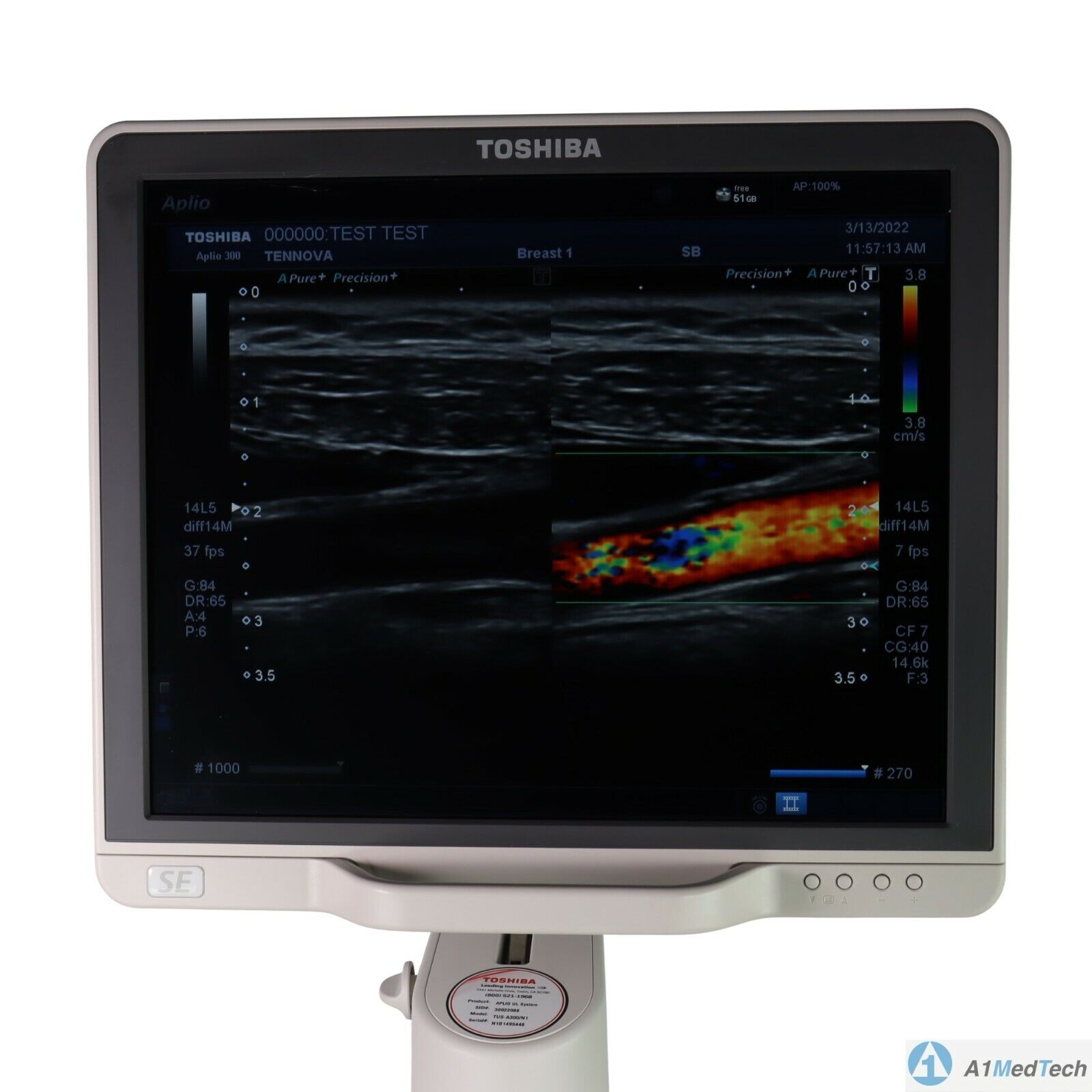
| Content | Toshiba Aplio 300 Ultrasound Machine
Toshiba Aplio 300 Ultrasound Machine Platinum and CV color Doppler ultrasound machines represent the more affordable versions of Canon’s high-end Aplio series of ultrasound machines. shared service machine or CV ultrasound includes Toshiba’s more advanced imaging innovations, including advanced compound imaging, speckle reduction, and tissue enhancement technologies. Additionally, the features 3D/4D imaging with high-density volume imaging.Toshiba Aplio 300 Ultrasound Machine For advanced performance, you can rely on day after day, look no further than . Cost-effective, productive and versatile, the system offers superior imaging with outstanding depth and detail for the widest range of clinical specialties. Lighter and with a smaller footprint, is designed to increase mobility, to optimize viewing, and to provide you with an ergonomic workplace. Products in general look like a technology oriented product, rather than a clinical oriented product. With this series, Canon has achieved fully digital beamforming technology, which is fully integrated into the Aplio Platinum series. The weight of each Platinum series is still high, above 320 lbs. The monitor size is 17-inches, but upgradable to 19-inches. Although the holds good quality and reputation in Radiology, they still need to be improved in cardiology and OB/GYN’s workflow, automation, and 4D quality, comparing GE or Samsung products to satisfy customers. - Shared Service
- Next Generation Precision Imaging
- ApliPure
- 3D/4D Imaging
- Advanced Dynamic Flow
- Contrast Harmonic Imaging
- Strain Elastography
- Gel Warmer
- Panoramic Option
- Stress Echo Option
- TDI Tissue Doppler Imaging
- HPRF High Pulsed Repetition Frequency
- DICOM Media Storage
- QuickScan
- 17″ LCD Monitor with 1280×1027 resolution and 170-degree viewing angle
- Trapezoidal Imaging
- TissuePure Speckle Reduction Imaging
- Compound Imaging
- IMT Intima-Media Thickness
- DICOM SR for Cardiac, Vascular, and OB/GYN
With the launch of the Toshiba Aplio 300 in 2011, the popularity with Toshiba is growing. Great for OB/Gyn to Cardiac, it offers the following:
• Differential Tissue Harmonics (D-THI): Enables uniformity in the near and far fields, especially while scanning at increased depth. • iStyleTM+ Productivity Suite: Delivers ergonomic relief by minimizing keystrokes and reducing exam times with the industry’s most customizable user interface and preset management tools.
• Ergonomics: Features a high-resolution LCD screen on a four-axis arm and handle, adjustable console, lighter weight and smaller footprint. | Siemens Acuson Sequoia 512 LCD Ultrasound Machine18″ Flat panel LCD Monitor–Progressive Media & Display II Package, Cardiac/OB/Vascular Calc. Package, Signature II Option, AUX, CPS Cardiology, CPS General Cadene Trigg Burst, Calc Data to MO, Card Cadence II Contrast Quant, Cornoary Flow reserve, Dicome Store/Print, DTI (Doppler Tissue Imaging), High HR ECG, MIS/MIF, Modality Worklist Native TEQ, Native TEQ, NTHI (Native Tissue Harmonics), Spectral TEQ, TEQ (Tissue Equalization), VVIFeatures- Shared Service
- Native Patient Specific Imaging
- Four Sight TEE view
- Axius quantitative strain rate imaging
- Cadence contrast pulse sequencing technology
- Native Tissue Harmonic Imaging
- Native TEQ ultrasound technology
- Color doppler, Color angio and b-color
- PW/CW Doppler
- M-Mode
- Folding Flat Panel Display
- DICOM capabilities
Applications- Abdominal
- Breast
- OB/GYN
- Vascular
- Pediatrics
- Cardiology
- Musculoskeletal
- Neonatal
Acuson Sequoia 512 Features:- Imaging: Gray-scale 2-D, M-mode, SST Color Doppler, Solo Spectral Doppler, 3-D Surface Rendering, Native Tissue Harmonic Imaging, Auto Doppler, M-Mode, PW Doppler, Color Doppler, Color Power Doppler
- 19" color CRT monitor
- Native tissue harmonic imaging (NTHI)
- DIMAQ: image management
- Advanced vascular analysis
- Advanced spatial compounding
- Native patient specific imaging
- Mitral valve assessment
- CPS contrast pulse sequencing technology
- Clarifying vascular enhancement technology
- High resolution color flow
- Vascular, and General Imaging calculation packages
- Coherent beamformer technology/spatial compounding
- Space/time resolution control
- Cine and zoom/RES
Compatible Ultrasound Probes and Transducers:- Acuson V5Ms TEE Probe
- Acuson 4C1 Curved Array Probe
- Acuson 6C2 Curved Array Probe
- Acuson 8C4 Curved Array Probe
- Acuson 4V1 Vector Array Probe
- Acuson EV-8C4 Endocavitary Probe
- Acuson EC-10C5 Endocavitary Probe
- Acuson 6L3 Linear Array Probe
- Acuson 8L5T Intraoperative Probe
- Acuson 8L5 Linear Array Probe
- Acuson 9L4 Linear Array Probe
- Acuson 15L8 Linear Array Probe
- Acuson 17L5 Linear Array Probe
The Sequoia 512 with LCD ultrasound machine uniquely minimizes automatic image optimization time by up to 80% to make way for an easier and speedier ultrasound test time as well as allow for a more confident diagnosis each time. As a patient-specific imaging ultrasound, it has a unique matched response capacity for accurate image quality, sharp clinical specificity, and optimized diagnostic outcomes that makes infinitesimal pathologies and other anatomical detail in the most difficult-to-image patients becomes more vivid and visible. | CLASSYS ULTRAFORMER III UTR3
You’ve heard about it, now see and feel the difference for yourself.Ultraformer III is the leading technology for facial lifting, tightening and contouring. Now with 7 interchangeable cartridges that offer your patients another dimension in non-surgical face and body treatments.Ultraformer III is widely used with over 10 million treatments performed worldwide. It has become the treatment of choice for patients who are not yet indicated or not willing to undergo surgery. This fills a huge gap in the market, and captures an audience of people from 30 years of age all the way to 80.ULTRAFORMER III FEATURESLess discomfort associated with treatments
Patented transducer design for more efficient energy delivery
Dual hand pieces with automatic recognition for ease of use
Faster shot and treatment speed to save time
Easy, user-friendly design and interface
Shot count information and voice assistance
Backed by clinical papersThe CLASSYS ULTRAFORMER III UTR3 advantage with slim transducer designimproves operator visibility
supports very narrow focus and precise treatment
fits facial contoursFast elliptic transducersmore efficient treatment timesUltraformer III’s 7 clip-in transducer cartridges project uniform ultrasound beams directly to multiple layers underneath the skin to promote tighter collagen formation, tissue contraction and reduction in the volume of adipocytes that form bulging areas of tissue.Patients notice an immediate benefit in improved skin tone and texture, with ongoing improvements over 3 to 6 months
Traditionally difficult areas such as eye lifting are made easier with Ultraformer III’s specialised cartridges. It is possible for patients to experience up to 6mm of eyebrow lift, depending on their particular situation.Ultraformer III’s 7 clip-in transducer cartridges project uniform ultrasound beams directly to multiple layers underneath the skin to promote tighter collagen formation, tissue contraction and reduction in the volume of adipocytes that form bulging areas of tissue.Patients notice an immediate benefit in improved skin tone and texture, with ongoing improvements over 3 to 6 months
Traditionally difficult areas such as eye lifting are made easier with Ultraformer III’s specialised cartridges. It is possible for patients to experience up to 6mm of eyebrow lift, depending on their particular situation. | GE Voluson E8 Ultrasound Machine15’’ LCD monitor [Rev BT06], 10.4” touchscreen, 3 Active Probe Ports, Floating Keyboard: Interactive back-lighting, Beta-View CrossXBeam, Virtual Convex, Extended View, ATO [Automatic Tissue Optimization], Coded Harmonic Imaging, High Resolution Zoom, Inversion Real-time automatic Doppler, OB/GYN; Vasc. Cardiac; Abd; Small-Parts; Urology; Ped; Ortho; Neuro reporting, Multigestational Calculations, HPRF [High Pulse Repetition Frequency], SonoVCAD [Matrix array volume technology], TruScan [On-board archive], VCI [Volume Contrast Imaging], SonoAVCfollicle [Sonography-based Automated Volume Count follicle], SonoVCADheart [Sonography-based Volume Computer Aided Display], TUI [Tomographic Ultrasound Imaging], CE [Coded Excitation], XTD, SRI III [Speckle reduction imaging], CRI [Compound Resolution Imaging], DICOM, Ethernet port, Video Out, 6 USB ports, Integrated HDD (160 GB) and DVD/CD-RW.
3D/4D, Endocrinology, Family Practice, Gastroenterology, Internal Medicine, Nephrology, Neurology, OB-GYN, Orthopedics, Otorhinolaryngology, Pain Management, Urology, Vascular Probes GE Voluson E8 Ultrasound Machine - 11L-D Linear Array
- 3S-D Cardiac Sector
- 4C-D Convex Array
- 9L-D Linear Array
- AB2-7 Convex
- IC5-9-D Intercavity
- M6C-D Convex
- P2D CWD Non-Imaging
- P6D CWD Non-Imaging
- PA6-8-D Cardiac
- RAB2-5-D 3D/ 4D Convex
- RAB4-8-D 3D/4D Convex
- RIC5-9-D 3D/4D Intracavity
- RIC6-12-D 3D/4D Intracavity
- RNA5-9-D 3D/4D Convex
- RRE6-10-D Endocavitary Micro-Convex
- RSP6-16-D 3D/D Linear
- SP10-16-D Linear
- 19″ monitor
- 4D Real Time
- CFM Doppler Mode
- Coded Contrast Imaging
- CrossBeam
- DICOM 4D
- HD flow
- Power Doppler Mode
- SonoVCAD
- STIC (Spacial-Temporal Image Correlation)
- T.U.I (Tomographic Ulrasound Imaging)
- VOCAL II VCI (Volume Contrast Imaging)
- Black and white printer
- Color printer
- DVD-Recorder
As your patient volume increases and your clinical cases become more complex, you will require an ultrasound system that enables you to deliver truly exceptional care – confidently and efficiently – every time. The Voluson E8 ultrasound system is designed to keep pace with busy practices – handling routine to complex women’s health exams with ease and precision. The premium image quality of the Voluson E8 combined with access to advanced capabilities, delivers the level of performance required by you and your patients. | Sonosite Titan Ultrasound Machine
The SonoSite Titan Portable Ultrasound is SonoSite’s second generation portable ultrasound system. This system improved upon the technology of the SonoSite 180 with a larger screen and keyboard packed into a laptop style system. This system is well suited for Vascular access, Musculoskeletal, Anesthesiology, Internal Medecine, Emergency rooms and Family Physicians. Veterinary configurations are available. Veterinary configurations are available.8.4″ LCD monitor, 2 probe ports, Vascular, OB/GYN, Cardiac reports, Soft keys, Split screen, Zoom, Storage via 64 or 256 MB flash cards, 220 image CINE loop, USB & Ethernet image transfer, Volume calculations via diameters, THI [Tissue Harmonic Imaging], SonoCalc [IMT analysis, PC-based], DICOM, S-video (in/out), RGB, Composite video.
Dimensions: 3″ (H) x 10.9″ (W) x 11.9″ (D) Weight: 7.7 lbs Manufacturer
Sonosite Application
Cardiology, Endocrinology, Family Practice, Gastroenterology, Internal Medicine, Nephrology, Neurology, OB-GYN, Orthopedics, Otorhinolaryngology, Pain Management, Surgery, Urology, Vascular Probes - C8 – prostate – 8-5 MHz 8-mm broadband intracavitary array – 8.5 cm – Available – MicroMaxx/ TITAN
- C11 – abdominal/ pediatric cardiology/ neonatal/ vascular – 8-5 MHz 11-mm broadband curved array – 10 cm – N/A – TITAN
- C15 – adult cardiology/ abdominal – 4-2 MHz 15-mm broadband tightly curved array – 25 cm – N/A – TITAN
- C60 – abdominal/ obstetrics/ gynecology – 5-2 MHz 60-mm broadband curved array – 22 cm – Available – TITAN
- ICTe – obstetrics/ gynecology – 8-5 MHz 11-mm broadband tightly curved array – 10 cm – Available – MicroMaxx/ TITAN
- L25 – nerve/ musculoskeletal/ vascular and superficial – 10-5 MHz 25-mm broadband linear array – 6 cm – Transverse Guide Available – TITAN
- L38 – small parts/ breast/ vascular/ nerve/ musculoskeletal/ superficial – 10-5 MHz 38-mm broadband linear array – 6.5 cm – Available – TITAN
Options - Extended cardiac package
- Pulsed wave (PW) doppler
- Continuous wave (CW) doppler
- Tissue Harmonic Imaging
Accessories - Cart
- Black and White Video Printer
- DVD Recorder
The SonoSite Titan is the earliest version of the PoC dedicated portable ultrasound machine from Sonosite. Though its form factor is compact, it provides high resolution imaging, rapid responsive time, and supports a broad range of applications including cardiac. | Samsung Medison SonoAce R3 Ultrasound MachineSonoAce R3 is a portable color ultrasound system with comprehensive diagnostic capabilities. Full Spectrum Imaging (FSI) combines with Speckle Reduction Filter (SRF) to deliver superior quality images, even in the challenging circumstances. The compact and user-friendly design features of the SonoAce R3 ensure portability and manoeuvrability in a lightweight unit. Sporting a 15-inch LCD monitor with Full-Featured Workflow, the SonoAceR3 offers your organisation flexibility, without compromising on precision and efficiency.Slim and Compact DesignThe compact design of SonoAce R3 offers Various-Featured Workflow with a thumbnail menu and shortcut, together with functional key groupings for increased efficiency. The user-friendly design combines a wide-view 15-inch LCD monitor with backlit control keys, customisable measurement package and body markers. The SonoAce R3 features 2 probe ports, 3 USB ports, rapid boot up capability and DICOM 3 compatible image filing. This slim, lightweight unit also has a convenient carrying handle for portability and flexibility.Full Spectrum Imaging (FSI) produces the penetration capabilities associated with the lower frequencies, while still maintaining the fine pixel uniformity associated with the higher frequencies. This combination consistently delivers superior quality images, even in the challenging diagnostic circumstances. The resulting clarity delivers improvements in accuracy and efficiency for greater confidence in your diagnostic procedures. Harmonic Imaging is a technique that records higher frequency information, resulting in greater resolution and fewer artifacts.Synthetic Aperture
The physical radio channel may present some limits to imaging which can be overcome by using Synthetic Aperture Control software. This method uses information from diverse sources to form a more better picture of the area under examination. It creates one single scan line by performing the TX and RX twice to create enhanced penetration and resolution. Synthetic Aperture Control technology delivers a comprehensive picture and to aid accurate diagnosis.QuickScan
QuickScan has been designed to increase workflow efficiency by automatically optimising key imaging parameters at the touch of a button. The resulting increase in efficiency allows your organisation to offer patients an improved service with faster and more accurate diagnoses. This reliable and safe system helps your clinic or hospital to deliver a superior patient experience.Features Samsung Medison SonoAce R3 Ultrasound Machine
Full Spectrum ImagingTM
Quick ScanTM
Color and Power Doppler
Harmonic imaging
Freehand 3D
3D Multi Planar Imaging
Sonoview Image Management
PW Doppler
15″ LCD monitor
Two probe ports
Lightweight |




Reviews
There are no reviews yet.