| Description | The weight of each Platinum series is still high, above 320 lbs. The monitor size is 17-inches, but upgradable to 19-inches. Although the holds good quality and reputation in Radiology, they still need to be improved in cardiology and OB/GYN’s workflow, automation, and 4D quality, comparing GE or Samsung products to satisfy customers. | 3D-4D ultrasound is a technique used during pregnancy that displays three-dimensional images and movement of a fetus (fetal face and other body parts). Other applications include multiplanar imaging of the pelvic organs. This developing technology is also being used in some diagnostic medical procedures such as biopsies and enhanced visualization of cardiac structures. National Ultrasound offers a variety of 3D-4D Ultrasound machines such as GE Voluson i, GE Voluson e, GE Voluson E8, Voluson 730 Expert, Medison Sonoace 8000, Sonoace 9900 or Toshiba with the Xario and Apilo. Our team of expert sales managers will help you find the right 3D-4D ultrasound machine to meet your diagnostic needs while staying within your budget. | Acuson Cypress cardiovascular system PLUS delivers advanced ergonomics and superior performance in a highly portable package. TheCypress System PLUSis a highly miniaturized, all digital, phased array echocardiography system that provides complete studies and outstanding images – even on patients who are difficult to image. | The Expert is the new flagship LOGIQ e. The imaging engine comes from GE leading console systems, delivering crisp images in a compact package. Providing ultrasound imaging with precise anatomical detail at a variety of depths. The system includes innovative features that help simplify proceedures. | Ultraformer III’s 7 clip-in transducer cartridges project uniform ultrasound beams directly to multiple layers underneath the skin to promote tighter collagen formation, tissue contraction and reduction in the volume of adipocytes that form bulging areas of tissue.Patients notice an immediate benefit in improved skin tone and texture, with ongoing improvements over 3 to 6 months
Traditionally difficult areas such as eye lifting are made easier with Ultraformer III’s specialised cartridges. It is possible for patients to experience up to 6mm of eyebrow lift, depending on their particular situation. | Acuson Cypress cardiovascular system PLUS delivers advanced ergonomics and superior performance in a highly portable package. The Cypress System PLUS is a highly miniaturized, all digital, phased array echocardiography system that provides complete studies and outstanding images – even on patients who are difficult to image. |
| Content | Toshiba Aplio 300 Ultrasound Machine
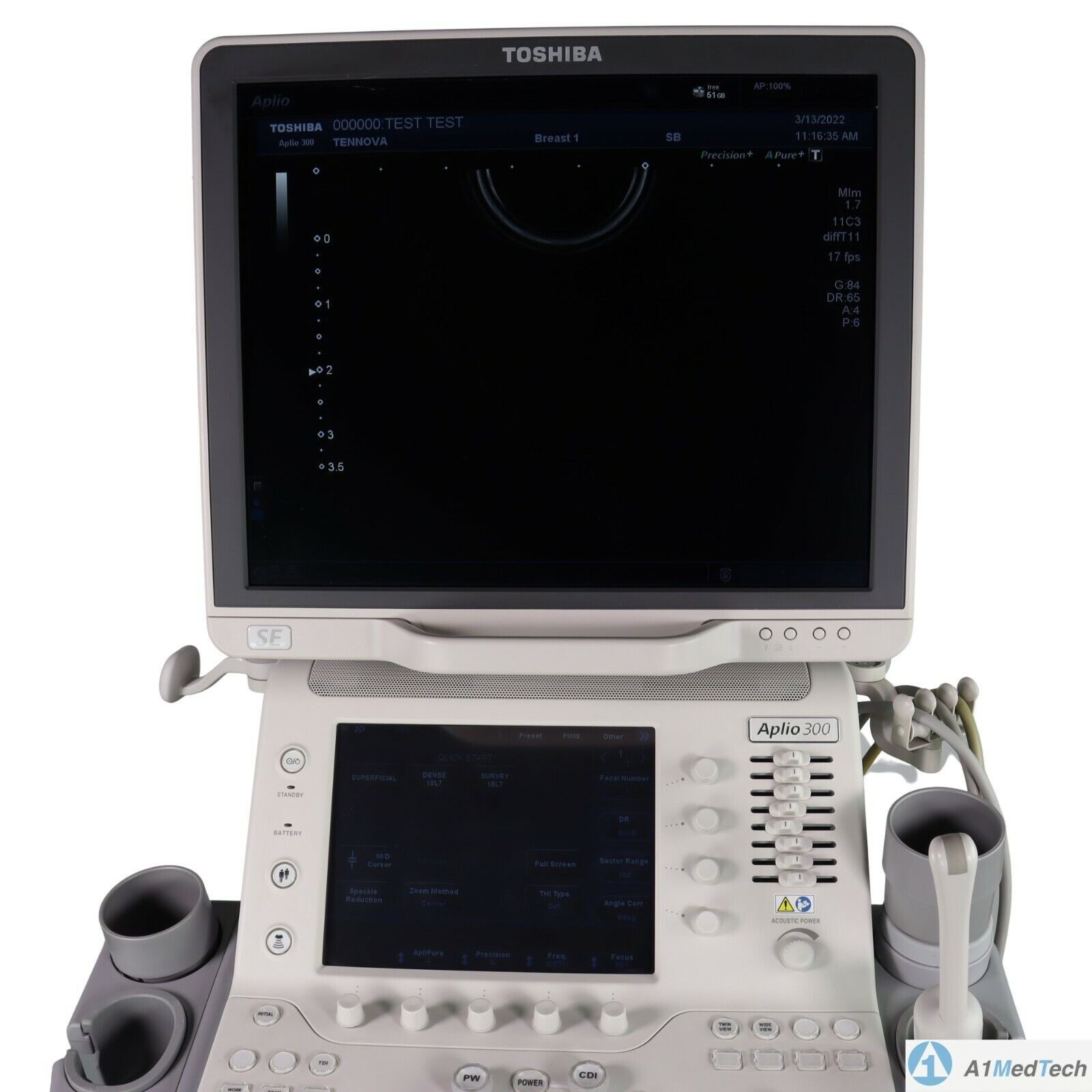
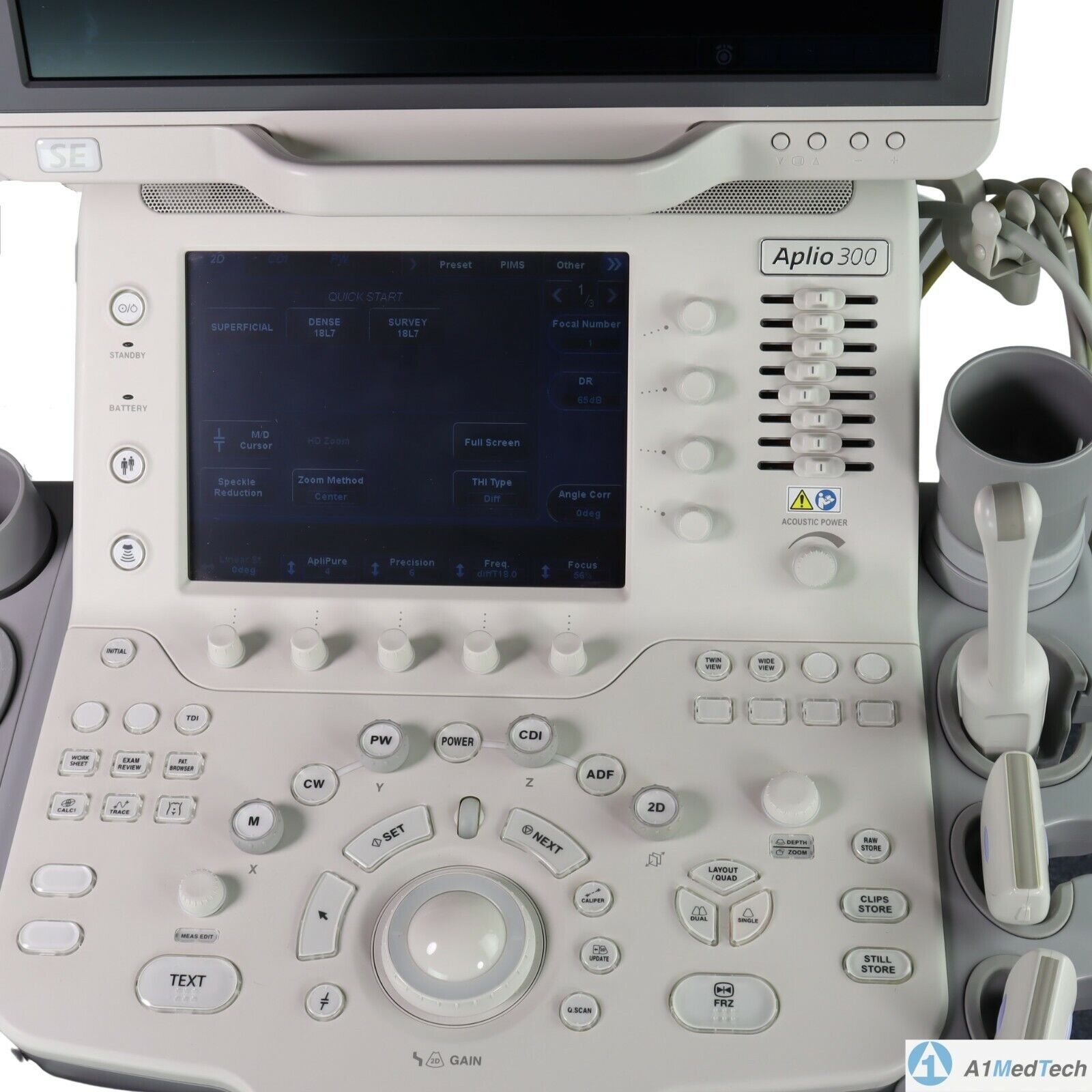
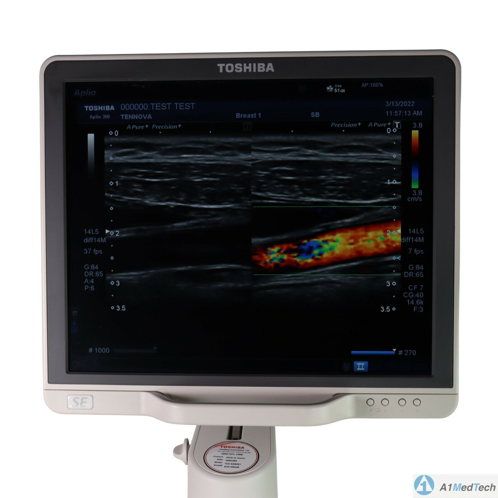
Toshiba Aplio 300 Ultrasound Machine Platinum and CV color Doppler ultrasound machines represent the more affordable versions of Canon’s high-end Aplio series of ultrasound machines. shared service machine or CV ultrasound includes Toshiba’s more advanced imaging innovations, including advanced compound imaging, speckle reduction, and tissue enhancement technologies. Additionally, the features 3D/4D imaging with high-density volume imaging.Toshiba Aplio 300 Ultrasound Machine For advanced performance, you can rely on day after day, look no further than . Cost-effective, productive and versatile, the system offers superior imaging with outstanding depth and detail for the widest range of clinical specialties. Lighter and with a smaller footprint, is designed to increase mobility, to optimize viewing, and to provide you with an ergonomic workplace. Products in general look like a technology oriented product, rather than a clinical oriented product. With this series, Canon has achieved fully digital beamforming technology, which is fully integrated into the Aplio Platinum series. The weight of each Platinum series is still high, above 320 lbs. The monitor size is 17-inches, but upgradable to 19-inches. Although the holds good quality and reputation in Radiology, they still need to be improved in cardiology and OB/GYN’s workflow, automation, and 4D quality, comparing GE or Samsung products to satisfy customers. - Shared Service
- Next Generation Precision Imaging
- ApliPure
- 3D/4D Imaging
- Advanced Dynamic Flow
- Contrast Harmonic Imaging
- Strain Elastography
- Gel Warmer
- Panoramic Option
- Stress Echo Option
- TDI Tissue Doppler Imaging
- HPRF High Pulsed Repetition Frequency
- DICOM Media Storage
- QuickScan
- 17″ LCD Monitor with 1280×1027 resolution and 170-degree viewing angle
- Trapezoidal Imaging
- TissuePure Speckle Reduction Imaging
- Compound Imaging
- IMT Intima-Media Thickness
- DICOM SR for Cardiac, Vascular, and OB/GYN
With the launch of the Toshiba Aplio 300 in 2011, the popularity with Toshiba is growing. Great for OB/Gyn to Cardiac, it offers the following:
• Differential Tissue Harmonics (D-THI): Enables uniformity in the near and far fields, especially while scanning at increased depth. • iStyleTM+ Productivity Suite: Delivers ergonomic relief by minimizing keystrokes and reducing exam times with the industry’s most customizable user interface and preset management tools.
• Ergonomics: Features a high-resolution LCD screen on a four-axis arm and handle, adjustable console, lighter weight and smaller footprint. | GE Voluson i Portable 3D/4D UltrasoundFeatures* Lightweight and portable
* Battery operation
* Real-Time 4D
* 3D Multiplanar Display
* 3D Power Doppler
* Automatic Optimization (AO)
* Speckle Reduction Imaging (SRI) and CrossXBeam deliver advanced computational power, allowing for simultaneous processing of CrossXBeam CRI and SRI together – enabling added speckle reduction, contrast resolution and image clarity.
* HD-Flow uses a bi-directional Doppler feature to achieve a more sensitive vascular study and reduce overwriting.
* Volume Contrast Imaging (VCI) allows for image quality in either a single plane or all three planes of a volume acquisition
* Spatio-Temporal Image Correlation (STIC) captures a full virtual fetal heart cycle in real time, and the volume can be saved for offline analysis.
* Tomographic Ultrasound Imaging (TUI) makes analysis and documentation of dynamic studies easier with a simultaneous view of multiple parallel slices of a volume data set.
* Virtual Organ Computer-aided AnaLysis (VOCAL) is a semi-automated volume measurement tool that utilizes computer technology to provide accurate volume calculations.Comes with:GE Voluson i Portable 3D/4D Ultrasound* 1x Voluson i Portable 3D/4D Ultrasound
* 1x RAB2-5RS (2-5MHz) 3/4D Convex Abdominal Probe
* 1x E8C-RS (4-10MHz) Wideband Microconvex Endocavity (Vaginal) Probe
* 1x RIC5-9-RS (5-9MHz) 2D/3D RealTime 4D Endocavitary ProbeThe GE Voluson i ultrasound machine is primarily used for OB-GYN applications, but is versatile and powerful enough to be used for a variety of other applications as well. The Voluson i ultrasound machine carries the power of the GE Voluson line with access to premium 2D and 4D imaging in a portable form factor. The GE Voluson i provides the extraordinary image quality needed to confidently scan for women’s health. The GE Voluson i portable ultrasound machine offers features such as speckle reduction imaging and CrossXBeamCRI, which both lend to the incredible image clarity and quality of this system. The Voluson I portable ultrasound system is known for ease of use, allowing the operator to scan patients faster and more efficiently with better accuracy. | Acuson Cypress PLUS UltrasoundAcuson Cypress cardiovascular system PLUS delivers advanced ergonomics and superior performance in a highly portable package. TheCypress System PLUSis a highly miniaturized, all digital, phased array echocardiography system that provides complete studies and outstanding images – even on patients who are difficult to image.The Cypress PLUS transitions easily from your small private office or hospital echo lab to the operating theatre or your patient’s bedside. It’s ultrasound where you need it.The Cypress PLUS delivers high performance in a complete range of cardiovascular capabilities.Designed with advanced ergonomics in mind, the Cypress system PLUS streamlines your workflow and increases your productivity.If you already own a Cypress system, keeping technologically current is simple with the Cypress Upgrade program.The Cypress system is the smallest, lightest, high performance, phased array echo system ever built. Weighing in at just 19 pounds, with digital storage capacity for over 100 studies and network capabilities to send studies from remote locations, clinicians can perform complete echocardiography studies from almost anywhere. TheCypress system was designed for portability, durability and reliability, and to withstand the rigors of constant transportation. Internal system interconnects are minimized. Key components are shock mounted and the entire system sits inside a robust chassis. From hospital to office or from airplane to mountain village, the Cypress system will perform.Full Range of Capabilities
The Cypress system is a complete, full-featured echo cardiography system that provides complete studies and outstanding image quality even on the most difficult to image patients. It offers a full range of features including:Acuson Cypress PLUS Ultrasound– 2D with Harmonics
– Patient Report with audio capture
– M-Mode
– Embedded DICOM conformance
– Color PW and CW Doppler
– High frame rate color flow imaging
– Stress echo
– Network output
– Vascular imaging
– Comprehensive calculations package
– Contrast agent imaging
– USB peripheral support (PC printer connectivity)Physical Characteristics
• Single unit houses ultrasound system with control panel, flat panel display and MO drive
• Weight: 8.6 Kg (19 pounds)
• Height: 35.6 cm (14 in)
• Width: 40.6 cm (16 in)
• Depth: 20.3 cm (8 in) – with keyboard folded up 34.3 cm (13.5 in) – with keyboard down
• Heat dissipation: 490 BTU/hour (nominal)User Interface
• Fold-up control panel for portability
• Direct access to system functions through dedicated keyboard controls and multifunctional soft keys
• Easy-access main controls, including multifunctional trackball, enter and select keys, for minimum hand movement while controlling the system
• Adjustable trackball speed
• Functional grouping of keys, rotational knobs, switches and sliding TGC controls for direct access to critical functions
• Tactile feedback on control activation
• Backlighting of control panel labels
• Mode-dependent soft keys
• User-interface tools
• Pre-defined and user-entered labeling textMonitor
• 10.4 inch/26.4 cm flat panel active matrix display screen
• Protective anti-glare, anti-reflective lensHard Drive
• Internal hard drive for image and data storage
• Internal storage of approximately 50 patient studies (25 – 30 loops per study)Transducer Ports
• One transducer connector (type IEC601BF)Operating/Display Modes
• 2-D, fundamental and harmonic imaging
• Color Doppler Velocity (CDV)
• Color Doppler Energy (CDE)
• M-mode, vertical split screen display with 2-D
• Pulsed Wave (PW) Spectral Doppler
• Continuous Wave (CW) Spectral Doppler
• Duplex Doppler (combined 2-D and Spectral Doppler display)2D Calipers – Generic Measurements and Calculations
• Measurements and calculations in 2D, M-mode, color Doppler, PW and CW spectral Doppler modes
• Basic measurements and report calculations
• Comprehensive patient calculations report
• On-screen “mini-reports”
• Complete or configured patient report
• Cardiac package
• Carotid package
• Arterial package
• Venous package
• Abdominal package
• User configurable Sites
• Selectable menu position
• Four cursor types
• Angle correction
• Distance
• Area
• Slope
• Time
• Velocity
• Velocity/pressure
• Mean velocity/pressure gradient
• Velocity Time Integral
• Pressure halftime
• Continuity Equation
• Ejection Fraction
• Left Ventricular Volumes, including End-Systolic and End-Diastolic volumes
• Cardiac output
• Cardiac index
• Fractional area changeCine Review
• 2D Cine memory up to 15 seconds of history
• Color Doppler cine memory up to 15 seconds of history
• M-mode strip cine along with PW and CW Doppler mode
• strip cine memory determined by sweep speed (selectable by the user) 10, 20, 30 or 40 secondsTransducer Technology
• 3V2c: 2-4 MHz wideband phased array
• 7V3c: 3-7 MHz wideband phased array
• 7L3: 5-7 MHz wideband linear array (Vascular)
• 4C1: 2-4 MHz wideband curved linear array (Abdominal)
• V5Ms: 3-6 MHz wideband phased array (micro-pinless (MP) connector) | GE Logiq e r7 Portable Ultrasound Machine
The LOGIQ e R7 provides even more impressive veterinary abdominal and cardiac images than the BT12 LOGIQe, combining the high performance of a console system with the portability of a laptop.Highlights of the GE LOGIQ e R7:- Simple. Fast. Precise
- Colour Flow Mode, Power Doppler Imaging (PDI), Pulse Wave Doppler (PWD), Continuous Wave Doppler
- 15” high resolution colour LCD screen
- Integrated solid state hard drive
- Automatic optimisation
- CrossXBeam
- Speckle Reduction Imaging (SRI-HD)
- Virtual Convex / Virtual Apex
- Fine Angle Steer
- HD Zoom (Write Zoom)
- Coded Harmonic Imaging (CHI) – Enhances near field reslolution for improved small parts imaging as well as far field penetration. Diminshes low frequency / amplitude noise and provides clarity to needle, anatomy and motion.
- Raw Data Processing
- Quicksave – Single button push sends single image or entire patient exam to memory stick or network
- On-board manual (Help)
- Loop storage – from live scanning and from memory
- Patient Information Database
- Customisable user interface
- Full M&A calculation package with real time Doppler calculations
- Report writer package – on board reporting package automates report writting
- Portable and battery operated so easy to move from room to room
- Export of stills and clips to USB.
More GE Logiq e r7 Portable Ultrasound MachineThe Expert is the new flagship LOGIQ e. The imaging engine comes from GE leading console systems, delivering crisp images in a compact package. Providing ultrasound imaging with precise anatomical detail at a variety of depths. The system includes innovative features that help simplify proceedures.GE NextGen LOGIQ e R7 Ultrasound System The GE NextGen LOGIQ e R7 Ultrasound System has an imaging engine that delivers crisp images in a compact package. Point-of-care-specific software and transducers help to see the needle to administer a block or perform an aspiration quickly. When productivity is crucial, you can control the console from the transducer, potentially eliminating the need to have a second person assist. Anatomy-specific presets and only displaying the functions that you need helps you to direct your focus on the patient rather than the control panel. Although the system features a large, user-friendly, high-res color display, it is also very durable and compact for easy portability. | CLASSYS ULTRAFORMER III UTR3
You’ve heard about it, now see and feel the difference for yourself.Ultraformer III is the leading technology for facial lifting, tightening and contouring. Now with 7 interchangeable cartridges that offer your patients another dimension in non-surgical face and body treatments.Ultraformer III is widely used with over 10 million treatments performed worldwide. It has become the treatment of choice for patients who are not yet indicated or not willing to undergo surgery. This fills a huge gap in the market, and captures an audience of people from 30 years of age all the way to 80.ULTRAFORMER III FEATURESLess discomfort associated with treatments
Patented transducer design for more efficient energy delivery
Dual hand pieces with automatic recognition for ease of use
Faster shot and treatment speed to save time
Easy, user-friendly design and interface
Shot count information and voice assistance
Backed by clinical papersThe CLASSYS ULTRAFORMER III UTR3 advantage with slim transducer designimproves operator visibility
supports very narrow focus and precise treatment
fits facial contoursFast elliptic transducersmore efficient treatment timesUltraformer III’s 7 clip-in transducer cartridges project uniform ultrasound beams directly to multiple layers underneath the skin to promote tighter collagen formation, tissue contraction and reduction in the volume of adipocytes that form bulging areas of tissue.Patients notice an immediate benefit in improved skin tone and texture, with ongoing improvements over 3 to 6 months
Traditionally difficult areas such as eye lifting are made easier with Ultraformer III’s specialised cartridges. It is possible for patients to experience up to 6mm of eyebrow lift, depending on their particular situation.Ultraformer III’s 7 clip-in transducer cartridges project uniform ultrasound beams directly to multiple layers underneath the skin to promote tighter collagen formation, tissue contraction and reduction in the volume of adipocytes that form bulging areas of tissue.Patients notice an immediate benefit in improved skin tone and texture, with ongoing improvements over 3 to 6 months
Traditionally difficult areas such as eye lifting are made easier with Ultraformer III’s specialised cartridges. It is possible for patients to experience up to 6mm of eyebrow lift, depending on their particular situation. | Acuson Cypress PLUS Ultrasound
Acuson Cypress cardiovascular system PLUS delivers advanced ergonomics and superior performance in a highly portable package. The Cypress System PLUS is a highly miniaturized, all digital, phased array echocardiography system that provides complete studies and outstanding images – even on patients who are difficult to image.The Cypress PLUS transitions easily from your small private office or hospital echo lab to the operating theatre or your patient’s bedside. It’s ultrasound where you need it.TheCypress PLUS delivers high performance in a complete range of cardiovascular capabilities.Designed with advanced ergonomics in mind, the Cypress system PLUS streamlines your workflow and increases your productivity.If you already own a Cypress system, keeping technologically current is simple with the Cypress Upgrade program.The Cypress system is the smallest, lightest, high performance, phased array echo system ever built. Weighing in at just 19 pounds, with digital storage capacity for over 100 studies and network capabilities to send studies from remote locations, clinicians can perform complete echocardiography studies from almost anywhere. The Cypress system was designed for portability, durability and reliability, and to withstand the rigors of constant transportation. Internal system interconnects are minimized. Key components are shock mounted and the entire system sits inside a robust chassis. From hospital to office or from airplane to mountain village, the Cypress system will perform.Full Range of Capabilities
TheCypress system is a complete, full-featured echo cardiography system that provides complete studies and outstanding image quality even on the most difficult to image patients. It offers a full range of features including:– 2D with Harmonics
– Patient Report with audio capture
– M-Mode
– Embedded DICOM conformance
– Color PW and CW Doppler
– High frame rate color flow imaging
– Stress echo
– Network output
– Vascular imaging
– Comprehensive calculations package
– Contrast agent imaging
– USB peripheral support (PC printer connectivity)Physical Characteristics Acuson Cypress PLUS Ultrasound• Single unit houses ultrasound system with control panel, flat panel display and MO drive
• Weight: 8.6 Kg (19 pounds)
• Height: 35.6 cm (14 in)
• Width: 40.6 cm (16 in)
• Depth: 20.3 cm (8 in) – with keyboard folded up 34.3 cm (13.5 in) – with keyboard down
• Heat dissipation: 490 BTU/hour (nominal)User Interface
• Fold-up control panel for portability
• Direct access to system functions through dedicated keyboard controls and multifunctional soft keys
• Easy-access main controls, including multifunctional trackball, enter and select keys, for minimum hand movement while controlling the system
• Adjustable trackball speed
• Functional grouping of keys, rotational knobs, switches and sliding TGC controls for direct access to critical functions
• Tactile feedback on control activation
• Backlighting of control panel labels
• Mode-dependent soft keys
• User-interface tools
• Pre-defined and user-entered labeling textMonitor
• 10.4 inch/26.4 cm flat panel active matrix display screen
• Protective anti-glare, anti-reflective lensHard Drive
• Internal hard drive for image and data storage
• Internal storage of approximately 50 patient studies (25 – 30 loops per study)Transducer Ports
• One transducer connector (type IEC601BF)Operating/Display Modes
• 2-D, fundamental and harmonic imaging
• Color Doppler Velocity (CDV)
• Color Doppler Energy (CDE)
• M-mode, vertical split screen display with 2-D
• Pulsed Wave (PW) Spectral Doppler
• Continuous Wave (CW) Spectral Doppler
• Duplex Doppler (combined 2-D and Spectral Doppler display)2D Calipers – Generic Measurements and Calculations
• Measurements and calculations in 2D, M-mode, color Doppler, PW and CW spectral Doppler modes
• Basic measurements and report calculations
• Comprehensive patient calculations report
• On-screen “mini-reports”
• Complete or configured patient report
• Cardiac package
• Carotid package
• Arterial package
• Venous package
• Abdominal package
• User configurable Sites
• Selectable menu position
• Four cursor types
• Angle correction
• Distance
• Area
• Slope
• Time
• Velocity
• Velocity/pressure
• Mean velocity/pressure gradient
• Velocity Time Integral
• Pressure halftime
• Continuity Equation
• Ejection Fraction
• Left Ventricular Volumes, including End-Systolic and End-Diastolic volumes
• Cardiac output
• Cardiac index
• Fractional area changeCine Review
• 2D Cine memory up to 15 seconds of history
• Color Doppler cine memory up to 15 seconds of history
• M-mode strip cine along with PW and CW Doppler mode
• strip cine memory determined by sweep speed (selectable by the user) 10, 20, 30 or 40 secondsTransducer Technology
• 3V2c: 2-4 MHz wideband phased array
• 7V3c: 3-7 MHz wideband phased array
• 7L3: 5-7 MHz wideband linear array (Vascular)
• 4C1: 2-4 MHz wideband curved linear array (Abdominal)
• V5Ms: 3-6 MHz wideband phased array (micro-pinless (MP) connector) |




Reviews
There are no reviews yet.