| Description | The Auto “Refkeratometer” RC-800 is the latest in economical, user-friendly, highly accurate auto “refkeratometry”. The RC-800 provides simple, easy measurement by the operator at an economical cost, without compromising accuracy or reliability. | Experience 6 analyses in 1 device: refraction, keratometry, topography, tonometry, pachymetry and dry eye analysis. | Mr Blue 2.0 is on the cutting edge of innovation – serving as a genuine ECP partner since its launch. Now featuring a whole range of new functions and even greater flexibility, Mr Blue is the perfect solution for demanding workshop. | The Tomey RC-5000 has highly accurate measurement combined with short examination time and ease of use, makes working with the Tomey RC-5000 Autorefractor Keratometer both professional and quick. The advanced touch-screen display allows you to perform both refraction and keratometry automatically with one simple movement, capture one eye on the display and the Tomey RC-5000 will do both eyes automatically. With the new features, the Tomey RC-5000 Autorefractor Keratometer will surely make your office more efficient. | The NIDEK OPD-Scan II provides information on corneal topography, wavefront, autorefraction, keratometry and pupillometry in one unit, utilizing state-of-the-art imaging and analysis technology developed specifically to measure normal to highly aberrated eyes. The system offers a variety of data maps to provide information on the total refractive error, wavefront, corneal shape, internal aberrations and visual quality of the eye, allowing highly accurate and reliable information for optic diagnostics. | Combination of auto and manual analysesAll three manual analysis methods can be performed on the same image and also on auto-analyzed images. The versatility of combining automated and manual analyses allows analysis of the range of pathology in a comprehensive practice.
The CEM-530 with new advanced software and enhanced image capture system allows rapid image acquisition. |
| Content | Tomey RC-800 Auto Refractor Keratometer
Refractometry: Simple handling of the fine adjustment and measurement by yourself. Fogging is applied automatically for each measurement for more accurate results.Keratometry: Short consecutive measurements and time provide reliable data. Measurements at φ3.0mm (R = 8.0mm) give you enough information for contact lens fitting.Diameter Measurement of Cornea and Pupil: Easy measurement by moving the two cursors on the display to the boundaries of Cornea or Pupil.Language Selection: RC-800 has a Multi-language function. You can select your required language from “English”, “Japanese”, “German”, “Chinese”, “Spanish”, “Spanish Latin America”, “Italian”, “Portuguese”, “French” and “Russian”.Features:• Highly Accurate
• Simple to Use
• Diameter Measurement of Cornea and Pupil
• High Speed Printer
• Auto Shot
• Color Touch Screen
• USB connection
• Large Patient Database
• Easy Database Operation
• Multi-Language Operation
• Fourier Refractive Analysis
• Quick Data Reference
• Built-in LCD Alignment
• Keratoconus Screening & Other ApplicationsMeasurement Ranges:Refraction:• Sphere: -25.00D to +22.00D (VD=12.0mm)
• Cylinder: 0D to ±10.00D (VD=12.0mm)
• Axis: 0° to 180°
• Minimum Pupil Dia: Ø2.0mm
• Measurement time: 0.2 second/single eye (data taking time)Keratometry:• Range: 5.00mm to 11.00mm
• Corneal Refraction: 30.68D to 67.50D (n=1.3375)
• Corneal Astigmatism: 0D to 10D (n=1.3375)
• Axis: 0° to 180°
• Measurement area: Ø3mm (at 8.00mm corneal curvature)
• Measurement time: 0.1 second/single eye (data taking time)Other features:Tomey RC-800 Auto Refractor Keratometer• PD Measurement range 50 to 86mm
• Corneal diameter measurement
• Measurement range: 10.0 to 14.0mm
• Display unit: 0.1mm
• Pupil diameter measurement
• Measurement range: 1.0 to 10.0 mm
• Display unit: 0.1mm
• Built-in Printer Thermal printer
• Data output type RS232C / USB-D
• Display 5.7 inch color liquid crystal display
• Dimensions and weight 297(W)x 500(D) x 448(H)mm
• Approx. 17kg
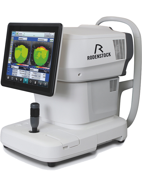
• Power Requirements AC 100V to 240V, 50 / 60 Hz, 80VA to 100VAThe Auto “Refkeratometer” RC-800 is the latest in economical, user-friendly, highly accurate auto “refkeratometry”. The RC-800 provides simple, easy measurement by the operator at an economical cost, without compromising accuracy or reliability. | Rodenstock Alino Multifunction Refractometer
Tune-up your businessExperience 6 analyses in 1 device: Rodenstock Alino Multifunction Refractometer
Are you ready for something that is all-in-one, timesaving, space-saving and suits your business perfectly?Have a look at how powerful ALINO can be.6 in 1 solution01 RefractionHaving a good starting value for subjective refraction is essential. The QUICK REF MODE supports this even for uncooperative patients.02 KeratometryKAI (Kerato-asymmetry index) & KRI (Kerato-regularity index) give the first hint of possible irregularities on the cornea. This supports evaluation of the best vision correction or best-fitting contact lens.03 TonometryThe very gentle air puff created by a new generation of air-flow technology means that patients more readily accept the need for regular check-ups of the IOP measurement.04 PachymetryCorrecting the IOP by measuring the central corneal thickness is essential in interpreting the intraocular pressure.05 TopographyVarious topographic maps support you in carrying out vision screenings, contact lens fittings and in patient education.06 Dry EyeObserve the customers’ eye health and evaluate the long-term tolerability of contact lenses.Take advantage of the combination of tests:Additional servicesTonometry + PachymetryContact lens fitting andtolerance controlKeratometry + Topography + Dry EyeEye healthcareTopography + Tonometry + Pachymetry + Dry EyeSpecifications Rodenstock Alino Multifunction Refractometer
REFRACTIVE POWER MEASUREMENTSpherical refractive power (S) -30.00 D to + 25.00 D (at VD = 12.0 mm)Cylindrical power (C) 0.00 D to ± 12.50 D (at VD = 12.0 mm)Astigmatic axis (A) 0° to 180°KERATOMETRY MEASUREMENTCorneal curvature radius 5.00 mm to 13.00 mmCorneal astigmatic axis 0° to 180°INTRAOCULAR PRESSURE MEASUREMENTMeasurement range 1 mmHg to 60 mmHG (1 hPa to 80 hPa)PACHYMETRY MEASUREMENTMeasurement range 300 μm to 800 μmTOPOGRAPHY MEASUREMENTCorneal curvature radius 5.50 mm to 10.00 mmCorneal astigmatic axis 0° to 180°AUXILIARY FUNCTIONInterpupillary distance Measurement range: 20 mm to 85 mmCorneal diameter and pupil diameter Measurement range: 1.00 mm to 14.00 mmDry eye observation Blinking Analysis, Tear Meniscus height, Hyperemia, Meibomian glandsDATA MANAGEMENTInternal Database Integrated SD CardPrinter Integrated thermal printerData Output Type 3x USB 2.0, 1x LAN, 1x SD Card-slot, 1x WLANExport Format CSV, XML, JPG, PDFDIMENSIONS & ELECTRIC REQUIREMENTSDimensions WDH 312 × 491 × 450 mmWeight Approx. 23 kgVoltage 100 VAC to 240 VACFrequency 50/60 HzPower consumption 110 VA | Essilor Mr Blue 2.0 Lens Edgers and TracerComplete Control of Lens Mounting
- Optimal monitoring of all trajectories, irrespective of finishing materials or shapes
- Absolute axis control thanks to hybrid edging technology: grinding + milling
- Ideally suited for quality finishing even on highest base curves
- Fast and accurate drilling with data acquired in a single operation
- Automatic centering and blocking for simple operation
Unrivalled Ergonomics- Completely integrated functions without external peripheral devices
- Vertical architecture with <Heads-up displays> for optimum comfort
- Storage features ensure all accessories are within reach.
- Intelligent interface adapts to the way you work.
Highest Quality Guaranteed Essilor Mr Blue 2.0 Lens Edgers and TracerPerfect Centering- Binocular 3-D tracing of full-rim frames up to base 9*
- Automatic adjustment of centering data based on the frames’ 30D parameters*
Aesthetic and Robust Mountings- FIT-4-Frame technology with dedicated small wheel*
- Drilling and grooving tools can be tilted up to 30o, making it safe to mount lenses of up to base 10. *Essilor patents and exclusives
Safe Process- Roughing cycle automatically adapts to the curves of lenses up to base 10, avoiding all risk of breakage
- Bevel placement and shelf depth optimized for safe mounting without undue stress of lens
Aesthetically Pleasing and Easy-to-Mount Bevels- The bevel heights are adjustable to perfectly adjust to the thinnest grooves
- The shelf bevel heights are optimized to follow the frame’s shape perfectly
- The step bevel is particularly suited for sport eyeglasses with deep, rectangular or uneven groove profile
Mr Blue 2.0 is on the cutting edge of innovation – serving as a genuine ECP partner since its launch. Now featuring a whole range of new functions and even greater flexibility, Mr Blue is the perfect solution for demanding workshop. | Tomey RC-5000 Auto Refractor Keratometer
Tomey RC-5000 RefractometryNormal Mode:-Fogging is applied automatically for each measurement for more Accurate ResultsQuick Mode:-Fogging is applied as needed for consecutive measurements.IOL/CAT Mode:-This mode is used to measure cataract or pseudophakic eyes.Keratometry:-Short consecutive measurements and time provide reliable data.
-Measurements at 6.00mm (R=8.0mm) as well as at 3.00mm give you enough information for contact lens fitting.
-base curves of contact lenses are stored, categorized and suggested manufacturers are automatically printed.Diameter Measurements of Cornea and Pupil:-Measurement can be done easily by moving the two cursors on the display to the boundary of cornea or pupil. This is useful for deciding the diameter of a contact lens and for other contact lens fitting practice.Power Joystick:-Move the joystick ring up/down and the instrument head moves up/down quickly.
-Move the joystick hand rest to the right/left or backward/forward and the instrument head moves to the right/left or backward/forward quickly.
-Rotate the joystick ring and the instrument head moves up/down slowly.
-Tilt the joystick and the instrument head moves in each direction slowly.Power Chinrest:-The chin rest can be moved by pushing the buttons on the front panel. No need to reach out to the chin rest for manual adjustments.Autorefractor Keratometer Features:Refractometory : Tomey RC-5000 Auto Refractor Keratometer-Sphere 25.00 to +22.00D (VD =12.00mm) Cylinder -10.00 to +10.00D (VD = 12.00mm)
-Axis 0 to 180 Degree
-Minimum Pupil Dia. 2.2mm
-Measurements Time 0.2 Sec.Keratometry :-Range 5.00mm to 11.00mm
-Corneal Refraction 30.68D to 67.50D (n = 1.3375)
-Corneal Astigmatism 0D to 10D (n = 1.3375)
-Axis 0 to 180 Degree
-Measured Area 3.0mm / 6.0 mm
-Measured Time 0.1 Sec PD
-Measurement Yes (50 to 86mm)
-Diameter Measuremnt of Cornea & Pupil 1.0 to 14.0mm
-Chinrest : Power Chin Rest
-Printer : High Speed Thermal Line Printer
-OutputPort RS-232C
-Power Requirements: AC 100V to 240V
-50/60Hz 130VA | Nidek OPD-Scan II ARK 10000 Autorefractor KeratometerThe OPD SCAN II is the only instrument that easily combines Wavefront, Topography and Refraction all into one unit that sits on a table top! This allows the isolation of any optical problem to cornea or crystalline lens making it easy to decide if lensectomy or corneal surgery is the procedure of choice. It is also very mobile if need be!The NIDEK OPD-Scan II provides information on corneal topography, wavefront, autorefraction, keratometry and pupillometry in one unit, utilizing state-of-the-art imaging and analysis technology developed specifically to measure normal to highly aberrated eyes. The system offers a variety of data maps to provide information on the total refractive error, wavefront, corneal shape, internal aberrations and visual quality of the eye, allowing highly accurate and reliable information for optic diagnostics.Our units are carefully refurbished, inspected and serviced by our trained technician prior to shipment. We also offer service on almost all Nidek equipment! ARK-10000 measures the refractive condition and analyzes corneal shape of the patient’s eye. The refractive condition is measured by weak infrared rays, and corneal shape is analyzedbased on projected placido rings onto the cornea. FinancingWe offer easy and flexible financing options on new and used ophthalmic equipment, including, Bausch & Lomb, Visx, Zeiss Humphrey, Topcon, Canon, Alcon and more! Maximize your budget and improve your bottom line with Laser Locators easy equipment financing plan. We have partnered with Stearns Bank to provide financing to our customers. Features Nidek OPD-Scan II ARK 10000 Autorefractor Keratometer
– OPD Map shows point by point aberration of patient’s eye in diopters
– Measurement Selection for Improved Reliability
– Fast Processing Speed
– Improved Accessibility to a Patient eye
– Wide Measurement Range
(Sphere -20.0 to +22.0D and Cylinder 0.0 to ±12.0D)
– Easy Data Maintenance with a Detachable HDD
– Network Capabilities
– Corneal NavigatorCorneal Navigator | Nidek CEM-530 Specular Microscope
The analysis results with graphic and color-coded cell images helps the clinician to rapidly and effectively evaluate the endothelial cell layer.In addition to conventional central and peripheral specular microscopy, the CEM-530 includes a unique function to capture paracentral images.- Paracentral specular microscopy
- Faster measurements and two-second auto analysis
- Comprehensive analysis
- Advanced manual analysis functions
- Easy operation
- Additional features with CEM Viewer for NAVIS-EX
Combination of auto and manual analysesAll three manual analysis methods can be performed on the same image and also on auto-analyzed images. The versatility of combining automated and manual analyses allows analysis of the range of pathology in a comprehensive practice. Features- Multi area specular microscopy
- Enhanced usability and quick analysis
- Advanced manual analysis functions
- Combination of auto and manual analyses
- Additional features with CEM Viewer for NAVIS-EX
Multi area specular microscopyIn addition to conventional central and peripheral specular microscopy, the CEM-530 includes a NIDEK original function that captures paracentral images. The combination of central, paracentral, and peripheral imaging provides a broader, overall view that can be used for detailed morphological and quantitative evaluation of the endothelial layer and individual cells.
Enhanced usability and quick analysis
The 3D auto tracking and auto shot functions result in a user friendly and patient friendly experience.
Data analysis within 2 seconds allows efficient patient flow.
  Detail analysis
Advanced manual analysis functions
Center point
Select the approximate center of a cell. The cells are detected based on the surrounding points. This method is effective for areas where groups of cells are clumped together.
Corner point
Trace the outlines of the cells to be analyzed by selecting the corners of each cell. This method is suitable for detailed identification of the size and dimension of isolated cells.
Pattern select
Select a hexagonal reference pattern that is similar to the cell size and drag it onto the cell to be analyzed. This method is effective for rough identification of the size and dimension of the cells.
Combination of auto and manual analyses All three manual analysis methods can be performed on the same image and on auto-analyzed images. The versatility of combining automated and manual analysis on the same image allows for better clinical interpretation of the diverse range of pathology in a comprehensive practice.
Additional features with CEM Viewer for NAVIS-EX* (optional)
CEM Viewer is software used for viewing and working with CEM-530 data via NAVIS-EX. This function enhances the capability of the CEM-530 with additional features and increases the efficiency of any clinic.
*NAVIS-EX is optional software and is required for use of the CEM Viewer.
Data management and endothelial cell count
Unlimited NAVIS-EX database is available for review on the CEM Viewer. The basic functions of the CEM-530 such as endothelial cell count are available on the CEM Viewer.
Progression follow-up and comparison
Multiple examination data sets are displayed in chronological order for follow-up. Additionally, two data sets are displayed for comparison. Endothelial changes can be monitored over time with this function.
Paracentral display with peripheralThe images and analyses of the paracentral and peripheral areas are displayed providing a comprehensive image of endothelial cells. |




Reviews
There are no reviews yet.