| Description | The flexible laser fiber always allows an optimum view of the preparation site. After each treatment, the laser fiber should be shortened with scissors. | The Waterlase express is the all-tissue laser that will change the way you practice dentistry. Waterlase Express has the power to solve dental problems beyond expectation. The versatile and gentle combination of laser energy and water has the power to relieve dental anxiety at the source, for you, your team, and your patients. The countertop all-tissue laser features a miniaturised laser engine that is 50% smaller than the Waterlase iPlus, offering the benefits of all-tissue laser technology in a way that is compact, simple and easy to use. | The APOLLO DI is our very latest camera. It impresses with its simple handling, small and lightweight camera and outstanding value for money. | The new highly accurate 0.5 mm milling tool reliably delivers first class clinical results. Switch to Extra Fine milling mode for a higher level of detail with occlusal fissures and interdental areas on bridges. | Designed especially for general dentists, the Sirona Orthophos XG3 combines top-tier image quality and exceptional ease-of-use in an affordable, digital panoramic x-ray solution. | With the integrated FaceScanner in GALILEOS you can now record the facial surface parallel to the x-ray image. And that, as opposed to traditional face scanners without the use of lasers, which is therefore gentler on the patients. Thanks to the simultaneous x-ray image and surface depiction, the fully automated overlaying of the data is extremely precise. In addition, texture and coloring in the software allow for a particularly realistic depiction of the soft tissue proportions – an ideal tool for patient consultation! |
| Content | Sirona Sirolaser Soft Tissue Laser
Contents Include:- · 2 laser handpieces
- · 6 tips for laser handpiece
- · 1 finger switch
- · 1 foot control
- · 2 laser fiber (200 μm/320 μm)
- · 1 pair of scissors
- · 3 laser protective goggles for the dentist/assistant/patient
- · 1 transport case/storage box
- · 50 patient information leaflet
These aplications are some of the things that Siro laser can do:1. Applications inendodontic:- · Endodontic treatment is a very delicate and complex therapy fora dentist.
- · The SIROLaser can support him with its programPulpotomy
- · Pulpotomy as adjunct to the root canal therapy
2. Applications in periodontology:· As an adjuvant to conventional treatment, the SIROLaser is usedduring periodontaltreatment for sulcular debridement and gingival incisions of granulation tissue.3. Applications in surgery:Sirona Sirolaser Soft Tissue Laser· The SIROLaser can be used in surgery for optimum treatment ofsubmerged implants and teeth, troughing, labial and lingual frena, for coagulation ofaphthae, herpes and for hemostasis. Other advantages of the SIROLaser in surgery are:- · High precision during surgery
- · Precise tissue removal
- · Minimum damage to the surrounding tissue
- · Minimum bleeding, ensuring a better view of the operating site
- · Minimizing of inflammation
- · Minimum scar formation
- · Lack of postoperative wound pain
There is a new diode dental laser on the market—the SIROLaser by Sirona. Diode lasers are not new to dentistry, but what separates this laser from other dental diode lasers is that the SIROLaser is much more compact than comparable diode lasers, while providing optimum performance in periodontal, surgical, and endodontic procedures. It has a weight of 450 grams, and dimensions of 79 mm x 54mm x 190mm. That is just slightly larger than the remote control for your cable television. This compactness makes for easy transfer from operatory to operatory, and is very space-effective. And because the SIROLaser is so compact, it can be placed just about anywhere the dentist wants it.SIROLaser has a wavelength of 970 nm +/- 10nm, power ranging from 0.5 watts-7 watts, with operating modes of continuous wave (CW), pulsated, and chopped. Continuous wave (CW) is a continuous, uninterrupted laser beam that provides very good power control. Pulsed power output is a very short treatment time (90-300 m) where the laser beam is pulsed (1,000-20,000 W) and has a longer cooling time. Chopped mode is where the laser beam is interrupted at regular intervals (50% ON and 50% OFF); the advantage of this mode is that it provides better thermal control. | 2019 BIOLASE Waterlase Express Dental Laser With Hand PieceRestorative
– Cavity Preparations
– Sub-gingival decay removal
– TroughingSoft-Tissue
– Achieving hemostasis
– Frenectomy techniques
– Biopsies and lesionsPerio
– REPAIR Perio protocol (all steps)
– Closed crown lengthening
– Flap surgeryImplant
– REPAIR Implant protocol (all steps)
– Implant debridement
– Flap surgeryEndo
– Access, cleaning, and shaping
– Pulpotomy, Pulp Cap
– Disinfection of Root Canal
– Removal of Smear LayerSpecifications:
– Wavelength: 2.78 µm (2780 nm)
– Max. Power Output: 4 W Pulse
– Energy: 200 mJ Pulse
– Rep Rate: 5-50 Hz Pulse
– Duration: H: 60 µsec, S: 700 µsec
– Laser Engine: Miniaturized Er,Cr:YSGG laser engine
– Fiber Optic Cable: White, highly flexible, reliable
– User Interface: Applications-based, removable tablet
– Touchscreen Size: 9.7”x 6” (24.64cm x 15.24cm)
– Pre-sets: 95 procedural presets, with simple slider bar to adjust settings within each procedure
– Additional Content: Procedure animations, case studies, best practices
– WiFi-enabled for BIOLASE Connect: Yes
– Weight: 27 Lbs (12.247 kg)The Waterlase express is the all-tissue laser that will change the way you practice dentistry. Waterlase Express has the power to solve dental problems beyond expectation. The versatile and gentle combination of laser energy and water has the power to relieve dental anxiety at the source, for you, your team, and your patients. The countertop all-tissue laser features a miniaturised laser engine that is 50% smaller than the Waterlase iPlus, offering the benefits of all-tissue laser technology in a way that is compact, simple and easy to use.2019 BIOLASE Waterlase Express Dental LaserWaterlase Express is the only laser to feature an in-depth, step-by-step, 4K HD animation for every step of every procedure. No matter your level of expertise, from beginner to advanced user, the Waterlase Express animation library is a dazzling, informative training resource.– 95 clinical presets for soft tissue, hard tissue and bone.
– Step-by-step procedure animations.
– Removable tablet with easy-to-use touchscreen interface.
– Loaded system menu features on-board reference material.
– H MODE – 60 microseconds – a cooler mode for cutting enamel and all other applications.
– S MODE – 700 microseconds – a hotter mode for coagulation.
– Stay up-to-date and connect with fellow users, clinical mentors and online training directly from the unit.
– Customer care access for direct help when needed.
– Sturdy handle for easy transport makes the Waterlase express conveniently portable.
– Wireless footswitch for easy integration.
– Light and flexible SureFire trunk fibre ensures minimal resistance and treatment fatigue.
– Over 2 hours of 4K-Ultra-HD learning content | Sirona Apollo DI Intra Oral Scanner
APOLLO DI:
The Sirona Apollo DI Intra Oral Scanner features easy handling, precise imaging, and the proven Sirona Connect workflow. The impression system includes an imaging unit, APOLLO Connect software, and the APOLLO DI intraoral
camera,with which users can make digital impressions of the clinical situation in a seamless workflow. To do this, a moisture-insensitive high-contrast spray is sprayed on the teeth very finely. Fine particles in the spray ensure high contrast and thus very precise images.The most cost-effective start to digital impression-taking.
APOLLO DI
APOLLO DI, the specially developed intraoral scanner for cost-efficient digital impressions.
Easy handling thanks to multitouch control
Small and lightweight camera
Export of scan-data in the laboratory
No follow-up costs
OPEN APOLLO DI*:
Export of digital impression data (captured with the APOLLO DI in the practice and received via the Sirona Connect Portal) in an open ST L format for processing in other CAD/CAM systems.Designed to provide an economical entry point into CAD/CAM dentistry, Sirona’s APOLLO DI digital impression system includes an imaging unit, APOLLO Connect software and the APOLLO DI intraoral camera.The system provides precise imaging with the use of a contrast spray, and digital impression data can be sent to dental labs via the Sirona Connect system where it can be turned into a digital restoration design in Sirona’s inLab software or compatible 3rd party software applications, according to a press release.The new APOLLO DI is a complement to the company’s existing CEREC Bluecam and CEREC Omnicam options which each use a different imaging technology, but send the digital impressions through the same workflow. APOLLO DI is designed for practices looking to take digital impressions but continue to work with a lab, while the Bluecam and Onmicam can each be purchased as stand-alone digital impression systems, or with a mill for complete chairside CAD/CAM dentistry. | DENTSPLY SIRONA CEREC PRIMEMILLSpeedEngineered for speed and precision, the new CEREC Primemill inherits the proven strength of its predecessor and raises its powerful performance to entirely new heights.You can now mill zirconia restorations in Super Fast milling mode in around 5 minutes, cutting processing times by more than half. The improved grinding mode for high quality glass ceramics also grinds the material faster than ever before. The result? Satisfied patients and high productivity.QualityDENTSPLY SIRONA CEREC PRIMEMILL A combination of new electronics, software, motors and mechanical components contributes to finer resolution and enhanced dynamics. This results in outstanding restoration margins and surface details – for extremely precise results.The new highly accurate 0.5 mm milling tool reliably delivers first class clinical results. Switch to Extra Fine milling mode for a higher level of detail with occlusal fissures and interdental areas on bridges.VersatilityDENTSPLY SIRONA CEREC PRIMEMILL Welcome to a world where you‘re spoiled for choice! The new CEREC Primemill combines wet and dry milling and wet grinding. No matter the indication, with CEREC Primemill you can select from a broad range of material options from validated partners providing the flexibility and predictability you need to offer patients the best possible treatment.Choose from the full spectrum of machining options, including dry and wet milled zirconia, or wet grinding of glass and hybrid ceramics.ConvenienceDENTSPLY SIRONA CEREC PRIMEMILL The new 7” touch interface with user guidance guides operators step-by-step through the workflow, key maintenance procedures and other routine tasks, so you can delegate milling with complete peace-of-mind. The integrated block scanner is a time-saving new feature that automatically scans material blocks with compatible data matrix codes and records the information, including type, size, color and zirconia enlargement factor. All CEREC Primemill tools are fitted with a color-coded RFID chip that is read by the RFID tool reader, feeding into the tool status display, alerting the user if a tool needs replacing. This ensures all tools are performing at optimal levels. A LED light strip indicates unit status and milling and grinding progress.The all-new CERECThe latest generation of our CEREC system sets new standards so you can offer your patients an unmatched combination of single-visit dentistry and excellent quality.A unique solutionThe CEREC system comprises world-class components that interact perfectly ensuring seamless workflows. With CEREC Primescan, CEREC Software 5 and CEREC Primemill, digital chairside dentistry is faster, easier and more reliable than ever before.Outstanding resultsAfter 35 years of continuous optimization, CEREC gives you the options you need to treat multiple indications with the confidence that comes from outstanding results.
Designed to stand out – and fit inThe new CEREC Primemill does not just communicate seamlessly with other CEREC devices, but its sleek design is also the perfect match – and the ideal look for a progressive dental practice. | SIRONA XG3 Digital Panoramic X-Ray
The ORTHOPHOS XG 3 is designed for the general practice dentist who would like to work with a basic X-ray system. The XG 3 offers you an outstanding price-performance ratio and provides the following benefits:- High image quality
- Extremely simple operation
- Solid workmanship
Accurate positioningThe ORTHOPHOS XG 3 uses proven, logical Sirona principles for exceptional imaging:- Exact, immediate positioning of the anterior teeth in the focal layer
- Effective, comfortable patient stabilisation with forehead and temple supports to prevent motion blurring
Proven TechnologyThe advanced technology from the XG product family enables the ORTHOPHOS XG 3 to produce high quality diagnostic images.
The benefits include:- Specific focal layers and orbital paths for anatomically optimised exposures
- Very detailed display of the anterior tooth region through automatic kV adaption as X-ray beam passes through spinal region
- 16 bit image acquisition technology provides more gray scales
- SIDEXIS XG imaging software with useful analysis tools
Features:SIRONA XG3 Digital Panoramic X-Ray- High-quality, digital panoramic and TMJ imaging power with advanced patient communication tools
- Outstanding price-performance ratio for seamless transition from film to direct digital imaging
- Easy operation with the Multipad; effective, three-point patient stabilization for sharp images
- Direct network capability for exceptional connectivity
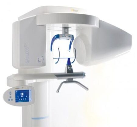
SIRONA ORTHOPHOS XG 3The horizontally and vertically reduced pediatric panoramic program offers outstanding image quality with the lowest dose. Ideal for practices that treat a lot of children. The unit’s easy operation and seamless integration into your practice’s daily routines will save you valuable time. The high image quality and proven Sidexis software guarantee that you will come to a safe diagnosis quickly and easily.Quality from the very startHigh quality. Durable. Safe: the Orthophos XG 3 X-ray unit is the perfect basic equipment for general dentists. The unit’s easy operation and seamless integration into your practice’s daily routines will save you valuable time. The high image quality and proven Sidexis software guarantees that you will reach a safe diagnosis quickly and easily.The positioning can be very easily reproduced since image parameters such as the bite block height are saved together with the image data. A bitewing program is available, which helps in special cases and patient situations where intraoral images cannot be used and a shorter panoramic program with a reduced dose, which is suitable for treating children, is also available. | Sirona GALILEOS Comfort/Compact| GALILEOS Comfort | GALILEOS Compact | | Oral and maxillofacial surgeons
Dental clinics
Private clinics
Orthodontists | General practioners
Dental implant practices
Oral surgeons |  |  | | Comparison Table of Galileos Comfort/ Compact & Orthophos XG 3D X-Ray systems | | Technical overview | GALILEOS Comfort | GALILEOS Compact | ORTHOPHOS
XG 3D | | Field of view | (15 x 15 x 15) cm³ | (12 x 15 x 15) cm³ | 8 cm x 8 cm (Ø x Höhe) | | 3D Resolution (isotropic voxel size) | 0.3/0.15 mm | 0.3 mm | 0,2 mm; 0,1 mm (available at the end of 201) | | Scan time / exposure time | 14 s / 2 – 6 s | 14 s / 2 – 6 s | 2–5 s | | Reconstruction time | 2.5 – 4.5 min | 4.5 min | 4,5 min | | Patient position | standing / seated | standing / seated | Standing/seated, chin rest/bite block, occlusal bite block for automatic patient positioning with 2D PAN images | | X-ray generator | | kV | 85 | 85 | 60–90 | | Effective dosage | 29 µSv / 68 µSv **
(21 mAs, 85 kV) | < 29 µSv / 68 µSv **
(21 mAs, 85 kV) | 43–175 μSv (Schulze)
(Standard: 100 μSv) | | Space requirements (minimum) | 1,6 x 1,6 x 2,25 m (d x w x h) | 1,6 x 1,6 x 2,25 m(d x w x h) | 1,5 m x 1,1 m x 2,25 m (Pan ), | | Space requirements (recommended) | >1,8 x 1,8 x 2,5 m (d x w x h) | 1,8 x 1,8 x 2,5 m (d x w x h) | 1,7 m x 1,3 m x 2,5 m (Pan ) | | User Interface | Full color touchscreen | Multipad | EasyPad | | Patient fixation | Occlusal bite block, forehead | Occlusal bite bock, forehead | | | Positioner | support, chin rest, head positioner *** | support, chin rest | | | Upgradable to Comfort version | |  | | | Optional floor stand | | | | | Wheelchair accessible | | | | | Remote control | | | | | Programs | | 150 µm resolution for close-up reconstruction | | | | | 300 µm resolution for standard volumes | | | | | High contrast mode for optimized hard tissue display | | | | | Views | PAN with 3D slice navigation | PAN with 3D slice navigation | Tiltable 2D slices, custom | | TSA, axial, sagittal, coronal, CEPH lat., CEPH pa/ap | TSA, axial, sagittal, coronal | 3D slicing, TSA, LSA, axial, sagittal, coronal | | 3D model, 1-click OP reports | 3D model, 1-click OP reports | 3D model, implant-oriented view, 1-Click OP reports |
With the integrated FaceScanner in GALILEOS you can now record the facial surface parallel to the x-ray image. And that, as opposed to traditional face scanners without the use of lasers, which is therefore gentler on the patients. Thanks to the simultaneous x-ray image and surface depiction, the fully automated overlaying of the data is extremely precise. In addition, texture and coloring in the software allow for a particularly realistic depiction of the soft tissue proportions – an ideal tool for patient consultation! |
|


Reviews
There are no reviews yet.