| Description | RIGHTON RETINOMAX-5 AUTO REFRACTOMETER / KERATOMETER
Automatic AxisCompensation and extended measurement range
Not only does the device let examiner know the cylinder axis angle, but it can
also be automatically adjusted if it is not level. | The NIDEK OPD-Scan II provides information on corneal topography, wavefront, autorefraction, keratometry and pupillometry in one unit, utilizing state-of-the-art imaging and analysis technology developed specifically to measure normal to highly aberrated eyes. The system offers a variety of data maps to provide information on the total refractive error, wavefront, corneal shape, internal aberrations and visual quality of the eye, allowing highly accurate and reliable information for optic diagnostics. | Experience 6 analyses in 1 device: refraction, keratometry, topography, tonometry, pachymetry and dry eye analysis. | The Tomey Refractometer Keratometer and Topographer only in 1 unit senior citizens and children can also get the inspection with out any trouble because they dont have to move the seat for three measurements. | Bison Dual ACCENTO is the unique device combination with Alexandrite laser (755nm) and Nd:YAG laser (1064nm Long pulse)
The combination of two different wavelengths makes DUAL ACCENTO a highly effective system for hair removal and pigmented lesions treatment. The 755nm penetrates deep into the skin and perfectly targets hair the follicle and absorbs melanin. | The Sequoia system’s advanced technologies are designed to make image acquisition easier, reduce exam time and increase overall efficiency. Along with many other included features, the Sequoia system offers Native, a patient specific imaging which adapts both phase and amplitude information of each unique patient’s properties which delivers a consistently better image across a broad range of patients. |
| Content | Righton Retinomax 5 Auto Refractor
The Retinomax Series, the world’s leading handheld autorefractor and autorefractor/keratometer, delivers wide measurement range on par with table type refr, now boasts greater mobility, stability and accuracy. The Retinomax series is widely used as a global standard device for pediatric, screening, medical care in remote areas, disaster area and wide range of ophthalmic scene.FEATURES- View Finder – With view finder observation, no limit of operator age. Diopter adjustable range is +/-8D the same as Retinomax3. View finder arm angle is changeable of 0-135 degrees, making measurement easy regardless of patient’s position or posture. Adjustable diopter +/- 8D.
- Wider Eye Piece – Eye piece relief is 48mm from 32.2mm so that enable to look inside image while even looking at outside. No need to stick eye to the eyepiece all the time.
- Improved battery life – At 180 minutes, the battery capacity is now twice that of conventional models
- Automatic Axis Compensation and Extended Measurement Range – Not only does the device let examiner know the cylinder axis angle, but it can also be automatically adjusted if it is not level
- New Child Mode – While the measurement is being taken, a melody plays continually to keep children’s attention. A constantly changing color display, both on the outside and inside target the device, also keeps children involved during the process.
- Light weight and easy-to-hold grip
View Finder Righton Retinomax 5 Auto RefractorWith view finder observation, no limit of operator age. Diopter adjustable rangeis +/-8D the same as Retinomax3. View finder arm angle is changeable of 0-135degrees, making measurement easy regardless of patient’s position or posture.Adjustable diopter +/- 8D.Improved battery lifeAt 180 minutes, the battery capacity is now twice that of conventional modelsWider Eye PieceEye piece relief is 48mm from 32.2mm so that enable to
look inside image while even looking at outside. No need
to stick eye to the eyepiece all the time.Focus AssistLets you know the exact focusing point with
the following color-coding system.New Child ModeWhile the measurement is being taken, a
melody plays continually to keep children’s
attention. A constantly changing color
display, both on the outside and inside of
the device, also keeps children involved
during the process.Automatic AxisCompensation and extended measurement range
Not only does the device let examiner know the cylinder axis angle, but it can
also be automatically adjusted if it is not level. | Nidek OPD-Scan II ARK 10000 Autorefractor KeratometerThe OPD SCAN II is the only instrument that easily combines Wavefront, Topography and Refraction all into one unit that sits on a table top! This allows the isolation of any optical problem to cornea or crystalline lens making it easy to decide if lensectomy or corneal surgery is the procedure of choice. It is also very mobile if need be!The NIDEK OPD-Scan II provides information on corneal topography, wavefront, autorefraction, keratometry and pupillometry in one unit, utilizing state-of-the-art imaging and analysis technology developed specifically to measure normal to highly aberrated eyes. The system offers a variety of data maps to provide information on the total refractive error, wavefront, corneal shape, internal aberrations and visual quality of the eye, allowing highly accurate and reliable information for optic diagnostics.Our units are carefully refurbished, inspected and serviced by our trained technician prior to shipment. We also offer service on almost all Nidek equipment! ARK-10000 measures the refractive condition and analyzes corneal shape of the patient’s eye. The refractive condition is measured by weak infrared rays, and corneal shape is analyzedbased on projected placido rings onto the cornea. FinancingWe offer easy and flexible financing options on new and used ophthalmic equipment, including, Bausch & Lomb, Visx, Zeiss Humphrey, Topcon, Canon, Alcon and more! Maximize your budget and improve your bottom line with Laser Locators easy equipment financing plan. We have partnered with Stearns Bank to provide financing to our customers. Features Nidek OPD-Scan II ARK 10000 Autorefractor Keratometer
– OPD Map shows point by point aberration of patient’s eye in diopters
– Measurement Selection for Improved Reliability
– Fast Processing Speed
– Improved Accessibility to a Patient eye
– Wide Measurement Range
(Sphere -20.0 to +22.0D and Cylinder 0.0 to ±12.0D)
– Easy Data Maintenance with a Detachable HDD
– Network Capabilities
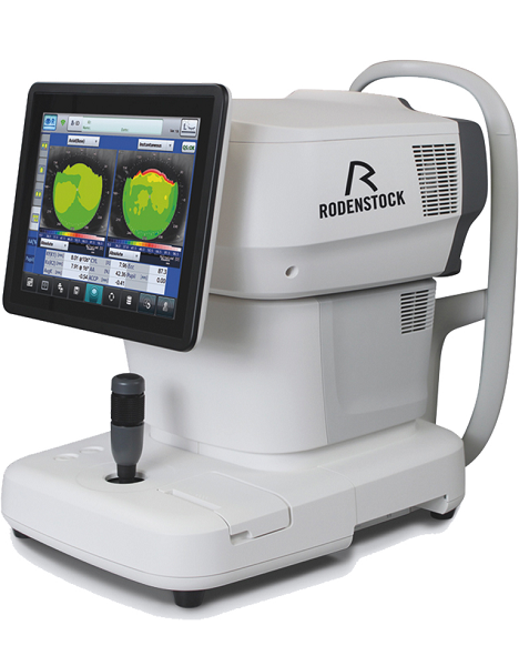
– Corneal NavigatorCorneal Navigator | Rodenstock Alino Multifunction Refractometer
Tune-up your businessExperience 6 analyses in 1 device: Rodenstock Alino Multifunction Refractometer
Are you ready for something that is all-in-one, timesaving, space-saving and suits your business perfectly?Have a look at how powerful ALINO can be.6 in 1 solution01 RefractionHaving a good starting value for subjective refraction is essential. The QUICK REF MODE supports this even for uncooperative patients.02 KeratometryKAI (Kerato-asymmetry index) & KRI (Kerato-regularity index) give the first hint of possible irregularities on the cornea. This supports evaluation of the best vision correction or best-fitting contact lens.03 TonometryThe very gentle air puff created by a new generation of air-flow technology means that patients more readily accept the need for regular check-ups of the IOP measurement.04 PachymetryCorrecting the IOP by measuring the central corneal thickness is essential in interpreting the intraocular pressure.05 TopographyVarious topographic maps support you in carrying out vision screenings, contact lens fittings and in patient education.06 Dry EyeObserve the customers’ eye health and evaluate the long-term tolerability of contact lenses.Take advantage of the combination of tests:Additional servicesTonometry + PachymetryContact lens fitting andtolerance controlKeratometry + Topography + Dry EyeEye healthcareTopography + Tonometry + Pachymetry + Dry EyeSpecifications Rodenstock Alino Multifunction Refractometer
REFRACTIVE POWER MEASUREMENTSpherical refractive power (S) -30.00 D to + 25.00 D (at VD = 12.0 mm)Cylindrical power (C) 0.00 D to ± 12.50 D (at VD = 12.0 mm)Astigmatic axis (A) 0° to 180°KERATOMETRY MEASUREMENTCorneal curvature radius 5.00 mm to 13.00 mmCorneal astigmatic axis 0° to 180°INTRAOCULAR PRESSURE MEASUREMENTMeasurement range 1 mmHg to 60 mmHG (1 hPa to 80 hPa)PACHYMETRY MEASUREMENTMeasurement range 300 μm to 800 μmTOPOGRAPHY MEASUREMENTCorneal curvature radius 5.50 mm to 10.00 mmCorneal astigmatic axis 0° to 180°AUXILIARY FUNCTIONInterpupillary distance Measurement range: 20 mm to 85 mmCorneal diameter and pupil diameter Measurement range: 1.00 mm to 14.00 mmDry eye observation Blinking Analysis, Tear Meniscus height, Hyperemia, Meibomian glandsDATA MANAGEMENTInternal Database Integrated SD CardPrinter Integrated thermal printerData Output Type 3x USB 2.0, 1x LAN, 1x SD Card-slot, 1x WLANExport Format CSV, XML, JPG, PDFDIMENSIONS & ELECTRIC REQUIREMENTSDimensions WDH 312 × 491 × 450 mmWeight Approx. 23 kgVoltage 100 VAC to 240 VACFrequency 50/60 HzPower consumption 110 VA | Tomey RT-7000 Autorefractor Keratometer Topographer
-Three Functions in One Instrument-Easy & Speedy Touch Screen Alignment-6.4 inches color TFT LCD-Dual CCD technology for Refractometry-IOL/ Cataract Mode-New incites for Keratometry KAI, KRI-Auto Alignment / Auto Shot of TopographyCorneal Topography– Auto Alignment / Auto ShotAny one can easily capture measurement with Auto Alignment and Auto Shot. The Measurement variation is significantly reduced no matter what the skill level is of the operator.CL Fitting Simulation-A pseudo-fluorescent pattern of the contact lens selected from the built-in data base can be confirmed and the fitting can be stimulated before the patient wears the trial lens.Corneal Eccentricity Index-The index of CEI (Corneal Eccentricity Index) indicates the ratio of flattering in percentage from the central cornea to the periphery.Three Function in One Instrument– Refractometer Keratometer and Topographer only in 1 unit senior citizens and children can also get the inspection with out any trouble because they don’t have to move the seat for three measurements. Switch from Ref-Kerato Mode to Corneal Topography Mode with only ONE TOUCH. The light come appears with only one button from the measuring head and the Ref-Keratometer is transformed to the topographer.Refractometry– Dual CCD technology for RefractometryTwo CCD cameras are used to capture images for observation and for measurement while providing highly accurate measurement data.Keratometory– New Indices for KeratometryThe Corneal Irregular astigmatism display function is installed to expand the possibility of Keratometer. This is the new function to measure the level of corneal irregular astigmatism which was difficult in the past.SPECIFICATIONSMeasurmentRangesRefraction MeasuremntSphere -25.00D to ±22.00DIVD=12.0mmCylinder 0D to ± 10.00DIVD=12.0mmAxis 0 to 180Corneal Curvature Measurement 5.00mm to 11.00mmMeasurement Zone 3mm R:8.00mmCorneal MappingDisplay Range 9 to 100 D-Normal Mode1.0 to 8.0mm (R:8.00mm)-Special Mode0.9 to 7.0 mm (R:8.00mm)Measurement TimeREF 0.2 SecondsKRT 0.1 SecondsMinimum Pupil Diameter 2.2mmMinimum Measurment Step Refraction MeasurementSphere 0.01DCylinder 0.01DAxis 1Corneal Curvature Measurement 0.01mmPD (Pupil Distance) 50mm to 86mmRecording Built-In Thermal PrinterVertex Distance 0mm, 12.0mm, 13.5mm, 14.0mm, 15.5mm, 16.0mmExterior Output (AN port / 4USB ports)Dimensions Tomey RT-7000 Autorefractor Keratometer TopographerHigh 466mmm or 18 InchesWidth 307mm or 12 InchesDepth 490mm or 19 InchesWeight Approx 20,000Kg / 44.4 PoundsDisplay 6.4 Inches color liquid crystalPower SupplyVoltage AC 100 to 240VFrequency 50/60 HzConsumption Power 120VA to 150VATemperature +10C to + 40CDisplay 30% to 75% | Bison Dual ACCENTO Nd YAG Laser
Bison Dual ACCENTOBison Dual ACCENTO is the unique device combination with Alexandrite laser (755nm) and Nd:YAG laser (1064nm Long pulse)
The combination of two different wavelengths makes DUAL ACCENTO a highly effective system for hair removal and pigmented lesions treatment. The 755nm penetrates deep into the skin and perfectly targets hair the follicle and absorbs melanin.The Dual Accento combined laser system is indicated for the following: 755 nm Long Pulsed Alexandrite Laser Temporary hair reduction. Stable long-term or permanent reduction through targeting of melanin in hair follicles. Treatment of bening pigmented lesions. Treatment of Wrinkles.The photocoagulation of dermatological vascular lesions (such as port of wine stains, hemangiomas, telangiectasis) 1064nm Long pulsed Nd:YAG laser. The removal of un-wanted hair reduction, for stable long term or permanent hair reduction and for treatment of PFB to all skin types Fitzpatrick I-VI including tanned skin.Pigmented lesions to reduce lesion size and patients with lesions. Reduction of red pigmentation in hypertrophic and keloid scars where vascularity is integral part of scar, treatment of wrinkle. Permanent hair reduction is defined as the long term, stable reduction in the number of hairs re- growing when measured at 6,9, and 12 months after the completion of treatment regime.The Dual Accento performs with high efficacy of the pigmentation problems without damaging tissue structures adjacent to the pigment. The wavelength of 1064nm Nd: YAG long pulse rejuvenates the skin by stimulating the collagen layer of the dermis and effectively treats vascular lesions and is appropriate for all skin types when used for hair removal.Bison Dual ACCENTO 1064nm NdYAG Laser With 755nm Alexandrite Laser
Bison Dual ACCENTO 1064nm NdYAG Laser with 755nm Alexandrite Laser is the unique device. Combining 755nm Long pulsed Alexandrite and 1064nm Long pulsed NdYAG Alexandrite 755nm long pulsed wavelength. Due to the high melanin absorption is effective for hair removal and pigmented lesions treatment. NdYAG 1064nm long pulsed wavelength rejuvenates skin by stimulating the dermis layer, effectively treating vascular lesions. | Siemens Acuson Sequoia 512 LCD Ultrasound Machine18″ Flat panel LCD Monitor–Progressive Media & Display II Package, Cardiac/OB/Vascular Calc. Package, Signature II Option, AUX, CPS Cardiology, CPS General Cadene Trigg Burst, Calc Data to MO, Card Cadence II Contrast Quant, Cornoary Flow reserve, Dicome Store/Print, DTI (Doppler Tissue Imaging), High HR ECG, MIS/MIF, Modality Worklist Native TEQ, Native TEQ, NTHI (Native Tissue Harmonics), Spectral TEQ, TEQ (Tissue Equalization), VVIFeatures- Shared Service
- Native Patient Specific Imaging
- Four Sight TEE view
- Axius quantitative strain rate imaging
- Cadence contrast pulse sequencing technology
- Native Tissue Harmonic Imaging
- Native TEQ ultrasound technology
- Color doppler, Color angio and b-color
- PW/CW Doppler
- M-Mode
- Folding Flat Panel Display
- DICOM capabilities
Applications- Abdominal
- Breast
- OB/GYN
- Vascular
- Pediatrics
- Cardiology
- Musculoskeletal
- Neonatal
Acuson Sequoia 512 Features:- Imaging: Gray-scale 2-D, M-mode, SST Color Doppler, Solo Spectral Doppler, 3-D Surface Rendering, Native Tissue Harmonic Imaging, Auto Doppler, M-Mode, PW Doppler, Color Doppler, Color Power Doppler
- 19" color CRT monitor
- Native tissue harmonic imaging (NTHI)
- DIMAQ: image management
- Advanced vascular analysis
- Advanced spatial compounding
- Native patient specific imaging
- Mitral valve assessment
- CPS contrast pulse sequencing technology
- Clarifying vascular enhancement technology
- High resolution color flow
- Vascular, and General Imaging calculation packages
- Coherent beamformer technology/spatial compounding
- Space/time resolution control
- Cine and zoom/RES
Compatible Ultrasound Probes and Transducers:- Acuson V5Ms TEE Probe
- Acuson 4C1 Curved Array Probe
- Acuson 6C2 Curved Array Probe
- Acuson 8C4 Curved Array Probe
- Acuson 4V1 Vector Array Probe
- Acuson EV-8C4 Endocavitary Probe
- Acuson EC-10C5 Endocavitary Probe
- Acuson 6L3 Linear Array Probe
- Acuson 8L5T Intraoperative Probe
- Acuson 8L5 Linear Array Probe
- Acuson 9L4 Linear Array Probe
- Acuson 15L8 Linear Array Probe
- Acuson 17L5 Linear Array Probe
The Sequoia 512 with LCD ultrasound machine uniquely minimizes automatic image optimization time by up to 80% to make way for an easier and speedier ultrasound test time as well as allow for a more confident diagnosis each time. As a patient-specific imaging ultrasound, it has a unique matched response capacity for accurate image quality, sharp clinical specificity, and optimized diagnostic outcomes that makes infinitesimal pathologies and other anatomical detail in the most difficult-to-image patients becomes more vivid and visible. |




Reviews
There are no reviews yet.