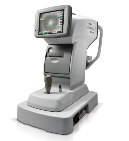
| Description | The Retinomax Series, the world’s leading handheld autorefractor and autorefractor/keratometer, delivers wide measurement range on par with table type refr, now boasts greater mobility, stability and accuracy. The Retinomax series is widely used as a global standard device for pediatric, screening, medical care in remote areas, disaster area and wide range of ophthalmic scene. Also known as: Retinomax K+, KPlus5 | The ZEISS Cirrus HD-OCT 4000 enable examination of the posterior and anterior of the eye at an extremely fine spatial scale, without surgical biopsy or even any contact with the eye. The Cirrus HD-OCT builds on and refines the retinal imaging technology first introduced with the ZEISS Stratus OCT™. HD-OCT stands for “high-definition optical coherence tomography.”Employing the advanced imaging technology of spectral domain optical coherence tomography, Cirrus HD-OCT acquires OCT data about 70 times faster (27,000 vs. 400 A-scans per second) and with better resolution (5 μm vs. ~10 μm axial resolution in tissue), compared to first-generation OCT technology. Cirrus acquires whole cubes of OCT image data, composed of hundreds of line scans, in about the same time as Stratus acquires a six-line scan. You can view these data cubes in three planes, or through three dimensions, giving you access to an extensive amount of retinal image data in one scan. | Laser-Like Precision
- Tactile control with minimal tissue drag
- Surface only thermal effect
- Predictable, char-free layer-by-layer dissection with optimal visualization of tissue planes
- Safe to use near nerves, vessels, and all delicate organs, including the heart | The Easyfield perimeter is operated via an external computer (notebook or PC). You can enjoy the full freedom of networking the examination data with the familiar user interface of the OCULUS programs. The use of translucent eye shields allows you to conduct measurements without the usual eye patch, thus saving you valuable time of preparing examinations. | Complete Control of Lens Mounting
- Optimal monitoring of all trajectories, irrespective of finishing materials or shapes
- Absolute axis control thanks to hybrid edging technology: grinding + milling
- Ideally suited for quality finishing even on highest base curves
- Fast and accurate drilling with data acquired in a single operation
- Automatic centering and blocking for simple operation
| The iCare IC200 tonometer is designed for professional use in the surgical operation room, the emergency room and also the clinic. It uses the same patented rebound measuring principle found in other iCare tonometers. There is no need for drops, air or specialised skills for its use. The IC200 gives you positional freedom with a new premium design and user interface. It allows measuring whether the patient is sitting, standing, half-sitting or in the supine or lateral recumbent position. |
| Content | Righton BON Retinomax K-Plus 5 (Autoref Keratometer)Retinomax Series, the world’s leading handheld Ref and Refract Keratometer, delivers wide measurement range on par with table type Ref, with greater mobility, stability and accuracy.The Retinomax series is widely used as a global standard device for pediatric, screening, medical care in remote areas, disaster area and wide range of ophthalmic scene.Current informatical society, eye fatigue, one of the typical modern diseases cause by eye accommodation issue is now easily discover by ACOMOREF2 series during screening basis. Accommodative Micro Fluctuation result will show in graph for easy reading, plus opacity detection function makes ACOMOREF2 the world first multi-functional Refractometer.Features:Righton BON Retinomax K-Plus 5 (Autoref Keratometer)View Finder – With view finder observation, no limit of operator age. Diopter adjustable range is +/-8D the same as Retinomax3. View finder arm angle is changeable of 0-135 degrees, making measurement easy regardless of patient’s position or posture. Adjustable diopter +/- 8D.Wider Eye Piece – Eye piece relief is 48mm from 32.2mm so that enable to look inside image while even looking at outside. No need to stick eye to the eyepiece all the time.Improved battery life – At 180 minutes, the battery capacity is now twice that of conventional modelsAutomatic Axis Compensation and Extended Measurement Range – Not only does the device let examiner know the cylinder axis angle, but it can also be automatically adjusted if it is not levelNew Child Mode – While the measurement is being taken, a melody plays continually to keep children’s attention. A constantly changing color display, both on the outside and inside target the device, also keeps children involved during the process.Even lighter and featuring an easy-to-hold gripRetinomax3 969g → Retinomax Screeen 960g -9gRetinomax K-plus3 999g → Retinomax K+Screeen 970g -29gREFRetinomax 5, Retinomax K+ Screeen −20D〜+23D | ZEISS Cirrus HD OCT 4000 Quad Core OCT
The ZEISS Cirrus HD-OCT 4000 (Cirrus HD-OCT or Cirrus) enable examination of the posterior and anterior of the eye at an extremely fine spatial scale, without surgical biopsy or even any contact with the eye. The Cirrus HD-OCT builds on and refines the retinal imaging technology first introduced with the ZEISS Stratus OCT™. HD-OCT stands for “high-definition optical coherence tomography.”Employing the advanced imaging technology of spectral domain optical coherence tomography, Cirrus HD-OCT acquires OCT data about 70 times faster (27,000 vs. 400 A-scans per second) and with better resolution (5 μm vs. ~10 μm axial resolution in tissue), compared to first-generation OCT technology. Cirrus acquires whole cubes of OCT image data, composed of hundreds of line scans, in about the same time as Stratus acquires a six-line scan. You can view these data cubes in three planes, or through three dimensions, giving you access to an extensive amount of retinal image data in one scan.Intended UseThe Cirrus HD-OCT with Retinal Nerve Fiber Layer (RNFL), Macular, Optic Nerve Head, and Ganglion Cell Normative Databases is indicated for in-vivo viewing, axial cross-sectional, and three-dimensional imaging and measurement of anterior and posterior ocular structures.Indications for UseThe Cirrus HD-OCT is a non-contact, high resolution tomographic and biomicroscopic imaging device. It is indicated for in-vivo viewing, axial cross-sectional, and three-dimensional imaging and measurement of anterior and posterior ocular structures, including cornea, retina, retinal nerve fiber layer, ganglion cell plus inner plexiform layer, macula, and optic nerve head. The Cirrus normative databases are quantitative tools for the comparison of retinal nerve fiber layer thickness, macular thickness, ganglion cell plus inner plexiform layer thickness, and optic nerve head measurements to a database of normal subjects. The Cirrus HD-OCT is intended for use as a diagnostic device to aid in the detection and management of ocular diseases including, but not limited to, macular holes, cystoid macular edema, diabetic retinopathy, age-related macular degeneration, and glaucoma.Note: The Cirrus HD-OCT is not intended to be used as the sole diagnostic for disease.Essential Performance ZEISS Cirrus HD OCT 4000 Quad Core OCTThe Essential Performance of the instrument is to provide accurate measurements of anterior and posterior ocular structure.Patient PopulationThe Cirrus HD-OCT may be used on all adults in need of diagnostic evaluation of the eye. This includes (but is not limited to) patients with the following disabilities or challenges:• Wheelchair user
• Very low or not measurable visual acuity
• Fixation problems
• Postural problems
• Deafness
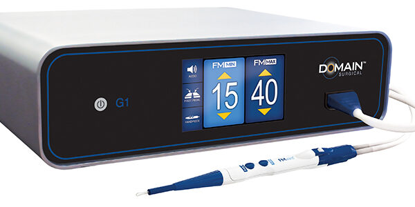
• Large body, but not those above 99th percentile based on anthropomorphic dataThere is a general requirement that the patient be able to sit upright and be able to place their face in the chin and forehead rest of the instrument (with or without supplemental human or mechanical support).Cirrus HD-OCT is designed for in-vivo viewing, axial cross-sectional, and three-dimensional imaging and measurement of anterior and posterior ocular structures. | Domain Surgical FMX and FMwand Ferromagnetic Surgical SystemThe FMX Generator intelligently delivers electrical energy to the surgical instruments. The advanced generator features a patented software control algorithm that continuously monitors and adjusts the delivery of energy, ensuring that the optimal amount of heat is delivered to the tissue at all times. The large touchscreen display provides the entire surgical team immediate feedback on power setting levels and changing settings is as easy as touching the display. The FMX Generator has earned the Type CF (“Cardiac Floating”) rating.Laser-Like Precision
- Tactile control with minimal tissue drag
- Surface only thermal effect
- Predictable, char-free layer-by-layer dissection with optimal visualization of tissue planes
- Safe to use near nerves, vessels, and all delicate organs, including the heartMinimal Thermal Injury
- As little as 1/10th the thermal injury of monopolar electrosurgery
- As few as 80 microns (0.08 mm) of thermal spread
- Clear margins for reliable pathology specimens
- Surgeons note less unintended damage to tissue, leading to reduced use of blood products during surgery, and less post-operative edema and drainageElectrical Silence
- No electrical current passes through tissue
- No grounding pad
- No spark, arcing, or stray currentPrepare the FMwand by following these simple steps:
- Plug the FMX Power Module into the front panel of the FMX Generator
- Snap the power module into the back of the FMwand Handpiece
- Set the power settings on the generator and the FMwand is readyFMX Generator Technical Specifications Domain Surgical FMX and FMwand Ferromagnetic
- Operating Environment Conditions
Temp: 10° to 30° C (50° to 86° F)
Humidity: 15% to 85% Non-condensing
- Storage Conditions
Temp: -20° to 65° C (-4° to 149° F)
Humidity: 15% to 85% Non-condensing
- Shipping Conditions
Temp: -20° to 65° C (-4° to 149° F)
Humidity: 15% to 85% Non-condensing
- Moisture Protection
Generator: IPX1
Foot Pedal: IPX7 minimum
- Generator Dimensions : 14″ x 4″ x 12″ 35.6 cm x 10.2 cm x 30.5 cm
- Generator Weight : Approximately 20 lbs. (9 kg)
- Power Requirements
Voltage: 110/220/230 Vac
Current: 4/2A rms max
Frequency: 50/60 Hz
- Power Output : 60W Max
- Fuses
For 110V: T 5A L 250V
For 220V/230V: T 2.5A L 250V
- Operating Frequency : 40.68 MHz
- Mode of Operation : Intermittent Operation 10 sec. ON, 30 sec. OFF
- Protection Class : Class I
- Output Type : Type CF | Oculus Easyfield Visual Field Analyzer Perimeter
KEY FEATURES
– Full-fledged compact perimeter capable of performing standard automated perimetry of the central visual field up to 30° eccentricity– Designed for the combined use as visual field screener and perimeter, offering features usually available only in large units– Spherical bowl with 30 cm (11.81â) radius is enclosed into an ergonomically movable cone equipped with a distance adapted lens– Conforms to the Goldmann standard and fulfills the ISO-12866 norm for perimeters– Measurements of the Easyfield are carried out using an LED grid with 135 fixed test locations, including the common 30-2, 24-2 and 10-2 patterns– Novel SPARK test strategy leads to faster and more stable threshold tests providing improved diagnostic capabilities– Besides the standard field indices the Easyfield delivers evaluations of the innovative Glaucoma Staging Program (GSP) and the classifications provided by the Glaucoma Staging SystemADVANTAGES Oculus Easyfield Visual Field Analyzer Perimeter
– Fast: Shorter examination times even for threshold tests– Compact: No completely dark room required thanks to the closed construction– Lightweight: Minimal footprint and maximal transportability– Full Threshold: The classical 4-2 dB staircase strategy using two reversals in the patients answer to deliver a threshold value– Fast Threshold: Bracketing strategy using variable steps and taking advantage of already measured locations– CLIP: Strategy using stimuli with continuously increasing luminance– Threshold value is assigned the moment the stimulus is perceived– SPARK: Fast and averaged threshold strategy based on statistical correlations between threshold values measured in different locationsThe Oculus Easyfield is a compact visual field that is packed with options with a very small footprint. This system is ideal for tight spaces or a smaller practice. Easy to use and very user friendly. The Oculus Easyfield is an exciting compact perimeter that performs static perimetry up to 30°. It has been designed as a visual field screener and perimeter, offering features usually available only in large units. The Easyfield’s integrated bowl with a 11.81” (30 cm) radius and distance corrected lens is adapted to the Goldmann standard and fulfills the ISO-norm 12866 for perimeters. The Easyfield has a fixed point grid with 135 test locations including 30-2. | Essilor Mr Blue 2.0 Sun & Sport Edition Patternless EdgerFrom Essilor Instruments, Mr Blue Sun & Sport Edition edging system expands your offer:- Produce Chemistrie clips as custom-made sunwear
- Achieve Half Jacket frame coverage for endurance and extreme sports
“Sun & Sport Edition” and its special features are available for every “Mr Blue Sun & Sport Edition” edger order, associated with a new orChemistrie clips :- Generate additional revenues through an alternative to both prescription sunwear and unsatisfactory clip-on sets..
- Magnetic lens layering system: a patented technology that attaches lenses to virtually any prescription eyewear using magnets embedded in the lenses.
- Base curve matched, lightweight, custom made.
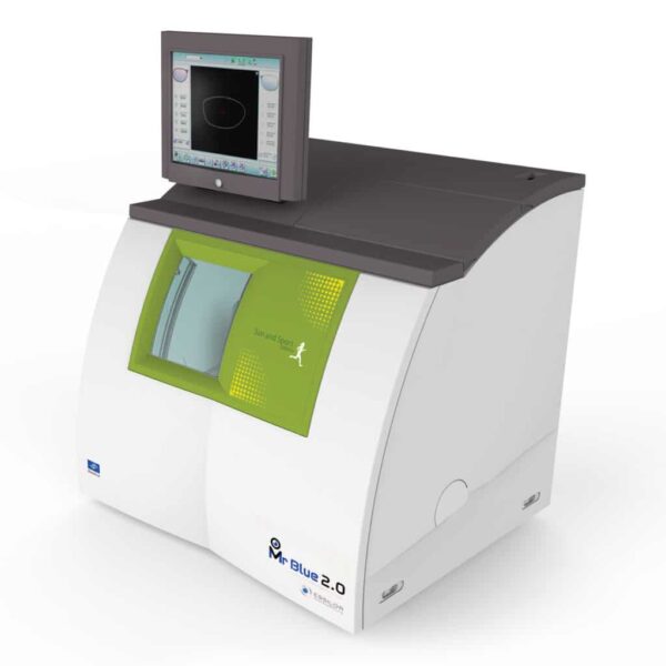
Half Jacket frames : Essilor Mr Blue 2.0 Sun & Sport Edition Patternless Edger- Capitalize on the rising popularity of endurance and extreme sports.
- Achieve a wider frames coverage with the partial step bevel inn addition to full high curve, asymmetric bevel, and step bevel.
- A dedicated process and assistance on your machine supported by new tools.
already owned Mr Blue Tracer or Mr Orange Tracer.Mr Blue 2.0 is on the cutting edge of innovation – serving as a genuine ECP partner since its launch. Now featuring a whole range of new functions and even greater flexibility than before, Mr. Blue 2.0 is the perfect solution for:- Working with cutting-edge technology
- Differentiating your offerings
- Optimizing your edging process
- Manufactured by Essilor | CE- and UL-certified
Chemistrie Clips to Bring You a New Innovative Sun Offer- Generate additional revenues through an alternative to both prescription sunwear and unsatisfactory clip-on sets
- Magnetic lens layering system: a patented technology that attaches lenses
to virtually any prescription eyewear using magnets embedded in the lenses
- Base curve matched, lightweight, and custom made
- A specifically-designed process on Mr Blue Sun & Sport Edition together with available materials and support
Half Jacket Frames to Excel in Your Sports Offer- Capitalize on the rising popularity of endurance and extreme sports
- Achieve a wider frames coverage with the partial step bevel(1) in addition to full high curve, asymmetric bevel, and step bevel
- A dedicated process and assistance on your machine supported by new tools
(1) Bevel a portion of the thick lens edge into a step to fit into frames so that only a part of the edge is inserted, and protrusions on the lens hook into indentations in the frame groove | Icare ic200 TonometerThe device is based on a rebound measuring principle that requires no drops, air or specialized skills for its use. The new premium design and user interface bring IOP measuring to a new level. The Icare ic200 tonometer is designed for professional use in the surgical operation room and emergency room as well as the clinic. The ic200 is fully portable, requires no anesthesia and its freedom of positioning allows measuring whether the patient is sitting, standing, half-sitting or in the supine or lateral recumbent position.A high-visibility indicator at the probe base confirms your positioning of the tonometer prior to measurement. A green light indicates measurement will be reliable, a red light indicates incorrect positioning. Features:
- Effortless probe loading with the same probe as for the Icare TA01i and ic100 tonometers
- The probe can’t accidentally drop from the device
- Easy menu in many languages
- Wireless (Bluetooth) printing and measurement transfer - Revolutionary design to allow for positional freedom when taking IOP measurements
- Innovative automatic measuring sequence allows for 6 automatic measurements or single measurements to be taken with the same button
- Intelligent positioning system for correct alignment of the tonometer
- Powered by AA batteries
Specifications:
- Dimensions : 43mm[W] x 104mm[H] x 214mm[L]
- Weight : 165 g (without batteries), 260 g (with 4 x AA batteries)
- Power supply : 4 x AA non-rechargeable batteries, 1.5V alkaline LR6
- Measurement range : 7 - 50 mmHg
- Accuracy : ±1.2 mmHg (≤20 mmHg) and
: ±2.2 mmHg (>20 mmHg)
- Repeatability (coefficient of variation) : <8% Package Includes:
- Icare ic200 tonometer
- 4 x AA batteries
- Aluminum case
- IOP pad
- Probe base cover
- Quick guide
- Screw driver
- Silicone grip
- Spare probe base
- Instruction manual
- Warranty card
- Wrist strap |

")
")
")
")


Reviews
There are no reviews yet.