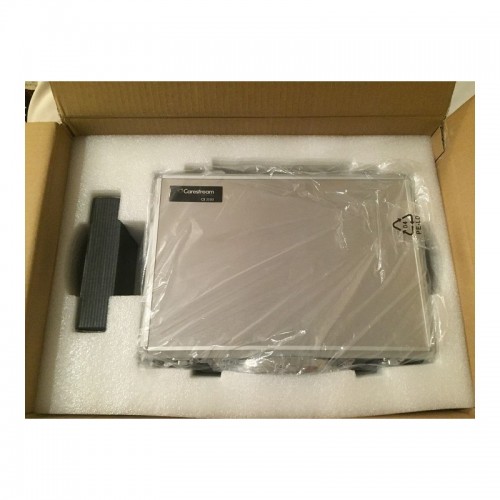
| Description | Includes Acquisition Computer. Monitor, keyboard and mouse are not included. | Are you paying too much for digital X-ray sensors and warranties? Consider QuickRay from Video Dental Concepts! A dental imaging leader and innovator since 1989, VDC has provided over 7,000 dental professionals with the best diagnostic imaging values and personalized support for over 20 years. | Caries diagnostic aid for surface caries.
TRIOS 4 has built-in fluorescent technology that aids in the identification of possible caries. Using TRIOS 4, dental professionals can now be aided in the early-detection of surface caries, without the need for an additional scanning device.
Caries diagnostic aid for interproximal caries.
TRIOS 4 will also feature a dedicated transillumination smart tip later this year. The smart tip will aid in the identification of possible interproximal caries undetectable to the eye, without emitting radiation. | Automatic Power Control: Epic Pro cuts four times faster1 than a classic diode laser with
minimal collateral damage. In this treatment mode, the laser in continuous wave mode converts light energy to thermal power at the tip. Laser energy is adjusted automatically in real time to maintain constant tip temperature. The result is extremely consistent and reliable cutting conditions, regardless of speed of movement, tissue type, etc. | The CS 3500 features a unique light guidance system that captures .STL data files during the image acquisition process. [I place a scan body abutment on the implant before the scanning process.] Scans acquired by the CS 3500 can be exported as open format .STL files and easily sent to the lab of your dentist’s choice to discuss making your new crown(s). Now your restorations are produced via the digital model utilizing CAD/CAM technology! | Perform a 3D body scan in seconds using just a simple turntable and 3D camera. In only 35 seconds, we extract millions of measurements with 2mm accuracy and reconstruct your body in 3D. |
| Content | Planmeca Promax Dimax 3 Digital Panoramic X RayPlanmeca ProMax offers a variety of imaging modalities in one intelligent X-ray unit.- Panoramic radiography for general diagnostics of the tooth arch and the jaw.
- Advanced panoramic radiography for specific diagnostics of the tooth arch, jaw maxillary sinuses and TMJ.
- Optional dental tomographic slices for detailed morphologic diagnostics of facial bones.
- Rotational freedom – the SCARA arm can produce any rotation pattern. Network Connectivity.
- Graphical user interface.
- Advanced collimation.
- Upgradable software.
- Dimax 3 Sensor
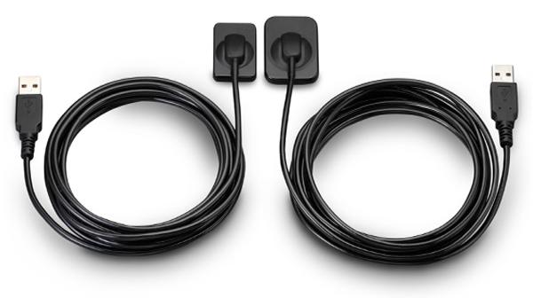
Planmeca is well-known for producing high-quality dental X-ray machines. With the Planmeca ProMax S3 pan-ceph with the DIMAX3, or previous generation 14-bit sensor, now orthodontists can add premium Planmeca imaging to their practices for a fraction of the price of new Planmeca dental equipment. Delivering standard and advanced 2D panoramic and cephalometric X-ray images, this versatile Planmeca X-ray machine can also be upgraded to medium field of view (8 x 8 cm) cone beam through the manufacturer, increasing your initial return on investment.PANORAMIC Planmeca Promax Dimax 3 Digital Panoramic X RayPlanmeca ProMax S3 includes the following panorex programs: standard, pediatric, lateral double TMJ, PA double TMJ, and PA rotational sinus. Optional advanced panorex programs include: horizontal segmenting, orthogonal, interproximal angulation, bitewings, lateral-PA double TMJ, PA multi-angle TMJ, lateral multi-angle TMJ, non-rotational sinus, and more.BITEWING PROGRAMWith the optional Planmeca extraoral bitewings program, you can replace conventional intraoral bitewing dental radiographs for sensitive patients with a fast and efficient alternative. (NOTE: Subject to availabilitySCARA TECHNOLOGYWith its advanced, exclusive robotic SCARA (Selectively Compliant Articulated Robot Arm) technology, the Planmeca ProMax S3 is triple-jointed to deliver precise, complex movements for optimized rotational maxillofacial digital denPEDO-FRIENDLYPlanmeca ProMax dental X-ray machines feature a pre-set "child" mode to reduce exposure area and minimize patient dose to help protect your smallest patients.tal X-ray radiography.DIMAX4 SENSORThis Planmeca ProMax dental X-ray machine includes the latest 16-bit DIMAX4 sensor, capturing the highest quality dental panoramic X-rays at up to 30-50% the price of new panoramic dental equipment. | QuickRay USB Size 1 And 2 X-Ray Sensor
QuickRay USB is a breakthrough in digital X-rays. The embedded electronics are located in the sensor case so there is no interface box! Simply connect the sensor cable via its USB connector to your computer and go! QuickRay USB is currently compatible with these Apteryx based software systems: XVa3, XVLite, Cliniview, Clinitouch, XDR Imaging, MPDX-DMD, Lightyear, DentiMax, ProfSuni, Dr. Suni, Sigma, AFP Digital and Belmont.Great value QuickRay USB digital X-ray sensors connect directly to USB— no interface box needed!Are you paying too much for digital X-ray sensors and warranties? Consider QuickRay from Video Dental Concepts! A dental imaging leader and innovator since 1989, VDC has provided over 7,000 dental professionals with the best diagnostic imaging values and personalized support for over 20 years.QuickRay USB Size 1 And 2 X-Ray Sensor For SaleQuickRay USB is a breakthrough in digital X-rays. The embedded electronics are located in the sensor case so there is no interface box! Simply connect the sensor cable via its USB connector to your computer and go! QuickRay USB is currently compatible with these Apteryx based software systems: XVa3, XVLite, Cliniview, Clinitouch, XDR Imaging, MPDX-DMD, Lightyear, DentiMax, ProfSuni, Dr. Suni, Sigma, AFP Digital and Belmont.The QuickRay sensor gives you the highest resolution and clarity! The thin (less than 1/4″) intraoral sensors include a CMOS image sensor chip, CsI Scintillator, electronic circuits, shielding foils and shock absorbers. Tough polyamide housings have durable replaceable polyurethane cables with ergonomically placed connectors. Their rounded corners and smooth edges optimize patient comfort.
And QuickRay is the fastest sensor on the market! View X-rays in USB 2.0 High Speed Mode in only 3 seconds. Operate in USB 2.0 Full Speed Mode, if necessary.Every sensor is covered by the VDC two year warranty with no annual fees. Technical support is available for an additional fee on an annual basis. Optional pro rata warranty is available from VDC after purchase. All VDC products are FDA registered. | 3Shape TRIOS 4 Intraoral ScannerOur most powerful intraoral scanner to date Take your dentistry beyond treatment
Our intraoral scanning solutions allow you to take your dentistry beyond. The new 3Shape TRIOS 4 combines our established superior scanning technology with groundbreaking caries diagnostic aid technology* for both surface and interproximal caries. No additional scanning device needed. Smart tips 3Shape TRIOS 4 Intraoral ScannerThe new generation of TRIOS smart tips feature instant-heat technology for optimized scanning.Be scan-ready in seconds with instant-heat technology.Scan 2-3 times as many patients with instant-heat in combination with 30% more battery life.Optimal scanning due to automatic use counter and tip-change alert.The TRIOS system is a complete in-house ecosystem including a scanner and extensive design software. This means there are plenty of options depending on your needs. Those who are just looking for a scanner and want to utilize labs may look into the basic, cheaper options. Those who want to design crowns, bridges and other prosthetics can opt for design modules. The only missing component is a milling machine, which if you want to mill restorations in-house, will have to be from a third party. This proves to be a weak link in the digital chain.At iDD, we have been using the TRIOS 3 for over four years in our clinic, spending literally thousands of hours learning the workflow. We have carried out all aspects of dentistry using the TRIOS system including same-day crowns, implant surgical guides, implant restorations and digital smile design.Disclaimer: This is an impartial review of the scanner and we do not have any ties to the company. The team at iDD remain unwaveringly committed to providing you with objective and trustworthy information and therefore do not seek any sponsorship. 3Shape kindly loaned us a TRIOS 4 scanner, due to a personal interest in upgrading our older TRIOS 3 model. Our team at iDD are constantly approached by scanner companies to review their scanners and it is always made clear that they will have no part in writing reviews or restricting any conclusions iDD makes in our thorough analysis and clinical use of these products. | Biolase Epic Pro Dental Soft Tissue LaserThe Epic Pro, which offers the most laser power of any diode laser in dentistry, is the first
product to be introduced resulting from BIOLASE’s strategic development agreement
with IPG Medical. The newest addition to the Epic family of dental soft-tissue lasers, Epic
Pro features several new innovations that are industry-firsts: - A new super pulse technology for more precise, enhanced laser tissue cutting
- Real-time automatic power control to enhance speed and consistency when performing surgery with pre-programed tip temperature
- Pre-initiated, bendable, disposable tips with new smart tip technology to ensure tip performance and quality
Automatic Power Control: Epic Pro cuts four times faster1 than a classic diode laser with
minimal collateral damage. In this treatment mode, the laser in continuous wave mode converts light energy to thermal power at the tip. Laser energy is adjusted automatically in real time to maintain constant tip temperature. The result is extremely consistent and reliable cutting conditions, regardless of speed of movement, tissue type, etc. Super Thermal Pulse: Epic Pro can achieve ten times the peak power of a standard diode (and up to three times that of the closest competitor) laser in short micro-pulses for the highest cutting efficiency and safety. In this treatment mode, the laser converts light energy to thermal power, which it emits as thermal power pulses, (i.e. multiple bursts of high power). Each of these thermal bursts cuts tissue highly efficiently. When the laser is activated, power bursts are emitted with a preset pulse width (duration), while maintaining a precise thermal level at the tip. The laser is powered on only about 3-5% of the time, giving tissue ample time to relax between pulses; creating a safer and gentler experience for patients. 
The Only Laser with Automatic Power ControlEpic Pro is the ONLY FDA-cleared laser with Automatic Power Control, an exclusive innovation from BIOLASE that enables the operator to lase with confidence and speed. 
Enhanced Clinical ApplicationsEpic Pro is the professional’s choice for managing soft tissue cases. With over 20 clinical presets, multiple treatment modes, and an advanced mode for expert users, Epic Pro provides unparalleled consistency and predictability. Unlike traditional diodes, Epic Pro moves quickly and with little snag-and-drag resistance. 
Greater Precision and ComfortEpic Pro brings more precision and comfort than traditional diodes due to innovations such as Super Thermal Pulse mode, which can pulse energy as quickly as ten-millionths of a second, resulting in efficient clinical cutting with less thermal damage than normal diode lasers. 
Introducing Smart Tip TechnologyEpic Pro uses durable new tip technology, available in uninitiated and pre-initiated formats depending on your clinical needs. Epic Pro tips work hand in hand with the laser’s Smart Tip technology to prompt you when a fresh tip is needed. 
With Epic Pro, ‘You Have to See It to Believe It’If you are a current diode laser or CO2 laser user, you have to see Epic Pro cut tissue live and compare to your current laser experience. When current Epic Pro customers were first shown Epic Pro, their response? “You have to see it cut to understand how much faster it is.” Fill out the contact form on this page to request a live demonstration for Epic Pro in your clinic.Technical Specifications Biolase Epic Pro Dental Soft Tissue Laser| Dimension | 7.25in (W) x 4.5in (L) x 6.5in (H) (18.4cm x 11.4cm x 16.5cm) | | Weight | 4 lbs (1.8 kg) | | Operating Voltage | 100V – 240V at 1.2A/0.5A | | Frequency | 50/60 Hz | | Main Control | Power Switch | | Remote Interruption | Remote Interlock | | Disable Control | Emergency Stop Button | | DC Power Supply Module | 12V DC, 5A | | Laser Classification | Class IV (4) | | Medium | Semi-conductor diode | | Wavelength | 940nm ± 20nm | | Power Modes | Continuous, Pulse Modulation | | Fiber Tip Diameter | 300µm, 400µm | | Pulse Duration | 10µm-100ms | | Pulse Interval | 0.01ms – 20ms | | Pulse Repetition Rate | Up to 20kHz (for reference) | | NOHD | 4.77 meters | | Beam Divergence | 0.22 | | Standard Fiber Cable Length | 6 feet (1.8 meters) |
| CARESTREAM CS3500 INTRAORAL DIGITAL SCANNERThe CS 3500 intraoral scanner includes a light guidance system that aides in the capture of the data during the image acquisition process, allowing the user to place more focus on the patient’s mouth rather than the monitor.The end result is a highly precise digital impression of your patients’ mouth in a matter of minutes.The CS 3500 was introduced as part of the CS Solutions portfolio, a unique open CAD/CAM system. This open system gives users the option to either use images captured by the scanner to design and mill crowns in-house or share digital impression files with preferred labs to create restorations or orthodontic appliances.Features of Carestream CS3500 Intraoral Digital Scanner :– High angulation scanning
– Unique light guidance system
– Direct USB connection
– Disposable and autoclavable tips
– Virtually automatic bite registration
– 1024×768 image resolutionFacts Carestream CS 3500 Intraoral Scanner– Handheld and truly portable requiring no trolley and can be plugged into any computer via USB cable
– No traditional impression material or powder required
– Easy to use with a light guidance system that tells you when a scan is successful
– Compact ergonomic shape and slim scanner headBenefits of Carestream CS3500 Intraoral Digital Scanner :– Can quickly acquire a full arch of information
– Eliminates the need to painstakingly maneuver the scanner head in patients’ mouths
– Compatible with open system CAD software like Dental Wings, 3Shape, and Exocad
– Comfortable and easy for users to navigate around patients’ mouths, especially during full arch acquisitionsWith the CS 3500 intraoral scanner, practitioners can easily acquire precise true color, 2D and 3D digital impressions. The CS 3500 works as part of the integrated CS Solutions CAD/CAM restoration portfolio, or a standalone solution in a practice’s restorative workflow. Requiring no external heater or trolley system, the CS 3500 features high-angulation scanning of up to 45 degrees and to a depth from -2 to +13mm. Additionally, the CS 3500 features a unique light guidance system that aids in the capture of the data during the image acquisition process.The Carestream CS3500 Intraoral Digital Scanner also offers unique dedicated scanning workflows for both implant-borne prosthetic planning and orthodontic digital models and appliance fabrication.* Laptop Acquisition Computer and/or Carestream Software and License available for an additional charge.Features:CARESTREAM CS3500 INTRAORAL DIGITAL SCANNER- Faster Scan Speed
- Superb Digital Impressions
- Light Guidance System
- Portable for Easy Sharing
- Lightweight Handpiece
- Disposable and Autoclavable Tips
- Flexible Open System
- Dedicated Scanning Implant Workflow
| Styku 3D Body scanner
Styku 3D is a professional, full-body 3D scanner. It is a high-precision measurement tool able to scan people in just 30 seconds. It uses Infrared Depth Sensor technology for high-resolution scanning results. It is composed of an automatic turntable and a sensor tower. Its small footprint and aluminum structure make it easy to move and reposition.This powerful scanner is simple and convenient to use. It can capture the full body of a person in three dimensions. The person only has to stand on the rotating plate for 30 seconds holding the pose. The scanner is able to extract millions of data points in no time thanks to the efficient Microsoft Kinect v2 3D camera. It lets you perform complete body scans, view 3D models, extract exact body measurements, track posture changes, and more. After generating the 3D avatar, the advanced scanning and feature recognition technology automatically detects specific landmarks, which are used to extract accurate data related to the volume, circumference, and surface of each body area.Styku is mainly used for fitness and professional health purposes. It consents to view and analyze the body shape, measurements, and other health indicators. It allows tracking the evolution of the body shape or posture of a person through several measurements. Find out more reading our complete review.Styku is often used to track body changes. For example, it is generally adopted by people who want to capture and compare before and after images of themselves in order to analyze their physical evolution.Specifications:- Scan accuracy
- Scan volume
- Scanner controls
- What’s in the box
- Look and feel
- Price
Scan accuracyStyku 3D body scanner is a professional, full-body 3D scanner. It features a depth data resolution of ~2mm (.08in). It grants a torso measurement precision of +- 2.5-5mm (.1-.2in) and can perform a complete scan in 30/40 seconds. It is engineered with advanced feature recognition and a 3D visualization system for automatic detection of landmark points and measurement extraction. It is capable of stitching more than 600 surfaces to create a three-dimensional avatar in seconds. The Kinect v2 3D camera has a depth image resolution of 512 x 424 pixels.Scan volume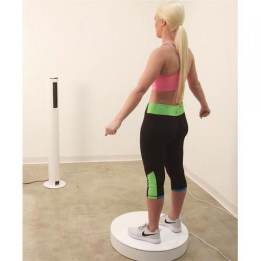 It features a minimum scanning area of 8.4 x 5.5 ft ( 251 x 168 cm). It works at a recommended distance of the sensor tower from the turntable of 46 inches (117 cm). It requires a minimum clearance of 21 inches (53 cm) around the turntable. In case of white walls or glossy surfaces, it might require more clearance.Scanner controlsThe scanner is controlled via an external computer. It features a minimum scanning area of 8.4 x 5.5 ft ( 251 x 168 cm). It works at a recommended distance of the sensor tower from the turntable of 46 inches (117 cm). It requires a minimum clearance of 21 inches (53 cm) around the turntable. In case of white walls or glossy surfaces, it might require more clearance.Scanner controlsThe scanner is controlled via an external computer.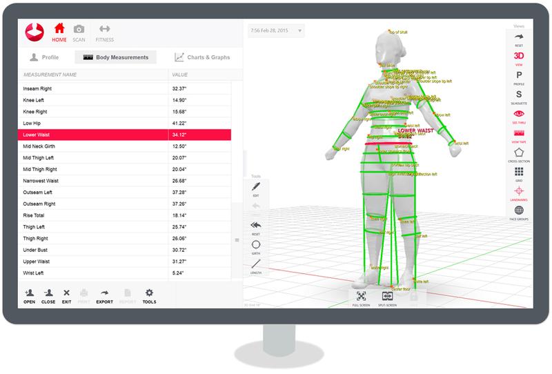 The proprietary Styku intuitive, multi-platform software allows performing a complete scan and all the measurements with ease. The generated avatar can be rotated, panned, and zoomed to evaluate each body part in detail. The collected data are strictly private. You can decide who can view them by setting appropriate export and sharing options. The software is compatible with Windows 10 and is optimized for touchscreens. The proprietary Styku intuitive, multi-platform software allows performing a complete scan and all the measurements with ease. The generated avatar can be rotated, panned, and zoomed to evaluate each body part in detail. The collected data are strictly private. You can decide who can view them by setting appropriate export and sharing options. The software is compatible with Windows 10 and is optimized for touchscreens.What’s in the box
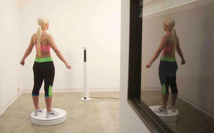 - Essential parts required to operate the Styku 3D body scanner
- Rotating platform (max. weight 250kg)
- Sensor tower
- Microsoft Kinect v2 3D camera
- Aluminum enclosure and stand to protect the 3D Camera
- All the necessary cables
For some items, package content may change. In case of any questions, please get in touch.Look and feel
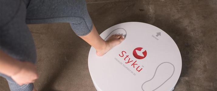 Styku 3D body scanner is available in white with red trims. Its stylish look makes it ideal for fitness clubs, gyms, healthcare facilities, and fashion companies.The scanner has an overall footprint of 101 x 66 inches ( 257cm x 168cm). The rotating platform measures 24 x 24 x 5 inches (66cm x 66cm x 12.5 cm) and weighs 23 lb (10.5 kg). It withstands a max load of 550 lb (250 kg). The sensor tower measures 10 x 10 x 46 inches ( 25.4 x 25.4 x 117 cm). It weighs 13 lb (6 kg). Styku 3D body scanner is available in white with red trims. Its stylish look makes it ideal for fitness clubs, gyms, healthcare facilities, and fashion companies.The scanner has an overall footprint of 101 x 66 inches ( 257cm x 168cm). The rotating platform measures 24 x 24 x 5 inches (66cm x 66cm x 12.5 cm) and weighs 23 lb (10.5 kg). It withstands a max load of 550 lb (250 kg). The sensor tower measures 10 x 10 x 46 inches ( 25.4 x 25.4 x 117 cm). It weighs 13 lb (6 kg). |





Reviews
There are no reviews yet.