| Description | Includes Acquisition Computer. Monitor, keyboard and mouse are not included. | As one of the most user-friendly Cone Beam Computed Tomography (CBCT) systems on the market, the 9000 3D Extraoral Imaging System offers several versatile solutions for your extraoral imaging needs. Combining focused-field 3D technology with dedicated panoramic imaging, this affordable 2-in-1 unit delivers the best of both worlds, offering the highest resolution and the lowest radiation dose. | Caries diagnostic aid for surface caries.
TRIOS 4 has built-in fluorescent technology that aids in the identification of possible caries. Using TRIOS 4, dental professionals can now be aided in the early-detection of surface caries, without the need for an additional scanning device.
Caries diagnostic aid for interproximal caries.
TRIOS 4 will also feature a dedicated transillumination smart tip later this year. The smart tip will aid in the identification of possible interproximal caries undetectable to the eye, without emitting radiation. | The Identica T300 dramatically improves the fundamental aspects of scanner performance and the latest software technology provides the clearest, sharpest data. Identica T300 is customized to deliver the finest data quality using its dual 2.0 MP cameras. Identica T300 will save valuable time and money by minimizing trial & error when adjusting restoration fits. | From general to littles, this machine has you covered. This digital system excels at both general and pediatric practices, with great strengths in cosmetic dentistry and orthodontics as well. | The Medit T710 desktop scanner is equipped with a fast scan engine and highly efficient software algorithm which work in tandem to produce a full-arch scan in just 8 seconds. |
| Content | Planmeca Promax Dimax 3 Digital Panoramic X RayPlanmeca ProMax offers a variety of imaging modalities in one intelligent X-ray unit.- Panoramic radiography for general diagnostics of the tooth arch and the jaw.
- Advanced panoramic radiography for specific diagnostics of the tooth arch, jaw maxillary sinuses and TMJ.
- Optional dental tomographic slices for detailed morphologic diagnostics of facial bones.
- Rotational freedom – the SCARA arm can produce any rotation pattern. Network Connectivity.
- Graphical user interface.
- Advanced collimation.
- Upgradable software.
- Dimax 3 Sensor
Planmeca is well-known for producing high-quality dental X-ray machines. With the Planmeca ProMax S3 pan-ceph with the DIMAX3, or previous generation 14-bit sensor, now orthodontists can add premium Planmeca imaging to their practices for a fraction of the price of new Planmeca dental equipment. Delivering standard and advanced 2D panoramic and cephalometric X-ray images, this versatile Planmeca X-ray machine can also be upgraded to medium field of view (8 x 8 cm) cone beam through the manufacturer, increasing your initial return on investment.PANORAMIC Planmeca Promax Dimax 3 Digital Panoramic X RayPlanmeca ProMax S3 includes the following panorex programs: standard, pediatric, lateral double TMJ, PA double TMJ, and PA rotational sinus. Optional advanced panorex programs include: horizontal segmenting, orthogonal, interproximal angulation, bitewings, lateral-PA double TMJ, PA multi-angle TMJ, lateral multi-angle TMJ, non-rotational sinus, and more.BITEWING PROGRAMWith the optional Planmeca extraoral bitewings program, you can replace conventional intraoral bitewing dental radiographs for sensitive patients with a fast and efficient alternative. (NOTE: Subject to availabilitySCARA TECHNOLOGYWith its advanced, exclusive robotic SCARA (Selectively Compliant Articulated Robot Arm) technology, the Planmeca ProMax S3 is triple-jointed to deliver precise, complex movements for optimized rotational maxillofacial digital denPEDO-FRIENDLYPlanmeca ProMax dental X-ray machines feature a pre-set "child" mode to reduce exposure area and minimize patient dose to help protect your smallest patients.tal X-ray radiography.DIMAX4 SENSORThis Planmeca ProMax dental X-ray machine includes the latest 16-bit DIMAX4 sensor, capturing the highest quality dental panoramic X-rays at up to 30-50% the price of new panoramic dental equipment. | Kodak 9000 3D Extraoral Imaging System
Add cephalometric imaging abilities to your 9000 3D System for even more abilities. Through our revolutionary one-shot technology, cephalometric image acquisition takes less than a second—thereby reducing exposure time and the risk of retakes due to patient movement. With the 9000C 3D System, you can perform a wider range of diagnoses and treatments right in your office, rather than referring patients to imaging centers. Reduce multiple visits, prevent rescheduling issues, and enhance patient care with three versatile imaging abilities right at your fingertips: panoramic, cephalometric, and 3D imaging.Superb image quality, all three modalitiesVariable focal trough adapts to patient’s jaw morphology – generating high-quality, true panoramic images Best-in-class cephalometric image quality with reduced risk of motion blur Ultra-high resolution, low-dose, focused-field CBCT images provide superior visualization of fine bony anatomy, such as radicular structures
Optimized for radiology workflowsDR technology reduces exam time and eliminates steps, such as film processing and cassette handling One-shot cephalometric technology captures images in less than a second, reducing exam time and risk of retakes Dedicated sensors for each modality save technologist time, reduce risk of breakage DICOM conformance ensures easy integration with PACS, RIS, and printing systems
Simple by design Kodak 9000 3D Extraoral Imaging SystemStreamlined user interface, preset programs and face-to-face positioning makes exams quick and simple Accommodates patients of all shapes and sizes and wheelchair accessible Intuitive, easy-to-use imaging software with improved features for reviewing, reporting and sharing images Includes a comprehensive implant planning model to help referring dentists identify exact measurements and create simulations of proposed treatments
A trusted company, proven productsProven product reliability – even in demanding, high-volume environments Broad range of Carestream equipment and professional services such equipment service agreements, site planning and DICOM integration services. - One-stop shopping for digital imaging and IT solutions from a company with over a century of medical and dental imaging expertise
| Panoramic Modality | Cephalometric Modality | Cone Beam CT Modality | Technology CCD – Optical | CCD – Optical Fiber Sensor | One-Shot with CCD Sensor with Optical Fiber | Cone Beam with CMOS Sensor with Optical Fiber | Gray Scale | 14 bit | 14 bit | 15 bit | 3D Field-of-View | n/a | n/a | • Single Volume: 5 cm x 3.75 cm • Stitched Volumes (up to 3): 9 cm x 7 cm x 3.75 cm | Exam Options | • Panoramic • Child Mode • Segmented Panoramic • Maxillary Sinus • LA TMJ x 2 • LA TMJ x 4 | • Cephalometric • Lateral • Oblique • Frontal (AP/PA) • Submento-Vertex • Carpus • Multiple Cephalometric Field Choices: 30 x 30, 24 x 30, 24 x 24, 18 x 24, 18 x 18 | • CBCT • Resolution Options: 76, 100, or 200 micrometers • Location of Exam: User-defined (mandible, maxilla, TMJ) | Input Voltage | 230/240 V – 60 Hz | 230/240 V – 60 Hz | 230/240 V – 60 Hz | Tube Voltage | 60-90 kV (max) | 60-90 kV (max) | 60-90 kV (max) | Tube Current | 2-15 mA (max | 2-15 mA (max | 2-15 mA (max | Frequency | 140 kHZ (max) | 140 kHZ (max) | 140 kHZ (max) | Total Filtration | > 3.5 mm eq. AL | > 3.5 mm eq. AL | > 3.5 mm eq. AL | Weight | 353 lb (160 kg) | 438 lb (199 kg) | 353 lb (160 kg) |
| 3Shape TRIOS 4 Intraoral ScannerOur most powerful intraoral scanner to date Take your dentistry beyond treatment
Our intraoral scanning solutions allow you to take your dentistry beyond. The new 3Shape TRIOS 4 combines our established superior scanning technology with groundbreaking caries diagnostic aid technology* for both surface and interproximal caries. No additional scanning device needed. Smart tips 3Shape TRIOS 4 Intraoral ScannerThe new generation of TRIOS smart tips feature instant-heat technology for optimized scanning.Be scan-ready in seconds with instant-heat technology.Scan 2-3 times as many patients with instant-heat in combination with 30% more battery life.Optimal scanning due to automatic use counter and tip-change alert.The TRIOS system is a complete in-house ecosystem including a scanner and extensive design software. This means there are plenty of options depending on your needs. Those who are just looking for a scanner and want to utilize labs may look into the basic, cheaper options. Those who want to design crowns, bridges and other prosthetics can opt for design modules. The only missing component is a milling machine, which if you want to mill restorations in-house, will have to be from a third party. This proves to be a weak link in the digital chain.At iDD, we have been using the TRIOS 3 for over four years in our clinic, spending literally thousands of hours learning the workflow. We have carried out all aspects of dentistry using the TRIOS system including same-day crowns, implant surgical guides, implant restorations and digital smile design.Disclaimer: This is an impartial review of the scanner and we do not have any ties to the company. The team at iDD remain unwaveringly committed to providing you with objective and trustworthy information and therefore do not seek any sponsorship. 3Shape kindly loaned us a TRIOS 4 scanner, due to a personal interest in upgrading our older TRIOS 3 model. Our team at iDD are constantly approached by scanner companies to review their scanners and it is always made clear that they will have no part in writing reviews or restricting any conclusions iDD makes in our thorough analysis and clinical use of these products. | Medit Identica T300 Dental 3D Scanner
Identica T300 to meet all of your needs. With powerful scan engine and affordable pricing, the new Identica T300 is a perfect fit for both performance enthusiasts and entry level users. Its evolutionary design and high tech features will help your lab rediscover productivity. Bundled colLab 2017 scan software makes scan data processing easy and efficient.Quality & affordability- 2.0 MP cameras, Blue LED
- Accuracy : 7 μm (ISO)
- Full arch scan time 24 sec
- 2-Axis Manual
Identica T300 dramatically improves the most important aspects of scanner performance. The latest software technology provides you with the cleanest and sharpest data. T300 is specially designed to bring out the finest data quality using its 2.0 MP cameras. T300 will save your precious time and money by minimizing trial & error to adjust restoration fits.Identica T300 is an upgrade version of Identica Blue, provided at lowest pricing. Giving better value for the affordable scanner users. Identica T300 has enhanced data quality compared to Identica Blue.Introducing the Next Generation of 3D Dental Scanners: Our T-Series Medit represents the apex in 3D dental scanning. With unsurpassed speed, accuracy, and reliability, Medit helps you rediscover your productivity. The T-Series’ new powerful scan engine and affordable pricing are the perfect fit for performance enthusiasts and entry-level users, helping your lab rediscovering productivity thanks to its convenient design and high-tech features. Medit T300 is the latest Medit scanner with an enhanced SW scan. It uses Blue Light technology, which enables quick scanning and provides excellent precision (7μm).The program is user-friendly, because it takes you through simple and fast individual steps, from inserting the model into the scanner to exporting the file to the CAD design program. FEATURES:Medit Identica T300 Dental 3D Scanner- Scanned printing: 2 manual axes.
- Touch sensor: no.
- Full arcade scan speed: 24 ´´
- Resolution of the camera: 2 x 2 MP camera.
- Scan volume: 90 x 72 x 60 mm.
- Scanning principle: Optical change phase.
- Dimensions: 290 x 290 x 340 mm.
- Weight: 12Kg.
- Light source: Blue LED.
- Connectivity: Type USB 3.0 B.
- Power: 100- 240 vac, 50-60 HZ.
- Warranty: 2 years.
ADVANCED DATA PROCESSING AND HIGHER RESOLUTION DUAL CAMERAS The Identica T300 dramatically improves the fundamental aspects of scanner performance and the latest software technology provides the clearest, sharpest data. Identica T300 is customized to deliver the finest data quality using its dual 2.0 MP cameras. Identica T300 will save valuable time and money by minimizing trial & error when adjusting restoration fits. | Soredex Cranex-D Panoramic X Ray
The Soredex Cranex D with cephalometric option provides a full range of digital panoramic and cephalometric dental x-ray examinations to meet the diagnostic needs of almost any general dental or specialty practice. With it’s minimal footprint, this Soredex option will help elevate the diagnostic functionality of any dental provider.Soredex Cranex D provides a full range of high quality digital panoramic x-ray examinations to meet the diagnostic needs of almost any dental practice – all at an affordable price.Acquisition computer includedFeatures Soredex Cranex-D Panoramic X Ray
CRANEX D is a direct digital panoramic and cephalometric x-ray system that provides superior image quality and more diagnostic value. Available as a single or dual sensor system, the CRANEX D grows with your practice, making it a valuable addition to meet the needs of your office. It combines quality images, ease-of-use, and flexibility to make your day-to-day diagnostics easy and accurate. Benefits
The system features superb anterior image quality in panoramic imaging with its patented Variable Panoramic Collimator (VPC) by expanding the focal trough by 50% compared to traditional techniques. Additional innovative features on the unit include Automatic Collimator Selector (ACS) to easily set proper collimation for the imaging program prescribed, as well as the Automatic Exposure Setting (AES) where the system recommends proper cephalometric exposure values based on the size of the patient. These automated and optimized exposure settings maximize the practice’s workflow. CRANEX D is built for contemporary, fast-paced dental care where reliability is key.The Soredex Cranex D with cephalometric option provides a full range of panoramic and cephalometric examinations to meet the needs of almost any practice.SOREDEX has over 30 years of expertise in state-of-the-art dental imaging systems. The Soredex® Cranex D with cephalometric option provides a full range of panoramic and cephalometric examinations to meet the needs of almost any practice. | Medit T710 Blue Light Dental Scanner
Introducing the New and Improved T-Series Lab Scanners
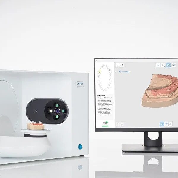
Medit have completely overhauled the design of their T-Series lab scanners. The result is a sleek and sophisticated dental scanner which not only does its work well, but in style.The Medit T710 desktop scanner is equipped with a fast scan engine and highly efficient software algorithm which work in tandem to produce a full-arch scan in just 8 seconds. The advanced, high-speed positioning system of the new T-Series is designed for optimal performance for your laboratory, speeding up your workflow and increasing productivity.The T710, with its new four 5.0MP-resolution cameras system and blue-light scanning technology, ensures your models and impressions are fully scanned, eliminating all blind spots.Flexible multi-die scanning
2 StepsT710 Specifications
Accuracy 4μm
Full Arch Scan 8 seconds
Full Arch Impression Scan Speed 45 seconds
Colour Texture
Auto-elevation
Let the scanner decide the scanning height for your object with our auto-elevation feature.
Wider Scan Area
Scan more objects at the same time thanks to the wider scan area of our T-Series tabletop scanners!
No Blind Spots
The 4 cameras in the T710 are positioned in a way to ensure that there are no blind spots in your scan data. It only takes one scan to get the full data!
Full-size articulator scanning
To reproduce the exact occlusion orientation, nothing beats scanning the occlusion in the articulator itself. We’ve designed our T-Series to accommodate any articulator available in the market, comfortably.
Replica Denture Included
Orthodontic Scanning Included
Impression Scanning IncludedMedit T710 Blue Light Dental Scanner- ultra-fast scanning of a dental arch in only 8 seconds
- scanning speed for a complete jaw impression is only 45 seconds
- color texture available
- accuracy of 4 µm
- open System
- 5.0 megapixel camera enables high-resolution and detailed scan data
- four-camera system for a large scanning area without blind spots
- automatic height adjustment for optimal scan Position
- flexible multi-die scanning
- complete scan in the articulator possible with the 3-in-1 KAS holding device for KaVo, Artex and SAM
- the AM holding device is compatible with the MARK330 and BIOART A7+ articulators - thus the use of the exocad software is possible
- orthodontic scanning and impression scanning possible
|




Reviews
There are no reviews yet.