| Description | Includes Acquisition Computer. Monitor, keyboard and mouse are not included. | Benefits:- Faster Scan Speed
- Superb Image Quality
- Light Guidance System
- Portable for Easy Sharing
- Lightweight Hand Piece
| The i500 intraoral scanner, exemplifies three qualities:
Value, Efficiency, and ProductivityPerfect software and hardware combination:
Value, Efficiency, and ProductivityOne Software to connect them all | The Identica T300 dramatically improves the fundamental aspects of scanner performance and the latest software technology provides the clearest, sharpest data. Identica T300 is customized to deliver the finest data quality using its dual 2.0 MP cameras. Identica T300 will save valuable time and money by minimizing trial & error when adjusting restoration fits. | Perform a 3D body scan in seconds using just a simple turntable and 3D camera. In only 35 seconds, we extract millions of measurements with 2mm accuracy and reconstruct your body in 3D. | From general to littles, this machine has you covered. This digital system excels at both general and pediatric practices, with great strengths in cosmetic dentistry and orthodontics as well. |
| Content | Planmeca Promax Dimax 3 Digital Panoramic X RayPlanmeca ProMax offers a variety of imaging modalities in one intelligent X-ray unit.- Panoramic radiography for general diagnostics of the tooth arch and the jaw.
- Advanced panoramic radiography for specific diagnostics of the tooth arch, jaw maxillary sinuses and TMJ.
- Optional dental tomographic slices for detailed morphologic diagnostics of facial bones.
- Rotational freedom – the SCARA arm can produce any rotation pattern. Network Connectivity.
- Graphical user interface.
- Advanced collimation.
- Upgradable software.
- Dimax 3 Sensor
Planmeca is well-known for producing high-quality dental X-ray machines. With the Planmeca ProMax S3 pan-ceph with the DIMAX3, or previous generation 14-bit sensor, now orthodontists can add premium Planmeca imaging to their practices for a fraction of the price of new Planmeca dental equipment. Delivering standard and advanced 2D panoramic and cephalometric X-ray images, this versatile Planmeca X-ray machine can also be upgraded to medium field of view (8 x 8 cm) cone beam through the manufacturer, increasing your initial return on investment.PANORAMIC Planmeca Promax Dimax 3 Digital Panoramic X RayPlanmeca ProMax S3 includes the following panorex programs: standard, pediatric, lateral double TMJ, PA double TMJ, and PA rotational sinus. Optional advanced panorex programs include: horizontal segmenting, orthogonal, interproximal angulation, bitewings, lateral-PA double TMJ, PA multi-angle TMJ, lateral multi-angle TMJ, non-rotational sinus, and more.BITEWING PROGRAMWith the optional Planmeca extraoral bitewings program, you can replace conventional intraoral bitewing dental radiographs for sensitive patients with a fast and efficient alternative. (NOTE: Subject to availabilitySCARA TECHNOLOGYWith its advanced, exclusive robotic SCARA (Selectively Compliant Articulated Robot Arm) technology, the Planmeca ProMax S3 is triple-jointed to deliver precise, complex movements for optimized rotational maxillofacial digital denPEDO-FRIENDLYPlanmeca ProMax dental X-ray machines feature a pre-set "child" mode to reduce exposure area and minimize patient dose to help protect your smallest patients.tal X-ray radiography.DIMAX4 SENSORThis Planmeca ProMax dental X-ray machine includes the latest 16-bit DIMAX4 sensor, capturing the highest quality dental panoramic X-rays at up to 30-50% the price of new panoramic dental equipment. | Carestream CS 3600 Intraoral ScannerAcquire true color:With the CS 3600 Intraoral Scanner, practitioners can access 2D and 3D images that can be used with CS Restore software to design restorations within their practice. Colored 3D scans allow practitioners to easily define the margin lines and identify the differences between natural tooth structure and existing restorations. This makes restoration planning more accurate for practitioners.No external heater, powder or trolley system needed:The CS 3600 does not require you to spray powder of liquid over the patient’s teeth or gingival tissue before scanning. It features high-angulation scanning of up to 45 degrees and to a depth from -2 to +13m.Features a unique light guidance systemYet another feature of the CS 3600 that assists it in the capture of the data during the image acquisition process. This feature allows the user to place more focus on the patient’s mouth rather than the monitor.This device is easily shared:The CS 3600 can be easily shared between operatories because there is no heavy trolley to push around. Additionally, the intraoral scanner connects via USB 2 cable* to any PC workstation or laptop. * Windows 7, 32-bit minimum; 64-bit recommendedEasy to use and hold:The CS 3600’s sleek, lightweight design (295 grams) makes it comfortable and easy for users to navigate the intraoral scanner around patients’ mouths, especially during full arch acquisitions.Benefits:Carestream CS 3600 Intraoral Scanner- Faster Scan Speed
- Superb Image Quality
- Light Guidance System
- Portable for Easy Sharing
- Lightweight Hand Piece
- Features
- High Speed Continuous Scanning
- Fast, Simple and Smooth user experience
- Freefill Missing Scan Data at your Leisure
- Scan History allows you to remove excess scanned tissue for more refined final digital impression
- Three Workflows
- user friendly interface
- Autoclavable reusable tips in two interchangeable styles
- Accurate full 3D HD Color scanning
| Medit i500 Intraoral ScannerOur Intraoral Scanner Delivers: Value, Efficiency, and Productivity
Together, these three qualities make it easy to incorporate our intraoral scanner into your workflow. Designed with quality in mind, we created the i500 to add value to your practice. Regardless of specialization the i500 ensures that professional needs are met, workflows are optimized, and flexibility is guaranteed.High ROI
An unparalleled performance combined with a competitive price delivers exceptionally fast return on investmentFlexibility
An open system for integrated CAD/CAM workflow allows for the export of STL files for easy file transfer and thus optimized collaborationImpressive Speed
Intelligent scan-detecting algorithm with two high-speed cameras for quick and efficient intraoral scanningPowderless
No need for powder in most regular cases, allowing for a seamless scanning process and increased patient comfortHigh Accuracy
Single crown* :Trueness (Accuracy) = 5.1 μm (±0.49 μm)
Precision (Consistency) = 3.2 μm (±0.74 μm)*Single crown accuracy test was conducted by Medit according to the methods in “Evaluation of the Accuracy of Six Intraoral Scanning Devices: An in-vitro Investigation. ADA Professional Product”
SPECIFICATION | Tip | 18 x 15.2 mm (WxH) | | Overall handpiece length | 266 mm | | Weight | 276g | | Imaging technology | 3D-in-motion video technology | | Color | 3D full color streaming capture | | Connectivity | USB 3.0 | | Scanning FOV | 14 x 13 mm |
The Medit i500 intraoral scanner delivers efficiency, productivity, and affordability.This scanner has been designed with quality in mind to add value to your practice. With its fast speed and powderless system, it allows for a smoother, more efficient scanning experience.The Medit Link software and hardware with workflow management and communication software will enhance and support your everyday CAD performance. Completed with integrated cloud storage and open data architecture. - Scanning for iOS in real time
- High-speed continuous scanning
- Color structure of the surface in the resolution of Full HD 3D
- Quick scan
- Natural color transfer
- Capturing UHD images (SR virtual viewer)
- Real-time 3D visualization (15 FPS 3D Data)
- Without using a spray
- Diagnostic image 2D
- Open file format (.stl)
- Scanner with the smallest size of the scanning module
| Medit Identica T300 Dental 3D Scanner
Identica T300 to meet all of your needs. With powerful scan engine and affordable pricing, the new Identica T300 is a perfect fit for both performance enthusiasts and entry level users. Its evolutionary design and high tech features will help your lab rediscover productivity. Bundled colLab 2017 scan software makes scan data processing easy and efficient.Quality & affordability- 2.0 MP cameras, Blue LED
- Accuracy : 7 μm (ISO)
- Full arch scan time 24 sec
- 2-Axis Manual
Identica T300 dramatically improves the most important aspects of scanner performance. The latest software technology provides you with the cleanest and sharpest data. T300 is specially designed to bring out the finest data quality using its 2.0 MP cameras. T300 will save your precious time and money by minimizing trial & error to adjust restoration fits.Identica T300 is an upgrade version of Identica Blue, provided at lowest pricing. Giving better value for the affordable scanner users. Identica T300 has enhanced data quality compared to Identica Blue.Introducing the Next Generation of 3D Dental Scanners: Our T-Series Medit represents the apex in 3D dental scanning. With unsurpassed speed, accuracy, and reliability, Medit helps you rediscover your productivity. The T-Series’ new powerful scan engine and affordable pricing are the perfect fit for performance enthusiasts and entry-level users, helping your lab rediscovering productivity thanks to its convenient design and high-tech features. Medit T300 is the latest Medit scanner with an enhanced SW scan. It uses Blue Light technology, which enables quick scanning and provides excellent precision (7μm).The program is user-friendly, because it takes you through simple and fast individual steps, from inserting the model into the scanner to exporting the file to the CAD design program. FEATURES:Medit Identica T300 Dental 3D Scanner- Scanned printing: 2 manual axes.
- Touch sensor: no.
- Full arcade scan speed: 24 ´´
- Resolution of the camera: 2 x 2 MP camera.
- Scan volume: 90 x 72 x 60 mm.
- Scanning principle: Optical change phase.
- Dimensions: 290 x 290 x 340 mm.
- Weight: 12Kg.
- Light source: Blue LED.
- Connectivity: Type USB 3.0 B.
- Power: 100- 240 vac, 50-60 HZ.
- Warranty: 2 years.
ADVANCED DATA PROCESSING AND HIGHER RESOLUTION DUAL CAMERAS The Identica T300 dramatically improves the fundamental aspects of scanner performance and the latest software technology provides the clearest, sharpest data. Identica T300 is customized to deliver the finest data quality using its dual 2.0 MP cameras. Identica T300 will save valuable time and money by minimizing trial & error when adjusting restoration fits. | Styku 3D Body scanner
Styku 3D is a professional, full-body 3D scanner. It is a high-precision measurement tool able to scan people in just 30 seconds. It uses Infrared Depth Sensor technology for high-resolution scanning results. It is composed of an automatic turntable and a sensor tower. Its small footprint and aluminum structure make it easy to move and reposition.This powerful scanner is simple and convenient to use. It can capture the full body of a person in three dimensions. The person only has to stand on the rotating plate for 30 seconds holding the pose. The scanner is able to extract millions of data points in no time thanks to the efficient Microsoft Kinect v2 3D camera. It lets you perform complete body scans, view 3D models, extract exact body measurements, track posture changes, and more. After generating the 3D avatar, the advanced scanning and feature recognition technology automatically detects specific landmarks, which are used to extract accurate data related to the volume, circumference, and surface of each body area.Styku is mainly used for fitness and professional health purposes. It consents to view and analyze the body shape, measurements, and other health indicators. It allows tracking the evolution of the body shape or posture of a person through several measurements. Find out more reading our complete review.Styku is often used to track body changes. For example, it is generally adopted by people who want to capture and compare before and after images of themselves in order to analyze their physical evolution.Specifications:- Scan accuracy
- Scan volume
- Scanner controls
- What’s in the box
- Look and feel
- Price
Scan accuracyStyku 3D body scanner is a professional, full-body 3D scanner. It features a depth data resolution of ~2mm (.08in). It grants a torso measurement precision of +- 2.5-5mm (.1-.2in) and can perform a complete scan in 30/40 seconds. It is engineered with advanced feature recognition and a 3D visualization system for automatic detection of landmark points and measurement extraction. It is capable of stitching more than 600 surfaces to create a three-dimensional avatar in seconds. The Kinect v2 3D camera has a depth image resolution of 512 x 424 pixels.Scan volume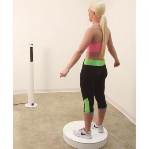 It features a minimum scanning area of 8.4 x 5.5 ft ( 251 x 168 cm). It works at a recommended distance of the sensor tower from the turntable of 46 inches (117 cm). It requires a minimum clearance of 21 inches (53 cm) around the turntable. In case of white walls or glossy surfaces, it might require more clearance.Scanner controlsThe scanner is controlled via an external computer. It features a minimum scanning area of 8.4 x 5.5 ft ( 251 x 168 cm). It works at a recommended distance of the sensor tower from the turntable of 46 inches (117 cm). It requires a minimum clearance of 21 inches (53 cm) around the turntable. In case of white walls or glossy surfaces, it might require more clearance.Scanner controlsThe scanner is controlled via an external computer.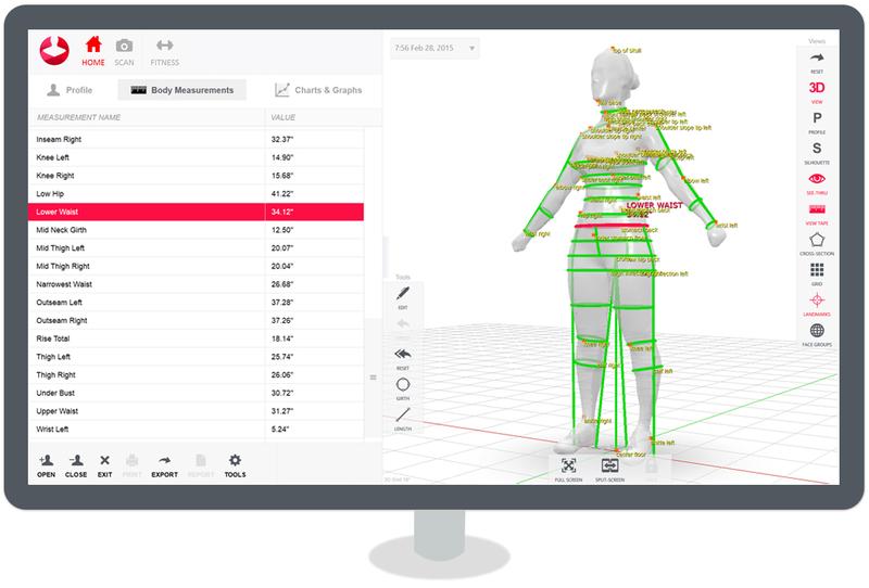 The proprietary Styku intuitive, multi-platform software allows performing a complete scan and all the measurements with ease. The generated avatar can be rotated, panned, and zoomed to evaluate each body part in detail. The collected data are strictly private. You can decide who can view them by setting appropriate export and sharing options. The software is compatible with Windows 10 and is optimized for touchscreens. The proprietary Styku intuitive, multi-platform software allows performing a complete scan and all the measurements with ease. The generated avatar can be rotated, panned, and zoomed to evaluate each body part in detail. The collected data are strictly private. You can decide who can view them by setting appropriate export and sharing options. The software is compatible with Windows 10 and is optimized for touchscreens.What’s in the box
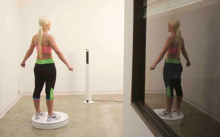 - Essential parts required to operate the Styku 3D body scanner
- Rotating platform (max. weight 250kg)
- Sensor tower
- Microsoft Kinect v2 3D camera
- Aluminum enclosure and stand to protect the 3D Camera
- All the necessary cables
For some items, package content may change. In case of any questions, please get in touch.Look and feel
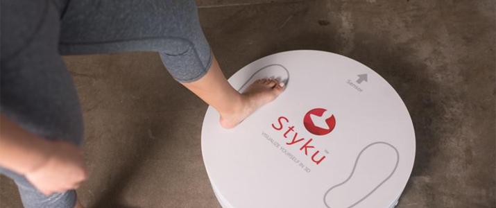 Styku 3D body scanner is available in white with red trims. Its stylish look makes it ideal for fitness clubs, gyms, healthcare facilities, and fashion companies.The scanner has an overall footprint of 101 x 66 inches ( 257cm x 168cm). The rotating platform measures 24 x 24 x 5 inches (66cm x 66cm x 12.5 cm) and weighs 23 lb (10.5 kg). It withstands a max load of 550 lb (250 kg). The sensor tower measures 10 x 10 x 46 inches ( 25.4 x 25.4 x 117 cm). It weighs 13 lb (6 kg). Styku 3D body scanner is available in white with red trims. Its stylish look makes it ideal for fitness clubs, gyms, healthcare facilities, and fashion companies.The scanner has an overall footprint of 101 x 66 inches ( 257cm x 168cm). The rotating platform measures 24 x 24 x 5 inches (66cm x 66cm x 12.5 cm) and weighs 23 lb (10.5 kg). It withstands a max load of 550 lb (250 kg). The sensor tower measures 10 x 10 x 46 inches ( 25.4 x 25.4 x 117 cm). It weighs 13 lb (6 kg). | Soredex Cranex-D Panoramic X Ray
The Soredex Cranex D with cephalometric option provides a full range of digital panoramic and cephalometric dental x-ray examinations to meet the diagnostic needs of almost any general dental or specialty practice. With it’s minimal footprint, this Soredex option will help elevate the diagnostic functionality of any dental provider.Soredex Cranex D provides a full range of high quality digital panoramic x-ray examinations to meet the diagnostic needs of almost any dental practice – all at an affordable price.Acquisition computer includedFeatures Soredex Cranex-D Panoramic X Ray
CRANEX D is a direct digital panoramic and cephalometric x-ray system that provides superior image quality and more diagnostic value. Available as a single or dual sensor system, the CRANEX D grows with your practice, making it a valuable addition to meet the needs of your office. It combines quality images, ease-of-use, and flexibility to make your day-to-day diagnostics easy and accurate. Benefits
The system features superb anterior image quality in panoramic imaging with its patented Variable Panoramic Collimator (VPC) by expanding the focal trough by 50% compared to traditional techniques. Additional innovative features on the unit include Automatic Collimator Selector (ACS) to easily set proper collimation for the imaging program prescribed, as well as the Automatic Exposure Setting (AES) where the system recommends proper cephalometric exposure values based on the size of the patient. These automated and optimized exposure settings maximize the practice’s workflow. CRANEX D is built for contemporary, fast-paced dental care where reliability is key.The Soredex Cranex D with cephalometric option provides a full range of panoramic and cephalometric examinations to meet the needs of almost any practice.SOREDEX has over 30 years of expertise in state-of-the-art dental imaging systems. The Soredex® Cranex D with cephalometric option provides a full range of panoramic and cephalometric examinations to meet the needs of almost any practice. |


Reviews
There are no reviews yet.