| Description | The Philips Affiniti 70, is a high-end shared service ultrasound machine that was developed in 2014 from the top Epiq 7. The Affinity 70 looks nearly identical to the Epiq 5 and 7 plus offers many of the Very Same attributes such as single crystal PureWave transducers | MedCorp is a trusted source for new and refurbished ultrasound systems with a combined 50 years experience in the industry and a detailed process for ensuring that each system they sell is as good as the day it was manufactured. | RealTime 4D imaging – continuous, 3D scanning with simultaneous visualization of the A, B and C planes.
The system allows you to explore images in any plane to reveal the smallest details with stunning clarity and apply sophisticated analytical tools to answer virtually any of your clinical questions.
• 3D Static/4D Real-time Imaging (15 FPS) State-of-the-art user interface with on screen menu’s Automatic Tissue Optimization (ATO) 15″ high resolution non-interlaced flat CRT 4 active probe ports Image Archive on CD, MOD, and Hard Drive (40 GB) B-Mode, M-Mode Tissue Harmonic Imaging Digital Beamformer with 512 system processing channel technology CINE memory: up to 256 MB (up to 3000 2D Images) Power Doppler Imaging (PDI) Tissue Doppler Imaging | The more patients you see, the more you need Voluson E8.
The Voluson E8 ultrasound system is designed to keep pace with busy practices that conduct a wide range of Women’s Health exams, from routine scanning to complex assessments.
The imaging excellence of Radiance System Architecture – your diagnostic confidence will be enhanced by the extraordinary image clarity and speed of Radiant System Architecture. | Acuson sequoia 512 ultrasound click images to enlarge item description acuson sequoia 512 ultrasoundsold as picturedincludes: 2 probes, 4c1 convex & 6l4 linearsony up 895 md printerunit will be cleaned, function checked and certified for electrical safety. Checked, cleaned, certified for electrical safetyguaranteed working when arrives on the guarantee working we need to be notified in 48 hrs. And the seals must be intact. | 15″ LCD monitor, 2 Hot-swappable Probe ports, Keyboard with programmable keys, HPRF (High Pulse Repetition Frequency), MBP (Multi-beam Processing), Trapezoidal Imaging, Panoramic Imaging, Advanced THI (Tissue Harmonic Imaging), TDI (Tissue Doppler Imaging) ,MicroScan (Noise & Artefact Reduction), Multi Parameter Compound Imaging, M Tuning (One Touch Image Optimization), Steerable M mode, uScan, Bi-Plane Transrectal Imaging, Multi-Plane Adult and Paediatric TEE/TOE, Endocavitary Imaging with Extended Field-of-View, Stress Echo, anatomic M mode, PDF report, Automatic flow volume analysis, IMT, HQNF (High Q Noise Filter), 2 USB ports, Video in/out ports, Li-ion battery. |
| Content | Philips Affiniti 70 Ultrasound system
Philips Affiniti 70 ultrasound system for women’s healthcarePhilips Affiniti 70 Ultrasound system delivers a powerful mix of performance and workflow for diagnosis. Engineered for efficiency and dependability and powered by Philips demonstrated performance, it gets users diagnostic pictures, quickly on the most technically difficult patients. Its layout and walk-up usability provide efficient maintenance every day.The Philips Affiniti 70 is a complete echocardiography alternative that addresses the needs of a busy office or department while incorporating those inventions that make Philips ultrasound the international pioneer in echocardiography.Philips Affiniti 70 precision beamforming, PureWave technology, Tissue-Specific Presets (TSP), and efficiency and automation tools provide both functionality and workflow for confident throughput. Designed around your workflow, the Philips Affiniti 70 provides mobility, ergonomics, and usability.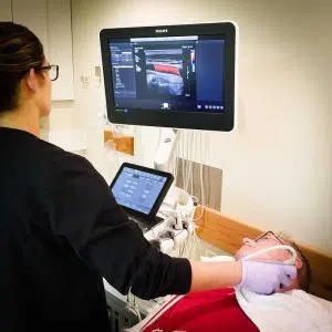 The Affiniti 70 also has many of the same technology as the Epiq series, including the amazing QLAB onboard quantification set and ShareWave elastography. The Philips Affiniti 70 is exceptional in the high-end price category in offering ShearWave. As a service ultrasound device that is shared the 70 excels at cardiology in addition to women’s health, vascular and radiology programs. Philips is well known for its imaging superiority over other brands, but the Philips Affiniti 70 is also rich with excellent image quality at its price level. Doctors who like the Affiniti 70, but do not need the transducers or ShearWave Elastography should consider the less costly Affiniti 50.The Philips Affiniti 70 combines outstanding performance and effective workflow and attributes innovative Philips technologies. The Precision Beamforming delivers outstanding tissue uniformity and contrast resolution, excellent spatial, few artifacts, and decreased image jumble. Tissue-Specific Presets (TSPs) mechanically adjust over 7500 parameters to optimize the transducer to your particular test type, making excellent picture quality with little if any demand for picture adjustment. The Affiniti 70 Machine can also be equipped with automation features, for example, AutoSCAN and automobile Doppler, which reduce repetitive tasks and make obtaining a picture. Exam efficiency is delivered by Philips SmartExam-guided workflow.Key Features: The Affiniti 70 also has many of the same technology as the Epiq series, including the amazing QLAB onboard quantification set and ShareWave elastography. The Philips Affiniti 70 is exceptional in the high-end price category in offering ShearWave. As a service ultrasound device that is shared the 70 excels at cardiology in addition to women’s health, vascular and radiology programs. Philips is well known for its imaging superiority over other brands, but the Philips Affiniti 70 is also rich with excellent image quality at its price level. Doctors who like the Affiniti 70, but do not need the transducers or ShearWave Elastography should consider the less costly Affiniti 50.The Philips Affiniti 70 combines outstanding performance and effective workflow and attributes innovative Philips technologies. The Precision Beamforming delivers outstanding tissue uniformity and contrast resolution, excellent spatial, few artifacts, and decreased image jumble. Tissue-Specific Presets (TSPs) mechanically adjust over 7500 parameters to optimize the transducer to your particular test type, making excellent picture quality with little if any demand for picture adjustment. The Affiniti 70 Machine can also be equipped with automation features, for example, AutoSCAN and automobile Doppler, which reduce repetitive tasks and make obtaining a picture. Exam efficiency is delivered by Philips SmartExam-guided workflow.Key Features:- 21.5” wide flat panel LCD display monitor
- Fully articulating arm
- 2” Tablet-like widescreen touchscreen
- 4 probe ports
- TDI [Tissue Doppler Imaging]
- THI [Tissue Harmonic Imaging]
- TAC [Tissue aberration correction]
- LVO [Left ventricular opacification]
- CPA and Directional CPA [Color Power Angio]
- Coded beamforming
- Steerable CW Doppler
- High-PRF PW Doppler
- Auto Doppler optimization
| SonoSite Nanomaxx Ultrasound MachineThe SonoSite NanoMaxx ultrasound system combines premium functionality and affordability with the versatility and portability that makes SonoSite a top competitor in the portable ultrasound market. The SonoSite NanoMaxx comes in a sleek, hand-held package that never compromises performance, with applications ranging from emergency medicine and anesthesia to vascular.MedCorp offers the refurbished SonoSite NanoMaxx for purchase. Used SonoSite NanoMaxx from MedCorp offer everything you’d expect from a newer machine, including PC/MAC export configurations, 2D, Color and Color Power Doppler modes, and 2GB of internal memory that can hold 50 images each for 40 patients, all in an 8” touch-screen interface. However, because this is a refurbished SonoSite NanoMaxx, the price is lower than the new models. - 8″ Touch-screen interface
- 2D, Color and Color Power Doppler modes
- 2GB internal memory that can hold 50 images each for 40 patients
- 2 hours of battery power
- PC/MAC export configuration
- Four preset pictograph screen positions
- Drop resistant magnesium case
System Description:SonoSite Nanomaxx Ultrasound Machine
The NanoMaxx ultrasound system offers excellent performance at an affordable price. It is easy to use and offers top of the line diagnostic imaging, color power Doppler and color-flow velocity. The NanoMaxx can aid users in making clinical decisions or guide interventional procedures. The unit uses proprietary technology which allows the optimization of many system settings by simply pressing a button. Application:
Abdominal, Cardiology, Gynecology, Nerve, Obstetrics, CIMT, Musculoskeletal, Small Parts, Superficial, Vascular, Venous | GE Voluson 730 Pro Ultrasound MachineThe Voluson 730 Pro is built on the same platform as GE’s leading Real-Time 4D system, the Voluson 730 Expert. With an easy to use interface and exceptional 2D image quality, in addition to GE’s exclusive 4D imaging technology, the Voluson 730 Pro is ideal for any clinical practice with a variety of patient workloads. Recognized world-wide for its 4D imaging capabilities, the Voluson 730 Pro also features exceptional 2D image quality.RealTime 4D imaging – continuous, 3D scanning with simultaneous visualization of the A, B and C planes.
The system allows you to explore images in any plane to reveal the smallest details with stunning clarity and apply sophisticated analytical tools to answer virtually any of your clinical questions.Performance:GE Voluson 730 Pro Ultrasound Machine
State-of-the-art user interface with high resolution 10.4 inch LCD touch panel
Automatic Tissue Optimization
Tissue Doppler
Coded Harmonic Imaging
Coded Excitation (CE)
HD-Flow
CrossXBeamCRI (Compound Resolution Imaging)
Static 3D Mode
B Mode
Focus & Frequency Composite (FFC)
High Resolution Zoom
Pan Zoom
Steering
Virtual Convex
Beta-View
Patient information database
Image Archive on MOD and hard drive
3D/4D data compression (lossy/lossless)
Inversion
Real-time automatic Doppler calcsCapabilities:
OB/GYN, Vascular, Cardio, Abdominal, Small-Parts, Urology, Pediatrics, Ortho, Neurology, Multigestational Calculations, B-Flow, VCI (Volume Contrast Imaging)Available Probes:
AB2-7 Wide Band Convex Probe
AC2-5 Wide Band Convex Probe
4C-A Wide Band Convex Probe
M7C-H Wide Band Convex Probe
IC5-9H Wide Band Convex Probe
PA2-5P Wide Band Phased Array Probe
PA6-8 Wide Band Phased Array Probe
SP10-16 Wide Band Linear Probe
SP4-10 Wide Band Linear Probe
SP6-12 Wide Band Linear Probe
M12L-H Wide Band Linear Probe (1.25D Array)
RAB2-5L Wide Band Convex Volume Probe
RAB4-8L Wide Band Convex Volume Probe
RIC5-9H Wide Band Convex Volume Probe
RIC5-9W Wide Band Convex Volume Probe
RRE6-10 Wide Band Convex Volume Probe
RSP6-16 Wide Band Linear Volume Probe
RNA5-9 Wide Band Convex Volume Probe
SCW 2.0; non imaging suprasternal CW-Pencil Probe
PCW 4.0; non imaging peripheral CW-Pencil Probe | GE Voluson E8 Ultrasound Machine15’’ LCD monitor [Rev BT06], 10.4” touchscreen, 3 Active Probe Ports, Floating Keyboard: Interactive back-lighting, Beta-View CrossXBeam, Virtual Convex, Extended View, ATO [Automatic Tissue Optimization], Coded Harmonic Imaging, High Resolution Zoom, Inversion Real-time automatic Doppler, OB/GYN; Vasc. Cardiac; Abd; Small-Parts; Urology; Ped; Ortho; Neuro reporting, Multigestational Calculations, HPRF [High Pulse Repetition Frequency], SonoVCAD [Matrix array volume technology], TruScan [On-board archive], VCI [Volume Contrast Imaging], SonoAVCfollicle [Sonography-based Automated Volume Count follicle], SonoVCADheart [Sonography-based Volume Computer Aided Display], TUI [Tomographic Ultrasound Imaging], CE [Coded Excitation], XTD, SRI III [Speckle reduction imaging], CRI [Compound Resolution Imaging], DICOM, Ethernet port, Video Out, 6 USB ports, Integrated HDD (160 GB) and DVD/CD-RW.
3D/4D, Endocrinology, Family Practice, Gastroenterology, Internal Medicine, Nephrology, Neurology, OB-GYN, Orthopedics, Otorhinolaryngology, Pain Management, Urology, Vascular Probes GE Voluson E8 Ultrasound Machine - 11L-D Linear Array
- 3S-D Cardiac Sector
- 4C-D Convex Array
- 9L-D Linear Array
- AB2-7 Convex
- IC5-9-D Intercavity
- M6C-D Convex
- P2D CWD Non-Imaging
- P6D CWD Non-Imaging
- PA6-8-D Cardiac
- RAB2-5-D 3D/ 4D Convex
- RAB4-8-D 3D/4D Convex
- RIC5-9-D 3D/4D Intracavity
- RIC6-12-D 3D/4D Intracavity
- RNA5-9-D 3D/4D Convex
- RRE6-10-D Endocavitary Micro-Convex
- RSP6-16-D 3D/D Linear
- SP10-16-D Linear
- 19″ monitor
- 4D Real Time
- CFM Doppler Mode
- Coded Contrast Imaging
- CrossBeam
- DICOM 4D
- HD flow
- Power Doppler Mode
- SonoVCAD
- STIC (Spacial-Temporal Image Correlation)
- T.U.I (Tomographic Ulrasound Imaging)
- VOCAL II VCI (Volume Contrast Imaging)
- Black and white printer
- Color printer
- DVD-Recorder
As your patient volume increases and your clinical cases become more complex, you will require an ultrasound system that enables you to deliver truly exceptional care – confidently and efficiently – every time. The Voluson E8 ultrasound system is designed to keep pace with busy practices – handling routine to complex women’s health exams with ease and precision. The premium image quality of the Voluson E8 combined with access to advanced capabilities, delivers the level of performance required by you and your patients. | Siemens / Acuson Sequoia 512 Ultrasound Machine
The ACUSON Sequoia 512 ultrasound is an ultra-premium system that utilizes revolutionary technology, dedicated to the advancement of diagnostic imaging. It represents some of the most advanced technology in ultrasound imaging. The Acuson Sequoia 512 has unparalleled imaging performance, greater image content, enhanced detail, superior contrast resolution & penetration, and excellent color Doppler sensitivity.One of the best selling ultrasounds in the United States, the Sequois 512 provides superior imaging with Native Patient Specific Imaging that detects, measures and adapts (in real time) to each patient’s individual acoustics.Performance:Reduce image optimization time by up to 80% and allow for an easier, faster and more confident diagnosis:- 2D
- SST Color Doppler
- Color Doppler Energy
- Color Doppler Velocity
- Solo Spectral Doppler
- M-mode and Color Doppler M-mode
- MultiHertz Multiple Frequency Imaging
- DELTA Differential Echo Amplification
- Home Base user interface
- One-Touch Image Control with exam and Image Presets
- Digital acquistion, storage, review and transfer of static images, dynamic clips, and cine
- Digital storage on an integrated hard disk
- Digital transfer via MO Disk
- Integrated Compression engine
- Embedded DICOM conformance
- Patient- oriented file structure
- Native Tissue Harmonic Imaging
- Tissue Equalization™ Technology (Optional)
- Signature II™ (Optional)
- Advanced Imaging package (Optional)
- Auto Doppler (Optional)
- Freestyle Compounding (Optional)
- FreeStyle Extended Imaging (Optional)
- 3D surface Rendering (Optional)
- Cadence™ ADI (Optional)
- Cadence CPS for 512 (Optional)
- Axius™ Edge Assisted EF (Optional)
- Doppler Tissue Imaging (Optional)
- DICOM Print And Store (Optional)
Capabilities:Siemens / Acuson Sequoia 512 Ultrasound MachineGeneral Radiology, Abdominal, Intraoperative Monitoring, OB/GYN, Neonatal, Vascular, Cardiac, Small PartsAvailable Transducers:4V1, 4V2, 8V5, 15L8W, 6L3, 8L5, 6C2, 5C2, 4C1, 3V2C, 5V2C,7V3C, EVC8-4V, EC-10C5, V5M TEE, V7B neo TEE, AcuNav Transducer, 2.0 Non Imaging CW Doppler probe.Acuson sequoia 512 ultrasound click images to enlarge item description acuson sequoia 512 ultrasoundsold as picturedincludes: 2 probes, 4c1 convex & 6l4 linearsony up 895 md printerunit will be cleaned, function checked and certified for electrical safety. Checked, cleaned, certified for electrical safetyguaranteed working when arrives on the guarantee working we need to be notified in 48 hrs. And the seals must be intact. | SonoScape S8 Portable UltrasoundSystem Description: The SonoScape S8 portable ultrasound is an award winning portable ultrasound system with outstanding image quality in all modalities. SonoScape has designed a fully featured, shared service ultrasound system producing image quality one would expect from a much more expensive console system. Frost & Sullivan “Product Quality Leadership” Award:
“The S8 is versatile in performance,” notes Frost & Sullivan Research Analyst Shriram Shanmugham. “It uses a 15 MHz linear transducer, 512 super high density transducers, and is the only echocardiography system that also offers 4D imaging capability. None of the company’s competitors offers such a combination of 4D and high-end echocardiography technology affording portability.” “The high frame rate and high definition image quality provide users better diagnostic experience than ever” Application:- Shared Service!
- Ob/Gyn
- Anesthesia
- Urology
- Vascular / Vascular Access
- MSK
- Breast Imaging
- Abdominal
- Small Parts
- Cardiac
Features:- 15” LCD Monitor
- 2 Hot Swap Probe Ports
- High Pulse Repetition Frequency [HPRF]
- Multi Beam Processing [MBP]
- Trapezoidal Imaging
- Panoramic
- Imaging
- Advanced Tissue Harmonic Imaging
- M-Tuning [One touch image optimization]
- uScan
- Multi Plane Adult & Pediatric TEE
- Intimae Media Thickness [IMT]
- High Q Nosie Filter [HQNF]
- B-Mode
- M-Mode
- Color Doppler
- Power Doppler
- Spectral Steered Doppler
- Real time Triplex: B, Flow & PW Doppler / B, Flow & Color M
- ECG
- 4D
- Tissue Doppler Imaging
- B-Color
- Anatomic M-Mode
- DICOM 3.0
- 2 USB Ports
- Video In/Out
| | Options:- Mobile Trolley
- DVD/CD-RW
- Printer
- Stress Echo
Transducers:- C344, 5-2MHz/R40mm, Abdominal Convex Probe
- C362, 5-2MHz/R60mm, Abdominal Convex probe
- C542, 7-4MHz/R40mm, Abdominal & Pediatric Probe
- C311, 4-2MHz/R15mm, Micro-convex Cardiac Probe
- C611, 8-4MHz/R11mm, Micro-convex Pediatric Cardiac Probe
- L741, 10-5MHz/46mm, Vascular Small part Linear Probe
- L742, 15-5MHz/38mm, Vascular Small part linear probe
- L743, 15-5MHz/46mm, Vascular Small part linear probe
- L541, 7-4MHz/38mm, Vascular Linear probe
- 10L1, 12-6MHz/36mm, Vascular Small Parts Probe
- 10I2, 15-5MHz/25mm, Linear Surgical Probe
- 6V1, 8-4MHz/R11mm, Endovaginal Probe (135 degree)
- 6V3, 9-5MHz/R10mm, Endovaginal probe (200 degree)
- EC9-5, 9-5MHz/R8mm, micro-convex Endocavity probe (150 degree)
- 2P1, 4-2MHz, Phased Array Cardiac Probe
- 5P1, 7-4MHz, Phased Array Pediatric Cardiac Probe
- MPTEE, 7-4MHz, Multi-plane Phased Array TEE Probe
- MPTEE Mini, 7-4MHz Pediatric Multi-plane Phased Array TEE Probe
- VC6-2, 6-2MHz/R40mm, Curved volumetric probe for Live 3D
Biopsy Guides:- C344, C362, L741, L742, L743, 10LI, L541, 6V1, 6V3, EC9-5, 2P1, 7U2
|
|


Reviews
There are no reviews yet.