| Description | The Philips Affiniti 70, is a high-end shared service ultrasound machine that was developed in 2014 from the top Epiq 7. The Affinity 70 looks nearly identical to the Epiq 5 and 7 plus offers many of the Very Same attributes such as single crystal PureWave transducers | The Expert is the new flagship LOGIQ e. The imaging engine comes from GE leading console systems, delivering crisp images in a compact package. Providing ultrasound imaging with precise anatomical detail at a variety of depths. The system includes innovative features that help simplify proceedures. | Solid imaging performance.
The sleek LOGIQ A5 can help meet your general imaging needs. This black and white ultrasound system has:- B-mode imaging capability and features that allow you to add advancements as your clinical needs grow.
- Auto TGC helps ensure a homogeneous picture from the near-to-far field.
- Virtual Convex provides an expanded field of view that enables you to see more anatomy with linear transducers.
- Color flow Doppler upgrade available to provide dynamic assessment of blood flow in the organs, as well as investigation of hypertension and resulting erectile dysfunction.
| Portable Color ultrasound System
This is a portable color ultrasound system that displays good image quality for diverse applications. This small and compact system has various diagnostic capabilities. | Highly Precise, Non-Surgical, Medical Procedure System for Professionals. Doublo HIFU’s Speed is Innovative Performance! Treatment time is dramatically reduced with Hironic’s High-capacity Technology and with Doublo HIFU System you can treat twice the amount of patients than beforeNew Standard of
MFU System
1.No. 1 MFU proven by the patents2.MFU proven by cumulative sales and clinical cases3.Fastest treatment by Two-way round shooting and Auto Shot mode | The GE Vivid q portable ultrasound machine is a more powerful version of the Vivid i that adds further quantitative features including ICE, Auto EF, AFI, TVI,TT, TSI, Blood Flow and Blood Flow imaging. The Vivid q is the pinnacle of GE’s portable cardiovascular ultrasounds. |
| Content | Philips Affiniti 70 Ultrasound system
Philips Affiniti 70 ultrasound system for women’s healthcarePhilips Affiniti 70 Ultrasound system delivers a powerful mix of performance and workflow for diagnosis. Engineered for efficiency and dependability and powered by Philips demonstrated performance, it gets users diagnostic pictures, quickly on the most technically difficult patients. Its layout and walk-up usability provide efficient maintenance every day.The Philips Affiniti 70 is a complete echocardiography alternative that addresses the needs of a busy office or department while incorporating those inventions that make Philips ultrasound the international pioneer in echocardiography.Philips Affiniti 70 precision beamforming, PureWave technology, Tissue-Specific Presets (TSP), and efficiency and automation tools provide both functionality and workflow for confident throughput. Designed around your workflow, the Philips Affiniti 70 provides mobility, ergonomics, and usability.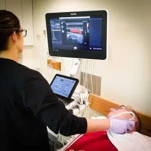 The Affiniti 70 also has many of the same technology as the Epiq series, including the amazing QLAB onboard quantification set and ShareWave elastography. The Philips Affiniti 70 is exceptional in the high-end price category in offering ShearWave. As a service ultrasound device that is shared the 70 excels at cardiology in addition to women’s health, vascular and radiology programs. Philips is well known for its imaging superiority over other brands, but the Philips Affiniti 70 is also rich with excellent image quality at its price level. Doctors who like the Affiniti 70, but do not need the transducers or ShearWave Elastography should consider the less costly Affiniti 50.The Philips Affiniti 70 combines outstanding performance and effective workflow and attributes innovative Philips technologies. The Precision Beamforming delivers outstanding tissue uniformity and contrast resolution, excellent spatial, few artifacts, and decreased image jumble. Tissue-Specific Presets (TSPs) mechanically adjust over 7500 parameters to optimize the transducer to your particular test type, making excellent picture quality with little if any demand for picture adjustment. The Affiniti 70 Machine can also be equipped with automation features, for example, AutoSCAN and automobile Doppler, which reduce repetitive tasks and make obtaining a picture. Exam efficiency is delivered by Philips SmartExam-guided workflow.Key Features: The Affiniti 70 also has many of the same technology as the Epiq series, including the amazing QLAB onboard quantification set and ShareWave elastography. The Philips Affiniti 70 is exceptional in the high-end price category in offering ShearWave. As a service ultrasound device that is shared the 70 excels at cardiology in addition to women’s health, vascular and radiology programs. Philips is well known for its imaging superiority over other brands, but the Philips Affiniti 70 is also rich with excellent image quality at its price level. Doctors who like the Affiniti 70, but do not need the transducers or ShearWave Elastography should consider the less costly Affiniti 50.The Philips Affiniti 70 combines outstanding performance and effective workflow and attributes innovative Philips technologies. The Precision Beamforming delivers outstanding tissue uniformity and contrast resolution, excellent spatial, few artifacts, and decreased image jumble. Tissue-Specific Presets (TSPs) mechanically adjust over 7500 parameters to optimize the transducer to your particular test type, making excellent picture quality with little if any demand for picture adjustment. The Affiniti 70 Machine can also be equipped with automation features, for example, AutoSCAN and automobile Doppler, which reduce repetitive tasks and make obtaining a picture. Exam efficiency is delivered by Philips SmartExam-guided workflow.Key Features:- 21.5” wide flat panel LCD display monitor
- Fully articulating arm
- 2” Tablet-like widescreen touchscreen
- 4 probe ports
- TDI [Tissue Doppler Imaging]
- THI [Tissue Harmonic Imaging]
- TAC [Tissue aberration correction]
- LVO [Left ventricular opacification]
- CPA and Directional CPA [Color Power Angio]
- Coded beamforming
- Steerable CW Doppler
- High-PRF PW Doppler
- Auto Doppler optimization
| GE Logiq e r7 Portable Ultrasound Machine
The LOGIQ e R7 provides even more impressive veterinary abdominal and cardiac images than the BT12 LOGIQe, combining the high performance of a console system with the portability of a laptop.Highlights of the GE LOGIQ e R7:- Simple. Fast. Precise
- Colour Flow Mode, Power Doppler Imaging (PDI), Pulse Wave Doppler (PWD), Continuous Wave Doppler
- 15” high resolution colour LCD screen
- Integrated solid state hard drive
- Automatic optimisation
- CrossXBeam
- Speckle Reduction Imaging (SRI-HD)
- Virtual Convex / Virtual Apex
- Fine Angle Steer
- HD Zoom (Write Zoom)
- Coded Harmonic Imaging (CHI) – Enhances near field reslolution for improved small parts imaging as well as far field penetration. Diminshes low frequency / amplitude noise and provides clarity to needle, anatomy and motion.
- Raw Data Processing
- Quicksave – Single button push sends single image or entire patient exam to memory stick or network
- On-board manual (Help)
- Loop storage – from live scanning and from memory
- Patient Information Database
- Customisable user interface
- Full M&A calculation package with real time Doppler calculations
- Report writer package – on board reporting package automates report writting
- Portable and battery operated so easy to move from room to room
- Export of stills and clips to USB.
More GE Logiq e r7 Portable Ultrasound MachineThe Expert is the new flagship LOGIQ e. The imaging engine comes from GE leading console systems, delivering crisp images in a compact package. Providing ultrasound imaging with precise anatomical detail at a variety of depths. The system includes innovative features that help simplify proceedures.GE NextGen LOGIQ e R7 Ultrasound System The GE NextGen LOGIQ e R7 Ultrasound System has an imaging engine that delivers crisp images in a compact package. Point-of-care-specific software and transducers help to see the needle to administer a block or perform an aspiration quickly. When productivity is crucial, you can control the console from the transducer, potentially eliminating the need to have a second person assist. Anatomy-specific presets and only displaying the functions that you need helps you to direct your focus on the patient rather than the control panel. Although the system features a large, user-friendly, high-res color display, it is also very durable and compact for easy portability. | GE Logiq A5 Ultrasound MachineThe lightweight, portable LOGIQ* A5 ultrasound system offers capabilities to help meet your imaging needs — particularly for urological applications.Affordable ultrasound.The LOGIQ A5 puts imaging capabilities at your fingertips in a portable, lightweight design. This system can go from your office to the surgery suite and helps meet a range of general imaging needs. Well-suited for urology, tools include soft tissue image analysis of the kidneys, bladder, penis, testicles, and prostate.Solid imaging performance.
The sleek LOGIQ A5 can help meet your general imaging needs. This black and white ultrasound system has:- B-mode imaging capability and features that allow you to add advancements as your clinical needs grow.
- Auto TGC helps ensure a homogeneous picture from the near-to-far field.
- Virtual Convex provides an expanded field of view that enables you to see more anatomy with linear transducers.
- Color flow Doppler upgrade available to provide dynamic assessment of blood flow in the organs, as well as investigation of hypertension and resulting erectile dysfunction.
Perform imaging with ease.
The LOGIQ A5 has user-friendly features with precision handling.- Automatic Optimization (AO), helps provide improved image quality at the touch of a button for B-mode, spectral and color Doppler imaging.
- Full-size keyboard for optimal ease of use.
- 15-inch adjustable LCD monitor allows a clear view in any position.
- Lightweight package easily travels with you.
Compatible Transducers:- 10L Linear Array
- 11L Wide Band Linear
- 12L Linear Array
- 3.5C Convex
- 3.5CS Convex (2-5 MHz)
- 3CRF Convex
- 3S Phased Array
- 3Sp Wide Band Phased Array Sector
- 4C Convex Array
- 4D3CL Real time 4D Convex
- 4D8C Convex Volume
- 4DE7C Real time 4D Endocavity
- 5CS Convex (2-5 MHz)
- 5S Cardiac Sector
- 5Sp Wide Band Phased Array Sector
- 7S Phased Array
- 8C Convex Array
- 8L Linear (3-7 MHz)
- 9L Wide Band Linear
- BE9C Transrectal
- BE9CS Micro convex Bi-plan
- E8C (4-10 MHz) Wide Band Microconvex
- E8CS Micro-convex (4-11 MHz)
- ERB Brachytherapy Transrectal
- i12L Linear Array
- i739 Intraoperative
- P2D CWD Non-Imaging
- P6D CWD Non-Imaging
- T739 Intra-operative
| Samsung Medison SonoAce R3 Ultrasound MachineSonoAce R3 is a portable color ultrasound system with comprehensive diagnostic capabilities. Full Spectrum Imaging (FSI) combines with Speckle Reduction Filter (SRF) to deliver superior quality images, even in the challenging circumstances. The compact and user-friendly design features of the SonoAce R3 ensure portability and manoeuvrability in a lightweight unit. Sporting a 15-inch LCD monitor with Full-Featured Workflow, the SonoAceR3 offers your organisation flexibility, without compromising on precision and efficiency.Slim and Compact DesignThe compact design of SonoAce R3 offers Various-Featured Workflow with a thumbnail menu and shortcut, together with functional key groupings for increased efficiency. The user-friendly design combines a wide-view 15-inch LCD monitor with backlit control keys, customisable measurement package and body markers. The SonoAce R3 features 2 probe ports, 3 USB ports, rapid boot up capability and DICOM 3 compatible image filing. This slim, lightweight unit also has a convenient carrying handle for portability and flexibility.Full Spectrum Imaging (FSI) produces the penetration capabilities associated with the lower frequencies, while still maintaining the fine pixel uniformity associated with the higher frequencies. This combination consistently delivers superior quality images, even in the challenging diagnostic circumstances. The resulting clarity delivers improvements in accuracy and efficiency for greater confidence in your diagnostic procedures. Harmonic Imaging is a technique that records higher frequency information, resulting in greater resolution and fewer artifacts.Synthetic Aperture
The physical radio channel may present some limits to imaging which can be overcome by using Synthetic Aperture Control software. This method uses information from diverse sources to form a more better picture of the area under examination. It creates one single scan line by performing the TX and RX twice to create enhanced penetration and resolution. Synthetic Aperture Control technology delivers a comprehensive picture and to aid accurate diagnosis.QuickScan
QuickScan has been designed to increase workflow efficiency by automatically optimising key imaging parameters at the touch of a button. The resulting increase in efficiency allows your organisation to offer patients an improved service with faster and more accurate diagnoses. This reliable and safe system helps your clinic or hospital to deliver a superior patient experience.Features Samsung Medison SonoAce R3 Ultrasound Machine
Full Spectrum ImagingTM
Quick ScanTM
Color and Power Doppler
Harmonic imaging
Freehand 3D
3D Multi Planar Imaging
Sonoview Image Management
PW Doppler
15″ LCD monitor
Two probe ports
Lightweight | DOUBLO GOLD HIFU SYSTEMWhy choose Doublo HIFU System?
HIFU (High-Intensity Focused Ultrasound System) is a highly precise medical procedure using high-intensity focused ultrasound to heat and destroy the pathogenic tissue rapidly. The earliest widespread use of HIFU was as a treatment for prostate cancer.HIFU hybrid system for Face & Body
Doublo Gold is the ultimate HIFU system for precise Face Lifting & Body Contouring. Offering the fastest treatment by Two-way round shooting and Auto Shot mode.Non Invasive – No Pain – No Bleeding – No Downtime
Faster shot speed : Efficacy improved by repetitious shooting with 300 shots in 8 minutes. Entertain more patients with a faster treatment procedure.Multiple treatment areas
By treating the upper dermis with wound healing effect, facial pores and skin tone is improved as the fibroblast is rebuilt and collagen is boosted.Dermal tissue elasticity and collagen increase as fibroblast is rebuilt by wound healing effect from damaging sub-cutaneous tissue and derma layer with thermal coagulation.
Skin tightening to reduce wrinkle and increase lifting effect is done by creating thermal coagulation at subcutaneous fat and SMASDOUBLO GOLD HIFU SYSTEM Highly Precise, Non-Surgical, Medical Procedure System for Professionals. Doublo HIFU’s Speed is Innovative Performance! Treatment time is dramatically reduced with Hironic’s High-capacity Technology and with Doublo HIFU System you can treat twice the amount of patients than beforeDOUBLO GOLD HIFU SYSTEM Accessories: Power cord for cart
Clear plastic tray insert
Large washer and small lock/spring washer
2x black attachements
2x treatment parameters guide
Simple user manual
Full user manual + CDHighly Precise, Non-Surgical, Medical Procedure System for Professionals. Doublo HIFU’s Speed is Innovative Performance! Treatment time is dramatically reduced with Hironic’s High-capacity Technology and with Doublo HIFU System you can treat twice the amount of patients than beforeNew Standard of
MFU System
1.No. 1 MFU proven by the patents2.MFU proven by cumulative sales and clinical cases3.Fastest treatment by Two-way round shooting and Auto Shot mode | GE Vivid Q Ultrasound Machine
The powerful GE Vivid Q ultrasound machine is the ultimate lightweight, compact high-performance cardiovascular system for clinicians needing a machine that combines user efficiency and reliable performance. This unique system performs consistently thanks to its TruScan architecture and can support up to 17 probes. The GE Vivid Q is recognized for its premium image quality, advanced image management, and high-performance quantification tools.This advanced GE ultrasound delivers improved patient care and enhanced user productivity with the new M4s-Matrix array technology transducer, which provides greatly enhanced image quality in electronic beamforming, raw data image acquisition and archiving, optimal onboard and post processing technology.This ultrasound system provides high clinical values for quantification and review of stored images, making it one of today’s most sought-after models. Some of its powerful features include compatibility with EchoPac clinical workstation and DICOM networking, speckle tracking strain, integrated stress echo, quantification tools, and reporting capabilities. Also of note is one of the Vivid Q’s best new features, which is its TEE transducer capability.A one-touch button optimization provides premium image quality for workflow efficiency, and maintains AFI- automatic function imaging with complete wallFeatures- Advanced QScan Imaging (incl TSI)
- AdvStress
- ATO
- Auto EF
- Automated Function Imaging
- Blood Flow Imaging
- CardiacApp
- Cardio Lab
- Color Doppler
- CW Doppler
- DICOM Networking
- DICOM: Print
- DICOM: Worklist
- DVD/RW
- ECG
- ECG Cable
- EchoDicom
- EchoPAC
- Evue
- ICE Imaging
- Intima Media Thickness (IMT)
- LVO Contrast
- M Mode
- M4S
- MPEGvue
- PW Doppler
- Small Depth
- TEE
- THI
- Tissue Harmonics Imaging (THI/NTHI)
- Tissue Velocity Imagine & Tissue Tracking
- TSI
- VascAbdContrast
- VascularApp
- VirtualPrinter
- Wide Aperture
GE Vivid Q FeaturesThe GE Vivid Qis a Portable Laptop-style Ultrasound that can be used to help diagnose cardiovascular anatomy. Features and tools based on the previous Vivid 7 and the Vivid E9 have been added to the new lightweight Vivid Q, mainly the M4S-RS transducer that allows for 3D image focusing and enables a higher level of image quality. The Vivid Q ultrasound is versatile and can additionally be used for Cardiac, vascular, Abdominal, Pediatric, Obstetrics, and Emergency medicine. The Vivid Q Ultrasound comes with a 15-inch color LCD monitor that can display a variety of imaging modes including; ICE, Auto EF, AFI, TVI, TT, TSI, and Blood Flow. Additional the Ultrasound system is comparable with 17 different transducers and probes to fit every imaging need. - Auto EF - Automated ejection fraction measurement program designed specifically for the GE Vivid q.
- Tissue Synchronization Imaging (TSI).
- Intracardiac Echo (ICE).
- 16 different compatible probes.
- Coded Octave imaging - enhances image color flow and Doppler imaging.
|


Reviews
There are no reviews yet.