| Description | The Philips Affiniti 70, is a high-end shared service ultrasound machine that was developed in 2014 from the top Epiq 7. The Affinity 70 looks nearly identical to the Epiq 5 and 7 plus offers many of the Very Same attributes such as single crystal PureWave transducers | GE Voluson 730 Expert is among the top-regarded ultrasounds in the industry. With up to 40 volumes per second rate of acquisition and construction of volumetric images in real time. The 730 Expert is loaded with the newest ultrasound technology including: CrossXBeam, Speckle Reduction Imaging, Tomographic Ultrasound Imaging (TUI), Spatio-Temporal Image Correlation (STIC), and much more. | The Samsung Medison SonoAce R3 portable ultrasound provides the image quality and features of a premium system at a fraction of the cost. The Samsung Medison SonoAce R3 portable ultrasound is brought to you by Samsung / Medison, a company whose name has been trusted in ultrasound since 1985. The Samsung Medison SonoAce R3 portable ultrasound is intuitive and easy to navigate with sophisticated software to increase workflow. | Acuson Cypress cardiovascular system PLUS delivers advanced ergonomics and superior performance in a highly portable package. The Cypress System PLUS is a highly miniaturized, all digital, phased array echocardiography system that provides complete studies and outstanding images – even on patients who are difficult to image. | Acuson sequoia 512 ultrasound click images to enlarge item description acuson sequoia 512 ultrasoundsold as picturedincludes: 2 probes, 4c1 convex & 6l4 linearsony up 895 md printerunit will be cleaned, function checked and certified for electrical safety. Checked, cleaned, certified for electrical safetyguaranteed working when arrives on the guarantee working we need to be notified in 48 hrs. And the seals must be intact. | The New Terason uSmart 3200T Ultrasound System, the first in Terason’s uSmart family of proprietary products. This revolutionary new tablet, deemed “disruptive technology” by industry leaders, will change the way healthcare is delivered in both hospital and outpatient sectors of point-of-care ultrasound. |
| Content | Philips Affiniti 70 Ultrasound system
Philips Affiniti 70 ultrasound system for women’s healthcarePhilips Affiniti 70 Ultrasound system delivers a powerful mix of performance and workflow for diagnosis. Engineered for efficiency and dependability and powered by Philips demonstrated performance, it gets users diagnostic pictures, quickly on the most technically difficult patients. Its layout and walk-up usability provide efficient maintenance every day.The Philips Affiniti 70 is a complete echocardiography alternative that addresses the needs of a busy office or department while incorporating those inventions that make Philips ultrasound the international pioneer in echocardiography.Philips Affiniti 70 precision beamforming, PureWave technology, Tissue-Specific Presets (TSP), and efficiency and automation tools provide both functionality and workflow for confident throughput. Designed around your workflow, the Philips Affiniti 70 provides mobility, ergonomics, and usability.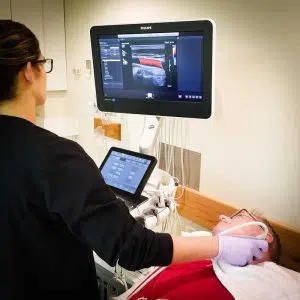 The Affiniti 70 also has many of the same technology as the Epiq series, including the amazing QLAB onboard quantification set and ShareWave elastography. The Philips Affiniti 70 is exceptional in the high-end price category in offering ShearWave. As a service ultrasound device that is shared the 70 excels at cardiology in addition to women’s health, vascular and radiology programs. Philips is well known for its imaging superiority over other brands, but the Philips Affiniti 70 is also rich with excellent image quality at its price level. Doctors who like the Affiniti 70, but do not need the transducers or ShearWave Elastography should consider the less costly Affiniti 50.The Philips Affiniti 70 combines outstanding performance and effective workflow and attributes innovative Philips technologies. The Precision Beamforming delivers outstanding tissue uniformity and contrast resolution, excellent spatial, few artifacts, and decreased image jumble. Tissue-Specific Presets (TSPs) mechanically adjust over 7500 parameters to optimize the transducer to your particular test type, making excellent picture quality with little if any demand for picture adjustment. The Affiniti 70 Machine can also be equipped with automation features, for example, AutoSCAN and automobile Doppler, which reduce repetitive tasks and make obtaining a picture. Exam efficiency is delivered by Philips SmartExam-guided workflow.Key Features: The Affiniti 70 also has many of the same technology as the Epiq series, including the amazing QLAB onboard quantification set and ShareWave elastography. The Philips Affiniti 70 is exceptional in the high-end price category in offering ShearWave. As a service ultrasound device that is shared the 70 excels at cardiology in addition to women’s health, vascular and radiology programs. Philips is well known for its imaging superiority over other brands, but the Philips Affiniti 70 is also rich with excellent image quality at its price level. Doctors who like the Affiniti 70, but do not need the transducers or ShearWave Elastography should consider the less costly Affiniti 50.The Philips Affiniti 70 combines outstanding performance and effective workflow and attributes innovative Philips technologies. The Precision Beamforming delivers outstanding tissue uniformity and contrast resolution, excellent spatial, few artifacts, and decreased image jumble. Tissue-Specific Presets (TSPs) mechanically adjust over 7500 parameters to optimize the transducer to your particular test type, making excellent picture quality with little if any demand for picture adjustment. The Affiniti 70 Machine can also be equipped with automation features, for example, AutoSCAN and automobile Doppler, which reduce repetitive tasks and make obtaining a picture. Exam efficiency is delivered by Philips SmartExam-guided workflow.Key Features:- 21.5” wide flat panel LCD display monitor
- Fully articulating arm
- 2” Tablet-like widescreen touchscreen
- 4 probe ports
- TDI [Tissue Doppler Imaging]
- THI [Tissue Harmonic Imaging]
- TAC [Tissue aberration correction]
- LVO [Left ventricular opacification]
- CPA and Directional CPA [Color Power Angio]
- Coded beamforming
- Steerable CW Doppler
- High-PRF PW Doppler
- Auto Doppler optimization
| GE Voluson 730 Expert Ultrasound MachineGE Voluson 730 Expert, 3D/4D Realtime Ultrasound System Color Doppler, PW Doppler, Tissue Harmonics, STIC, VOCAL DICOM, M-mode, Angio, ATO, TUI, SonoView II, Crossbeam (CRI) MO Drive, Cineloop, High Resoluotion Interlaced Monitor Probe Options: RAB4-8 4D Convex RIC5-9 4D Transvaginal AB2-7 2D Convex IC5-9 2D Transvaginal SP6-12 2D Linear PA2-5 Cardiac Sector
Two Probes included:- RAB4-8L – Live 4D Abdominal Transducer
- RIC5-9 – Live 4D Transvaginal Transducer
Options:
About the Voluson 730:GE Voluson 730 Expert Ultrasound MachineWith GE Voluson 730, you can now acquire and construct volumetric images in real time – up to 40 volumes per second. The system allows you to explore images in any plane to reveal the smallest details with stunning clarity and apply sophisticated analytical tools to answer virtually any of your clinical questions. Volume Ultrasound not only improves your 3D/4D capabilities, it enhances your 2D imaging as well, for even greater diagnostic confidence in your obstetrical, gynecologic, breast and general imaging studies.The used GE Voluson 730 Expert was among the top-rated and biggest breakthrough ultrasounds in the industry. With up to 40 volumes per second rate of acquisition and construction of volumetric images in real time. The 730 Expert is loaded with the newest ultrasound technology including: CrossXBeam, Speckle Reduction Imaging, Tomographic Ultrasound Imaging (TUI), Spatio-Temporal Image Correlation (STIC), and much more.The Voluson 730 Expert is the more advanced version of the Voluson 730 Pro and was replaced by the GE Voluson e8. Compared to the 730 Pro, the Voluson 730 Expert features a touchscreen interface, higher frame rates in 2D and 4D, and more advanced imaging options such as STIC and TUI.Providian Medical Voluson 730 Expert Review:The Voluson 730 is a workhorse of a 4D ultrasound machine. It remains a popular system in many OB/GYN/Women’s Health offices, as well as 4D “Babyface” studios around the world. It is a durable machine with very good image quality for all women’s health needs. | Samsung Medison SonoAce R3 Ultrasound Machine
SonoAce R3 is a portable color ultrasound system with comprehensive diagnostic capabilities. Full Spectrum Imaging™ (FSI™) combines with Speckle Reduction Filter™ (SRF™) to deliver superior quality images, even in the challenging circumstances. The compact and user-friendly design features of the SonoAce R3 ensure portability and manoeuvrability in a lightweight unit. Sporting a 15-inch LCD monitor with Full-Featured Workflow, the SonoAceR3 offers your organisation flexibility, without compromising on precision and efficiency.Slim and Compact DesignThe compact design of SonoAce R3 offers Various-Featured Workflow with a thumbnail menu and shortcut, together with functional key groupings for increased efficiency. The user-friendly design combines a wide-view 15-inch LCD monitor with backlit control keys, customisable measurement package and body markers. The SonoAce R3 features 2 probe ports, 3 USB ports, rapid boot up capability and DICOM 3 compatible image filing. This slim, lightweight unit also has a convenient carrying handle for portability and flexibility.Samsung Medison SonoAce R3 Ultrasound MachineFull Spectrum Imaging™ (FSI™) produces the penetration capabilities associated with the lower frequencies, while still maintaining the fine pixel uniformity associated with the higher frequencies. This combination consistently delivers superior quality images, even in the challenging diagnostic circumstances. The resulting clarity delivers improvements in accuracy and efficiency for greater confidence in your diagnostic procedures. Harmonic Imaging is a technique that records higher frequency information, resulting in greater resolution and fewer artifacts.Synthetic Aperture
The physical radio channel may present some limits to imaging which can be overcome by using Synthetic Aperture Control software. This method uses information from diverse sources to form a more better picture of the area under examination. It creates one single scan line by performing the TX and RX twice to create enhanced penetration and resolution. Synthetic Aperture Control technology delivers a comprehensive picture and to aid accurate diagnosis.QuickScan
QuickScan has been designed to increase workflow efficiency by automatically optimising key imaging parameters at the touch of a button. The resulting increase in efficiency allows your organisation to offer patients an improved service with faster and more accurate diagnoses. This reliable and safe system helps your clinic or hospital to deliver a superior patient experience.Features
Full Spectrum ImagingTM
Quick ScanTM
Color and Power Doppler
Harmonic imaging
Freehand 3D
3D Multi Planar Imaging
Sonoview Image Management
PW Doppler
15″ LCD monitor
Two probe ports
Lightweight | Acuson Cypress PLUS Ultrasound
Acuson Cypress cardiovascular system PLUS delivers advanced ergonomics and superior performance in a highly portable package. The Cypress System PLUS is a highly miniaturized, all digital, phased array echocardiography system that provides complete studies and outstanding images – even on patients who are difficult to image.The Cypress PLUS transitions easily from your small private office or hospital echo lab to the operating theatre or your patient’s bedside. It’s ultrasound where you need it.TheCypress PLUS delivers high performance in a complete range of cardiovascular capabilities.Designed with advanced ergonomics in mind, the Cypress system PLUS streamlines your workflow and increases your productivity.If you already own a Cypress system, keeping technologically current is simple with the Cypress Upgrade program.The Cypress system is the smallest, lightest, high performance, phased array echo system ever built. Weighing in at just 19 pounds, with digital storage capacity for over 100 studies and network capabilities to send studies from remote locations, clinicians can perform complete echocardiography studies from almost anywhere. The Cypress system was designed for portability, durability and reliability, and to withstand the rigors of constant transportation. Internal system interconnects are minimized. Key components are shock mounted and the entire system sits inside a robust chassis. From hospital to office or from airplane to mountain village, the Cypress system will perform.Full Range of Capabilities
TheCypress system is a complete, full-featured echo cardiography system that provides complete studies and outstanding image quality even on the most difficult to image patients. It offers a full range of features including:– 2D with Harmonics
– Patient Report with audio capture
– M-Mode
– Embedded DICOM conformance
– Color PW and CW Doppler
– High frame rate color flow imaging
– Stress echo
– Network output
– Vascular imaging
– Comprehensive calculations package
– Contrast agent imaging
– USB peripheral support (PC printer connectivity)Physical Characteristics Acuson Cypress PLUS Ultrasound• Single unit houses ultrasound system with control panel, flat panel display and MO drive
• Weight: 8.6 Kg (19 pounds)
• Height: 35.6 cm (14 in)
• Width: 40.6 cm (16 in)
• Depth: 20.3 cm (8 in) – with keyboard folded up 34.3 cm (13.5 in) – with keyboard down
• Heat dissipation: 490 BTU/hour (nominal)User Interface
• Fold-up control panel for portability
• Direct access to system functions through dedicated keyboard controls and multifunctional soft keys
• Easy-access main controls, including multifunctional trackball, enter and select keys, for minimum hand movement while controlling the system
• Adjustable trackball speed
• Functional grouping of keys, rotational knobs, switches and sliding TGC controls for direct access to critical functions
• Tactile feedback on control activation
• Backlighting of control panel labels
• Mode-dependent soft keys
• User-interface tools
• Pre-defined and user-entered labeling textMonitor
• 10.4 inch/26.4 cm flat panel active matrix display screen
• Protective anti-glare, anti-reflective lensHard Drive
• Internal hard drive for image and data storage
• Internal storage of approximately 50 patient studies (25 – 30 loops per study)Transducer Ports
• One transducer connector (type IEC601BF)Operating/Display Modes
• 2-D, fundamental and harmonic imaging
• Color Doppler Velocity (CDV)
• Color Doppler Energy (CDE)
• M-mode, vertical split screen display with 2-D
• Pulsed Wave (PW) Spectral Doppler
• Continuous Wave (CW) Spectral Doppler
• Duplex Doppler (combined 2-D and Spectral Doppler display)2D Calipers – Generic Measurements and Calculations
• Measurements and calculations in 2D, M-mode, color Doppler, PW and CW spectral Doppler modes
• Basic measurements and report calculations
• Comprehensive patient calculations report
• On-screen “mini-reports”
• Complete or configured patient report
• Cardiac package
• Carotid package
• Arterial package
• Venous package
• Abdominal package
• User configurable Sites
• Selectable menu position
• Four cursor types
• Angle correction
• Distance
• Area
• Slope
• Time
• Velocity
• Velocity/pressure
• Mean velocity/pressure gradient
• Velocity Time Integral
• Pressure halftime
• Continuity Equation
• Ejection Fraction
• Left Ventricular Volumes, including End-Systolic and End-Diastolic volumes
• Cardiac output
• Cardiac index
• Fractional area changeCine Review
• 2D Cine memory up to 15 seconds of history
• Color Doppler cine memory up to 15 seconds of history
• M-mode strip cine along with PW and CW Doppler mode
• strip cine memory determined by sweep speed (selectable by the user) 10, 20, 30 or 40 secondsTransducer Technology
• 3V2c: 2-4 MHz wideband phased array
• 7V3c: 3-7 MHz wideband phased array
• 7L3: 5-7 MHz wideband linear array (Vascular)
• 4C1: 2-4 MHz wideband curved linear array (Abdominal)
• V5Ms: 3-6 MHz wideband phased array (micro-pinless (MP) connector) | Siemens / Acuson Sequoia 512 Ultrasound Machine
The ACUSON Sequoia 512 ultrasound is an ultra-premium system that utilizes revolutionary technology, dedicated to the advancement of diagnostic imaging. It represents some of the most advanced technology in ultrasound imaging. The Acuson Sequoia 512 has unparalleled imaging performance, greater image content, enhanced detail, superior contrast resolution & penetration, and excellent color Doppler sensitivity.One of the best selling ultrasounds in the United States, the Sequois 512 provides superior imaging with Native Patient Specific Imaging that detects, measures and adapts (in real time) to each patient’s individual acoustics.Performance:Reduce image optimization time by up to 80% and allow for an easier, faster and more confident diagnosis:- 2D
- SST Color Doppler
- Color Doppler Energy
- Color Doppler Velocity
- Solo Spectral Doppler
- M-mode and Color Doppler M-mode
- MultiHertz Multiple Frequency Imaging
- DELTA Differential Echo Amplification
- Home Base user interface
- One-Touch Image Control with exam and Image Presets
- Digital acquistion, storage, review and transfer of static images, dynamic clips, and cine
- Digital storage on an integrated hard disk
- Digital transfer via MO Disk
- Integrated Compression engine
- Embedded DICOM conformance
- Patient- oriented file structure
- Native Tissue Harmonic Imaging
- Tissue Equalization™ Technology (Optional)
- Signature II™ (Optional)
- Advanced Imaging package (Optional)
- Auto Doppler (Optional)
- Freestyle Compounding (Optional)
- FreeStyle Extended Imaging (Optional)
- 3D surface Rendering (Optional)
- Cadence™ ADI (Optional)
- Cadence CPS for 512 (Optional)
- Axius™ Edge Assisted EF (Optional)
- Doppler Tissue Imaging (Optional)
- DICOM Print And Store (Optional)
Capabilities:Siemens / Acuson Sequoia 512 Ultrasound MachineGeneral Radiology, Abdominal, Intraoperative Monitoring, OB/GYN, Neonatal, Vascular, Cardiac, Small PartsAvailable Transducers:4V1, 4V2, 8V5, 15L8W, 6L3, 8L5, 6C2, 5C2, 4C1, 3V2C, 5V2C,7V3C, EVC8-4V, EC-10C5, V5M TEE, V7B neo TEE, AcuNav Transducer, 2.0 Non Imaging CW Doppler probe.Acuson sequoia 512 ultrasound click images to enlarge item description acuson sequoia 512 ultrasoundsold as picturedincludes: 2 probes, 4c1 convex & 6l4 linearsony up 895 md printerunit will be cleaned, function checked and certified for electrical safety. Checked, cleaned, certified for electrical safetyguaranteed working when arrives on the guarantee working we need to be notified in 48 hrs. And the seals must be intact. | Terason uSmart 3200t Ultrasound
The new light-weight, power-packed uSmart 3200T Ultrasound System is a fusion of cutting-edge technology with an intuitive interface that defines simplicity. The newest member of the Terason family weighs less than 5 lbs., provides razor-sharp image quality, and is fully equipped with all the features and functionality users have come to expect from Terason products. Developed by clinicians for clinicians, the uSmart 3200T provides the best tools for busy practices, including a 128GB Solid State hard drive, Smart Gestures, an Adaptive Touch Screen, uConnect remote capabilities, and a fast boot-up time.The Terason uSmart 3200T Ultrasound is truly a unique and revolutionary product in the world of portable ultrasound. In our fast-paced technology environment, healthcare providers require products that can keep up with the ever-changing demands of their hospital or practice. We have developed the first tablet-based system where high technology meets superior performanceThe Terason uSmart 3200T is a tablet-style portable color Doppler ultrasound machine designed for the point-of-care bedside ultrasound market.This handheld ultrasound weighs less than 5lbs and uses gestures and touch operation much like a tablet or touchscreen computer. With Terason’s well-known unique beamforming microprocessor technology, the Terason uSmart 3200T Ultrasound provides excellent image quality with an efficient workflow and easy use.Gold Standard, Crystal Clear Images you have to see to believe! As an industry pioneer, Terason developed and is the first to patent color portable ultrasound. Since then, our razor-sharp image quality has earned a reputation for excellence.
The uSmart 3200T captures every detail, subtlety, and nuance that others overlook. The AHVA LCD provides consistent image quality from multiple viewing angles.Grab-And-Go Portability
As part of one of the most comprehensive product families in the industry, the uSmart 3200T weighs in at just under 5 pounds. The touchscreen interface, small footprint an space-saving power dock can go anywhere you need quickly and easily. It’s ready when you are! The Best Tools For Your Practice- Small Gestures
- Adaptive touch screen
- uConnect remote capabilities
- Multi-Function docking station
- Solid State Hard Drive
- Application-Specific presets
- Fast boot-up time
Enhanced Needle Visualization (ENV)
With ENV- AS a leader in ultrasound, Terason is pleased to introduce ENV. This feature provides needle visualization unlike any other. - Frame rate stability
- One-button operation
- Works with all needle gauges
- 15L transducer solution
- Easy to use
Terason uSmart 3200T Ultrasound Specifications- Dimensions: 22.4 cm x 3. 2 cm x 3.21 cm
(8.82″ x 1.26″ x 12.64″)
- 12.5″ x 1.25″ x 8.75″ High-Res AHVA LCD – Touch Screen
- 128 GB Hard Drive
- Input / Output: 3 USB-A, Ethernet & DVI
- Weight: 4.5 pounds
- Export: to USB, Network Folder
- Operates on AC or internal battery power
Imaging Modes:- B- mode
- M-mode
- Color Doppler
- Color Power Doppler
- Directional Color Power Doppler
- PW Doppler
|


Reviews
There are no reviews yet.