| Description | The OPD-Scan III is an automated 5 in 1 instrument that combines topography, wavefront, autorefraction, keratometry, and pupillometry — allowing accurate and reliable analysis of corneal aberration. | The image can then be displayed on a monitor, helping you to educate your patients The HRK can provide peripheral keratometry measurement data which is very useful when fitting contact lenses. The eye can be displayed in full colour mode on a tilting touch screen which enables you to see the patient’s contact lens fitting status, previously only possible using a slit lamp. As the HRK has the world’s first contact lens fitting function in an auto keratometer, you can now see the flourescein liquid with blue illumination along with automatic calculation and recommendation for contact lens fitting. | The Tomey RC-5000 has highly accurate measurement combined with short examination time and ease of use, makes working with the Tomey RC-5000 Autorefractor Keratometer both professional and quick. The advanced touch-screen display allows you to perform both refraction and keratometry automatically with one simple movement, capture one eye on the display and the Tomey RC-5000 will do both eyes automatically. With the new features, the Tomey RC-5000 Autorefractor Keratometer will surely make your office more efficient. | Experience 6 analyses in 1 device: refraction, keratometry, topography, tonometry, pachymetry and dry eye analysis. | The HUMPHREY 599 is the best combination refractor-keratometer to offer subjective refinement of sphere, cylinder, and axis as well as isolated acuity lines, children’s targets, near testing, and low contrast sensitivity testing. | CIRRUS HD-OCT 5000 is configured with the highest resolution visualization capabilities and sophisticated clinical applications such as Advanced RPE analysis to track retinal pigment epithelial integrity and Ganglion Cell Analysis to assess glaucomatous loss in the macula that may not be evident in the peripapillary region. The new FastTrac retinal tracking reduces eye motion artifacts without sacrificing patient throughput with a proprietary scan acquisition strategy, high speed 20 Hz LSO camera, and single-pass alignment scanning. This feature enables the highest resolution B-Scans to be captured in identical locations from visit to visit providing precise assessment of change in targeted pathologies. |
| Content | Nidek OPD-Scan III Corneal AnalyzerComprehensive vision analysis
OPD-Scan III VS is a device that mainly measures corneal shape (topographer) and refractive error (refractometer).
It clarifies causes of vision difficulties through wavefront analysis of information measured over a wide area.
Solutions for face-to-face consultingTablet viewer provides four kinds of easy-to-understand reports for explanation and consulting.
*The tablet is not included or sold with the OPD-Scan III VS.
Basic information reportBasic information to generally judge the patient’s eye conditions.
Topography reportIntuitive maps and numerical data for comprehending the corneal surface.
Simulation reportVisual performance simulations and MTF graphs for a variety of conditions.
Eye diagram image reportEye model to visually understand eye conditions ranging from eye fundus to cornea.
Wide range of display patterns
  Placido image
 Axial map (Additional indication)
 Visual Acuity map (Image charts)
Features Nidek OPD-Scan III Corneal Analyzer- Five-in-One true refractive workstation for all practitioners
- Overview summary for optimal clinical decisions
- A number of summaries
- Wider measurement area
- Greater topography resolution, blue placido rings
The OPD-Scan lll is the Five-in-One true refractive workstation combining - Wavefront Aberrometer
- Topographer
- Auto Refractometer
- Auto Keratometer
- Pupillometer and Pupillographer
- Irregularity helps determine the best strategy for vision correction. Separation into Total, Corneal and Internal components allows determination of the source of the optical pathology.
- PSF images of OPD, Axial, and Internal OPD map simulate objective retinal visual quality from each component of the eye for easy clinical assessment and patient education.
- Corneal Spherical Aberration aids in the selection of aspheric IOLs and contact lenses.
- Color coded Classification Indices help identify post-LASIK corneas and Keratoconus.
- The Astigmatism index aids the implantation of toric IOLs such as incision placement and lens alignment.
- A retroillumination image of cataracts captured during the OPD exam allows better understanding of pupillary effects on vision and in patient education.
| Huvitz HRK-8000A Auto Refractor KeratometerHuvitz Auto Ref – Keratometer HRK-8000A- Touch and tilting 7″ LCD Screen that allows for free and smooth tilting for viewing at any angle.
- Optimized Optical System
- Micro Lens Array
- Aberration Measurement which also can be displayed in a graphical Zernike Refraction Map
- PSF & Image Simulation
- Colour View Mode
- Peripheral Keratometry Measurement
- Ultra High Precision KER Data
- Contact Lens Fitting Assistance Guide
- Contact Lens Prescription Guide
- Auto Tracking
- Animated Guide
- Faster Measurement Speed
- Quiet and speedy auto cutting printer
- More data on aberration measurement
With its extreme precision and accuracy, the HRK8000A uses the most advanced wavefront technology to measure the light reflected from the retina and helps you to diagnose any refractive error. With a micro lens array developed by Huvitz, the HRK creates a number of focal points, the pattern of which can give a valuable insight into the patients ocular system. The HRK not only displays the conventional data such as spherical, cylinder and axis, the high order aberration data is displayed in a graphical Zernike refractive map, giving you a deeper understanding of your patient’s eyes.Optimized Optical System Huvitz HRK-8000A Auto Refractor Keratometer
A wavefront is a physical representation of the optical quality of a light beam. Using various methods, aberrometers map how light rays travel through the eye. The HR-8000A is engineered according to the Hartmann-Shack wavefront measurement technique, which is widely preferred for its speed and repeatability.Touch & Tilting Color Display Screen
A bright, high contrast 7″ VGA color TFT LCD screen provides high resolution video images. A smooth, free tilting function also offers you a comfortable and clear view at any angle.The image can then be displayed on a monitor, helping you to educate your patients The HRK can provide peripheral keratometry measurement data which is very useful when fitting contact lenses. The eye can be displayed in full colour mode on a tilting touch screen which enables you to see the patient’s contact lens fitting status, previously only possible using a slit lamp. As the HRK has the world’s first contact lens fitting function in an auto keratometer, you can now see the flourescein liquid with blue illumination along with automatic calculation and recommendation for contact lens fitting. | Tomey RC-5000 Auto Refractor Keratometer
Tomey RC-5000 RefractometryNormal Mode:-Fogging is applied automatically for each measurement for more Accurate ResultsQuick Mode:-Fogging is applied as needed for consecutive measurements.IOL/CAT Mode:-This mode is used to measure cataract or pseudophakic eyes.Keratometry:-Short consecutive measurements and time provide reliable data.
-Measurements at 6.00mm (R=8.0mm) as well as at 3.00mm give you enough information for contact lens fitting.
-base curves of contact lenses are stored, categorized and suggested manufacturers are automatically printed.Diameter Measurements of Cornea and Pupil:-Measurement can be done easily by moving the two cursors on the display to the boundary of cornea or pupil. This is useful for deciding the diameter of a contact lens and for other contact lens fitting practice.Power Joystick:-Move the joystick ring up/down and the instrument head moves up/down quickly.
-Move the joystick hand rest to the right/left or backward/forward and the instrument head moves to the right/left or backward/forward quickly.
-Rotate the joystick ring and the instrument head moves up/down slowly.
-Tilt the joystick and the instrument head moves in each direction slowly.Power Chinrest:-The chin rest can be moved by pushing the buttons on the front panel. No need to reach out to the chin rest for manual adjustments.Autorefractor Keratometer Features:Refractometory : Tomey RC-5000 Auto Refractor Keratometer-Sphere 25.00 to +22.00D (VD =12.00mm) Cylinder -10.00 to +10.00D (VD = 12.00mm)
-Axis 0 to 180 Degree
-Minimum Pupil Dia. 2.2mm
-Measurements Time 0.2 Sec.Keratometry :-Range 5.00mm to 11.00mm
-Corneal Refraction 30.68D to 67.50D (n = 1.3375)
-Corneal Astigmatism 0D to 10D (n = 1.3375)
-Axis 0 to 180 Degree
-Measured Area 3.0mm / 6.0 mm
-Measured Time 0.1 Sec PD
-Measurement Yes (50 to 86mm)
-Diameter Measuremnt of Cornea & Pupil 1.0 to 14.0mm
-Chinrest : Power Chin Rest
-Printer : High Speed Thermal Line Printer
-OutputPort RS-232C
-Power Requirements: AC 100V to 240V
-50/60Hz 130VA | Rodenstock Alino Multifunction Refractometer
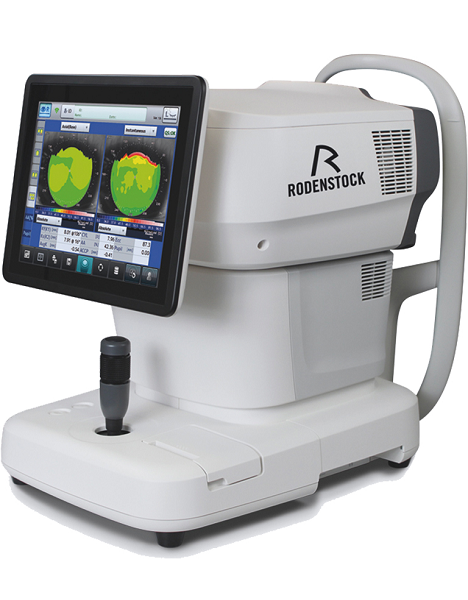
Tune-up your businessExperience 6 analyses in 1 device: Rodenstock Alino Multifunction Refractometer
Are you ready for something that is all-in-one, timesaving, space-saving and suits your business perfectly?Have a look at how powerful ALINO can be.6 in 1 solution01 RefractionHaving a good starting value for subjective refraction is essential. The QUICK REF MODE supports this even for uncooperative patients.02 KeratometryKAI (Kerato-asymmetry index) & KRI (Kerato-regularity index) give the first hint of possible irregularities on the cornea. This supports evaluation of the best vision correction or best-fitting contact lens.03 TonometryThe very gentle air puff created by a new generation of air-flow technology means that patients more readily accept the need for regular check-ups of the IOP measurement.04 PachymetryCorrecting the IOP by measuring the central corneal thickness is essential in interpreting the intraocular pressure.05 TopographyVarious topographic maps support you in carrying out vision screenings, contact lens fittings and in patient education.06 Dry EyeObserve the customers’ eye health and evaluate the long-term tolerability of contact lenses.Take advantage of the combination of tests:Additional servicesTonometry + PachymetryContact lens fitting andtolerance controlKeratometry + Topography + Dry EyeEye healthcareTopography + Tonometry + Pachymetry + Dry EyeSpecifications Rodenstock Alino Multifunction Refractometer
REFRACTIVE POWER MEASUREMENTSpherical refractive power (S) -30.00 D to + 25.00 D (at VD = 12.0 mm)Cylindrical power (C) 0.00 D to ± 12.50 D (at VD = 12.0 mm)Astigmatic axis (A) 0° to 180°KERATOMETRY MEASUREMENTCorneal curvature radius 5.00 mm to 13.00 mmCorneal astigmatic axis 0° to 180°INTRAOCULAR PRESSURE MEASUREMENTMeasurement range 1 mmHg to 60 mmHG (1 hPa to 80 hPa)PACHYMETRY MEASUREMENTMeasurement range 300 μm to 800 μmTOPOGRAPHY MEASUREMENTCorneal curvature radius 5.50 mm to 10.00 mmCorneal astigmatic axis 0° to 180°AUXILIARY FUNCTIONInterpupillary distance Measurement range: 20 mm to 85 mmCorneal diameter and pupil diameter Measurement range: 1.00 mm to 14.00 mmDry eye observation Blinking Analysis, Tear Meniscus height, Hyperemia, Meibomian glandsDATA MANAGEMENTInternal Database Integrated SD CardPrinter Integrated thermal printerData Output Type 3x USB 2.0, 1x LAN, 1x SD Card-slot, 1x WLANExport Format CSV, XML, JPG, PDFDIMENSIONS & ELECTRIC REQUIREMENTSDimensions WDH 312 × 491 × 450 mmWeight Approx. 23 kgVoltage 100 VAC to 240 VACFrequency 50/60 HzPower consumption 110 VA | Zeiss Humphrey Hark 599 Autorefractor / Keratometer
The HUMPHREY 599 allows for immediate patient feedback on the prescription and can be linked to the Humphrey Lens Analyzer to allow patients to compare their current RX to the new refraction results. Precise keratometer readings can be instantly obtained for all clinical needs.Fast, accurate and objective measurements with complete printout
Automatic cycle for objective measurements
Complete automatic alignment and tracking
Acuity line isolation
Cylinder and axis refinement
Duochrome target
Jackson Cross Cylinder Test
Near vision testing
Glare testing
Spherical equivalents
PD displayed on printout
Children’s acuity targets
Recall for old and new Rx comparisons
Communicom Interface
Keratometry Reading including average and delta K’s - Fast, accurate and objective measurements with complete printout
- Automatic cycle for objective measurements
- Complete automatic alignment and tracking
- Acuity line isolation
- Cylinder and axis refinement
- Duochrome target
- Jackson Cross Cylinder Test
- Near vision testing
- Spherical equivalents
- PD displayed on printout
- Children’s acuity targets
- Recall for old and new Rx comparisons
- Communicom Interface
- Keratometry Reading including average and delta K’s
Features:Fast, accurate and objective measurements with complete printout
Automatic cycle for objective measurements
Complete automatic alignment and tracking
Acuity line isolation
Cylinder and axis refinement
Duochrome target
Jackson Cross Cylinder Test
Near vision testing
Glare testing
Spherical equivalents
PD displayed on printout
Children’s acuity targets
Recall for old and new Rx comparisons
Communicom Interface
Keratometry Reading including average and delta K’s in the Humphrey HARK 599About us:Zeiss Humphrey Hark 599 Autorefractor / Keratometer
ALT Medical Device is your complete eye care equipment solution. Is comprehensive company offering only high end, quality equipment. As a company, ALT Medical Device combines outstanding business practices with an unparalleled approach to customer service.
We have 100% customer satisfaction by delivering quality equipment and full customer service with every transaction. | Carl Zeiss Cirrus HD-OCT 5000 Smart Advanced Topography HDAdvancing Smart OCTfor Anterior Segment, Glaucoma and Retina- Anterior Segment Premier Module
- PanoMap Wide-Field Display
- Smart HD Scans
- En face report
Visualization at the speed of CIRRUSAnalyzing a single pathology from multiple views provides comprehensive insight and analysis of the clinical situation. How this helps you: - Spot small areas of pathology. Tightly spaced B-scans, (either 30 or 47 μm apart), in the cube ensure that small areas of pathology are imaged. For reference, a human hair is about 40-120 μm in diameter.
- Visualize the fovea. Scans that are spaced further apart than in the CIRRUS cube may miss the central fovea.
- Fuel for analysis. Millions of data points from the cube are fed into the Zeiss proprietary algorithms for accurate segmentation, reproducible measurements and registration for change analysis.
- Take the pressure off the operator. As long as the scan is placed in the vicinity of the fovea or optic nerve, the software automatically centers the measurements after the capture.
- See the tissue from different perspectives. View the cube data from all angles, with 3D rendering, OCT fundus images and Advanced Visualization™.
- Future ready. Previously captured CIRRUS cubes can be analyzed using new analyses.
Tracking at the speed of CIRRUSFastTrac™ reduces eye motion artifacts without sacrificing patient throughput with a proprietary scan acquisition strategy, high speed 20 Hz LSO camera, and single-pass alignment scanning
With FastTrac, scan at the highest resolution at the same location at each visit. Assessment at the speed of CIRRUSMeasurement centering with FoveaFinder and AutoCenterOn the macula, the unique FoveaFinder technology ensures the ETDRS and ganglion cell plus inner plexiform layer measurement frameworks are centered on the fovea.AutoCenter™ function automatically centers the 3.4 mm diameter peripapillary RNFL calculation circle around the disc for precise placement and repeatable registration. The placement of the circle is not operator dependent. Accuracy, registration and reproducibility are assured. Manage at the speed of CIRRUS Carl Zeiss Cirrus HD-OCT 5000 Smart Advanced Topography HDGuided Progression Analysis (GPA™) for Assessing RNFL and Optic Disc ChangeGPA™ compares RNFL thickness and optic disc measurements over time and determines if statistically significant change has occurred.Focal Progression Analysis – Up to 6 progression maps are compared to two baselines. Areas of statistically significant change are color-coded yellow when first noted and then red when the change is sustained over consecutive visits.
Trend Analysis with Rate of Change – RNFL thickness values are plotted over time. Trends are calculated and confidence intervals are shaded when rate of change is statistically significant. |




Reviews
There are no reviews yet.