| Description | The image can then be displayed on a monitor, helping you to educate your patients The HRK can provide peripheral keratometry measurement data which is very useful when fitting contact lenses. The eye can be displayed in full colour mode on a tilting touch screen which enables you to see the patient’s contact lens fitting status, previously only possible using a slit lamp. As the HRK has the world’s first contact lens fitting function in an auto keratometer, you can now see the flourescein liquid with blue illumination along with automatic calculation and recommendation for contact lens fitting. | RIGHTON RETINOMAX-5 AUTO REFRACTOMETER / KERATOMETER
Automatic AxisCompensation and extended measurement range
Not only does the device let examiner know the cylinder axis angle, but it can
also be automatically adjusted if it is not level. | The Auto “Refkeratometer” RC-800 is the latest in economical, user-friendly, highly accurate auto “refkeratometry”. The RC-800 provides simple, easy measurement by the operator at an economical cost, without compromising accuracy or reliability. | Experience 6 analyses in 1 device: refraction, keratometry, topography, tonometry, pachymetry and dry eye analysis. | CIRRUS HD-OCT 5000 is configured with the highest resolution visualization capabilities and sophisticated clinical applications such as Advanced RPE analysis to track retinal pigment epithelial integrity and Ganglion Cell Analysis to assess glaucomatous loss in the macula that may not be evident in the peripapillary region. The new FastTrac retinal tracking reduces eye motion artifacts without sacrificing patient throughput with a proprietary scan acquisition strategy, high speed 20 Hz LSO camera, and single-pass alignment scanning. This feature enables the highest resolution B-Scans to be captured in identical locations from visit to visit providing precise assessment of change in targeted pathologies. | Eclipse Evolves Pulse Wave Market with eVive and Improved Maintenance Program |
| Content | Huvitz HRK-8000A Auto Refractor KeratometerHuvitz Auto Ref – Keratometer HRK-8000A- Touch and tilting 7″ LCD Screen that allows for free and smooth tilting for viewing at any angle.
- Optimized Optical System
- Micro Lens Array
- Aberration Measurement which also can be displayed in a graphical Zernike Refraction Map
- PSF & Image Simulation
- Colour View Mode
- Peripheral Keratometry Measurement
- Ultra High Precision KER Data
- Contact Lens Fitting Assistance Guide
- Contact Lens Prescription Guide
- Auto Tracking
- Animated Guide
- Faster Measurement Speed
- Quiet and speedy auto cutting printer
- More data on aberration measurement
With its extreme precision and accuracy, the HRK8000A uses the most advanced wavefront technology to measure the light reflected from the retina and helps you to diagnose any refractive error. With a micro lens array developed by Huvitz, the HRK creates a number of focal points, the pattern of which can give a valuable insight into the patients ocular system. The HRK not only displays the conventional data such as spherical, cylinder and axis, the high order aberration data is displayed in a graphical Zernike refractive map, giving you a deeper understanding of your patient’s eyes.Optimized Optical System Huvitz HRK-8000A Auto Refractor Keratometer
A wavefront is a physical representation of the optical quality of a light beam. Using various methods, aberrometers map how light rays travel through the eye. The HR-8000A is engineered according to the Hartmann-Shack wavefront measurement technique, which is widely preferred for its speed and repeatability.Touch & Tilting Color Display Screen
A bright, high contrast 7″ VGA color TFT LCD screen provides high resolution video images. A smooth, free tilting function also offers you a comfortable and clear view at any angle.The image can then be displayed on a monitor, helping you to educate your patients The HRK can provide peripheral keratometry measurement data which is very useful when fitting contact lenses. The eye can be displayed in full colour mode on a tilting touch screen which enables you to see the patient’s contact lens fitting status, previously only possible using a slit lamp. As the HRK has the world’s first contact lens fitting function in an auto keratometer, you can now see the flourescein liquid with blue illumination along with automatic calculation and recommendation for contact lens fitting. | Righton Retinomax 5 Auto Refractor
The Retinomax Series, the world’s leading handheld autorefractor and autorefractor/keratometer, delivers wide measurement range on par with table type refr, now boasts greater mobility, stability and accuracy. The Retinomax series is widely used as a global standard device for pediatric, screening, medical care in remote areas, disaster area and wide range of ophthalmic scene.FEATURES- View Finder – With view finder observation, no limit of operator age. Diopter adjustable range is +/-8D the same as Retinomax3. View finder arm angle is changeable of 0-135 degrees, making measurement easy regardless of patient’s position or posture. Adjustable diopter +/- 8D.
- Wider Eye Piece – Eye piece relief is 48mm from 32.2mm so that enable to look inside image while even looking at outside. No need to stick eye to the eyepiece all the time.
- Improved battery life – At 180 minutes, the battery capacity is now twice that of conventional models
- Automatic Axis Compensation and Extended Measurement Range – Not only does the device let examiner know the cylinder axis angle, but it can also be automatically adjusted if it is not level
- New Child Mode – While the measurement is being taken, a melody plays continually to keep children’s attention. A constantly changing color display, both on the outside and inside target the device, also keeps children involved during the process.
- Light weight and easy-to-hold grip
View Finder Righton Retinomax 5 Auto RefractorWith view finder observation, no limit of operator age. Diopter adjustable rangeis +/-8D the same as Retinomax3. View finder arm angle is changeable of 0-135degrees, making measurement easy regardless of patient’s position or posture.Adjustable diopter +/- 8D.Improved battery lifeAt 180 minutes, the battery capacity is now twice that of conventional modelsWider Eye PieceEye piece relief is 48mm from 32.2mm so that enable to
look inside image while even looking at outside. No need
to stick eye to the eyepiece all the time.Focus AssistLets you know the exact focusing point with
the following color-coding system.New Child ModeWhile the measurement is being taken, a
melody plays continually to keep children’s
attention. A constantly changing color
display, both on the outside and inside of
the device, also keeps children involved
during the process.Automatic AxisCompensation and extended measurement range
Not only does the device let examiner know the cylinder axis angle, but it can
also be automatically adjusted if it is not level. | Tomey RC-800 Auto Refractor Keratometer
Refractometry: Simple handling of the fine adjustment and measurement by yourself. Fogging is applied automatically for each measurement for more accurate results.Keratometry: Short consecutive measurements and time provide reliable data. Measurements at φ3.0mm (R = 8.0mm) give you enough information for contact lens fitting.Diameter Measurement of Cornea and Pupil: Easy measurement by moving the two cursors on the display to the boundaries of Cornea or Pupil.Language Selection: RC-800 has a Multi-language function. You can select your required language from “English”, “Japanese”, “German”, “Chinese”, “Spanish”, “Spanish Latin America”, “Italian”, “Portuguese”, “French” and “Russian”.Features:• Highly Accurate
• Simple to Use
• Diameter Measurement of Cornea and Pupil
• High Speed Printer
• Auto Shot
• Color Touch Screen
• USB connection
• Large Patient Database
• Easy Database Operation
• Multi-Language Operation
• Fourier Refractive Analysis
• Quick Data Reference
• Built-in LCD Alignment
• Keratoconus Screening & Other ApplicationsMeasurement Ranges:Refraction:• Sphere: -25.00D to +22.00D (VD=12.0mm)
• Cylinder: 0D to ±10.00D (VD=12.0mm)
• Axis: 0° to 180°
• Minimum Pupil Dia: Ø2.0mm
• Measurement time: 0.2 second/single eye (data taking time)Keratometry:• Range: 5.00mm to 11.00mm
• Corneal Refraction: 30.68D to 67.50D (n=1.3375)
• Corneal Astigmatism: 0D to 10D (n=1.3375)
• Axis: 0° to 180°
• Measurement area: Ø3mm (at 8.00mm corneal curvature)
• Measurement time: 0.1 second/single eye (data taking time)Other features:Tomey RC-800 Auto Refractor Keratometer• PD Measurement range 50 to 86mm
• Corneal diameter measurement
• Measurement range: 10.0 to 14.0mm
• Display unit: 0.1mm
• Pupil diameter measurement
• Measurement range: 1.0 to 10.0 mm
• Display unit: 0.1mm
• Built-in Printer Thermal printer
• Data output type RS232C / USB-D
• Display 5.7 inch color liquid crystal display
• Dimensions and weight 297(W)x 500(D) x 448(H)mm
• Approx. 17kg
• Power Requirements AC 100V to 240V, 50 / 60 Hz, 80VA to 100VAThe Auto “Refkeratometer” RC-800 is the latest in economical, user-friendly, highly accurate auto “refkeratometry”. The RC-800 provides simple, easy measurement by the operator at an economical cost, without compromising accuracy or reliability. | Rodenstock Alino Multifunction Refractometer
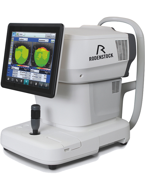
Tune-up your businessExperience 6 analyses in 1 device: Rodenstock Alino Multifunction Refractometer
Are you ready for something that is all-in-one, timesaving, space-saving and suits your business perfectly?Have a look at how powerful ALINO can be.6 in 1 solution01 RefractionHaving a good starting value for subjective refraction is essential. The QUICK REF MODE supports this even for uncooperative patients.02 KeratometryKAI (Kerato-asymmetry index) & KRI (Kerato-regularity index) give the first hint of possible irregularities on the cornea. This supports evaluation of the best vision correction or best-fitting contact lens.03 TonometryThe very gentle air puff created by a new generation of air-flow technology means that patients more readily accept the need for regular check-ups of the IOP measurement.04 PachymetryCorrecting the IOP by measuring the central corneal thickness is essential in interpreting the intraocular pressure.05 TopographyVarious topographic maps support you in carrying out vision screenings, contact lens fittings and in patient education.06 Dry EyeObserve the customers’ eye health and evaluate the long-term tolerability of contact lenses.Take advantage of the combination of tests:Additional servicesTonometry + PachymetryContact lens fitting andtolerance controlKeratometry + Topography + Dry EyeEye healthcareTopography + Tonometry + Pachymetry + Dry EyeSpecifications Rodenstock Alino Multifunction Refractometer
REFRACTIVE POWER MEASUREMENTSpherical refractive power (S) -30.00 D to + 25.00 D (at VD = 12.0 mm)Cylindrical power (C) 0.00 D to ± 12.50 D (at VD = 12.0 mm)Astigmatic axis (A) 0° to 180°KERATOMETRY MEASUREMENTCorneal curvature radius 5.00 mm to 13.00 mmCorneal astigmatic axis 0° to 180°INTRAOCULAR PRESSURE MEASUREMENTMeasurement range 1 mmHg to 60 mmHG (1 hPa to 80 hPa)PACHYMETRY MEASUREMENTMeasurement range 300 μm to 800 μmTOPOGRAPHY MEASUREMENTCorneal curvature radius 5.50 mm to 10.00 mmCorneal astigmatic axis 0° to 180°AUXILIARY FUNCTIONInterpupillary distance Measurement range: 20 mm to 85 mmCorneal diameter and pupil diameter Measurement range: 1.00 mm to 14.00 mmDry eye observation Blinking Analysis, Tear Meniscus height, Hyperemia, Meibomian glandsDATA MANAGEMENTInternal Database Integrated SD CardPrinter Integrated thermal printerData Output Type 3x USB 2.0, 1x LAN, 1x SD Card-slot, 1x WLANExport Format CSV, XML, JPG, PDFDIMENSIONS & ELECTRIC REQUIREMENTSDimensions WDH 312 × 491 × 450 mmWeight Approx. 23 kgVoltage 100 VAC to 240 VACFrequency 50/60 HzPower consumption 110 VA | Carl Zeiss Cirrus HD-OCT 5000 Smart Advanced Topography HDAdvancing Smart OCTfor Anterior Segment, Glaucoma and Retina- Anterior Segment Premier Module
- PanoMap Wide-Field Display
- Smart HD Scans
- En face report
Visualization at the speed of CIRRUSAnalyzing a single pathology from multiple views provides comprehensive insight and analysis of the clinical situation. How this helps you: - Spot small areas of pathology. Tightly spaced B-scans, (either 30 or 47 μm apart), in the cube ensure that small areas of pathology are imaged. For reference, a human hair is about 40-120 μm in diameter.
- Visualize the fovea. Scans that are spaced further apart than in the CIRRUS cube may miss the central fovea.
- Fuel for analysis. Millions of data points from the cube are fed into the Zeiss proprietary algorithms for accurate segmentation, reproducible measurements and registration for change analysis.
- Take the pressure off the operator. As long as the scan is placed in the vicinity of the fovea or optic nerve, the software automatically centers the measurements after the capture.
- See the tissue from different perspectives. View the cube data from all angles, with 3D rendering, OCT fundus images and Advanced Visualization™.
- Future ready. Previously captured CIRRUS cubes can be analyzed using new analyses.
Tracking at the speed of CIRRUSFastTrac™ reduces eye motion artifacts without sacrificing patient throughput with a proprietary scan acquisition strategy, high speed 20 Hz LSO camera, and single-pass alignment scanning
With FastTrac, scan at the highest resolution at the same location at each visit. Assessment at the speed of CIRRUSMeasurement centering with FoveaFinder and AutoCenterOn the macula, the unique FoveaFinder technology ensures the ETDRS and ganglion cell plus inner plexiform layer measurement frameworks are centered on the fovea.AutoCenter™ function automatically centers the 3.4 mm diameter peripapillary RNFL calculation circle around the disc for precise placement and repeatable registration. The placement of the circle is not operator dependent. Accuracy, registration and reproducibility are assured. Manage at the speed of CIRRUS Carl Zeiss Cirrus HD-OCT 5000 Smart Advanced Topography HDGuided Progression Analysis (GPA™) for Assessing RNFL and Optic Disc ChangeGPA™ compares RNFL thickness and optic disc measurements over time and determines if statistically significant change has occurred.Focal Progression Analysis – Up to 6 progression maps are compared to two baselines. Areas of statistically significant change are color-coded yellow when first noted and then red when the change is sustained over consecutive visits.
Trend Analysis with Rate of Change – RNFL thickness values are plotted over time. Trends are calculated and confidence intervals are shaded when rate of change is statistically significant. | Eclipse Evive
Get Your Patients Back in the GameeVive is the first pulse wave therapy device with the specific purpose of making men’s health treatments affordable, comfortable and efficacious.About eViveeVive uses acoustic sound waves which diffuses as it travels away from the applicator tip, creating a therapeutic effect as it passes through the target tissue.Patients benefit from improved blood flow to the penis by opening existing blood vessels and stimulating the growth of new blood vessels, without the use of injections or oral medications.Using eViveMultiple studies have proven that eVive’s use of Pulse Wave Therapy helps to repair aged blood vessels, stimulate growth of new blood vessels and remove decades of micro-plaque.The applied pulse propagates radially within the tissue and has a therapeutic effect on areas of the tissue. This provides patients with a simple treatment option with no downtime, and lasting results.Treatment with eViveIn 6 to 12 sessions each taking between 15 to 20 minutes, eVive treatments:Stimulate the release of growth factors in penile tissue, resulting in the generation of new blood vessels
Break up plaque formation in existing blood vessels
Activate dormant stem cells, leading to new cell growthEclipse eVIVE Pulse Wave Therapy Device Specifications(1) eVIVE console with touchscreen
(2) eVIVE handpiece with standard 15mm transmitter tip
(2) 36mm radial transmitter
(2) 6 mm focus transmitter
(1) cleansing brush
(1) mobile cart
(1) box single-use treatment covers (50/box)
operators manualAbout eViveeVive uses acoustic sound waves which diffuses as it travels away from the applicator tip, creating a therapeutic effect as it passes through the target tissue.Patients benefit from improved blood flow to the penis by opening existing blood vessels and stimulating the growth of new blood vessels, without the use of injections or oral medications. Using eViveMultiple studies have proven that eVives use of Pulse Wave Therapy helps to repair aged blood vessels, stimulate growth of new blood vessels and remove decades of micro-plaque.The applied pulse propagates radially within the tissue and has a therapeutic effect on areas of the tissue. This provides patients with a simple treatment option with no downtime, and lasting results. Treatment with eViveIn 6 to 12 sessions each taking between 15 to 20 minutes, eVive treatments: - Stimulate the release of growth factors in penile tissue, resulting in the generation of new blood vessels
- Break up plaque formation in existing blood vessels
- Activate dormant stem cells, leading to new cell growth
|




Reviews
There are no reviews yet.