| Description | The affordable Gendex 9200 DDE dental radiography system offers standard panoramic and cephalometric x-ray projections, as well as a wide range of advanced programs to support most dental imaging and exams. | The Clio Digital X-ray Sensor is equipped with a patented, Kevlar reinforced cable to provide maximum durability. Unlike other products, the USB cable of the Clio is fully replaceable if damaged. Constructed with special shock absorbers inside the sensor housing to protect components from exterior damage, the Clio is also one of the few fully water submersible sensors, sealed with a unique rubber polymer interior housing. | Automatic Power Control: Epic Pro cuts four times faster1 than a classic diode laser with
minimal collateral damage. In this treatment mode, the laser in continuous wave mode converts light energy to thermal power at the tip. Laser energy is adjusted automatically in real time to maintain constant tip temperature. The result is extremely consistent and reliable cutting conditions, regardless of speed of movement, tissue type, etc. | TRIOS MOVE makes it simple to show digital impressions to your patients in order to explain their treatment plans and improve communication. The system features the same scanning technology as the TRIOS 3, including the option of a wireless model, and can capture full-color digital impressions, provide shade matching and more quickly and easily. As an open system, it can send digital impressions to almost any dental CAD/CAM software platform for the fabrication of almost any fixed, removable or orthodontic solution. | The Identica T300 dramatically improves the fundamental aspects of scanner performance and the latest software technology provides the clearest, sharpest data. Identica T300 is customized to deliver the finest data quality using its dual 2.0 MP cameras. Identica T300 will save valuable time and money by minimizing trial & error when adjusting restoration fits. | Identica Hybrid is our fastest and most accurate scanner. When designing Identica Hybrid we focused on the requirements of your lab to have a precise, fast, and reliable machine that is easy to learn and easy to use. |
| Content | Gendex Orthoralix 9200 DDE Digital Cephalometric + Panoramic X Ray
The versatile Orthoralix 9200 system offers standard panoramic projections, as well as a full array of advanced programs to satisfy the demanding needs of dental specialists. Plus, with both ?lm-based and direct digital models and the optional cephalometric arm, the capabilities of the 9200 can expand with your practice.Discover all of the benefits and options this imaging system has to offer.Wide Range of Projections – The Orthoralix 9200 offers 10 standard projections, with the ability to add 5 Cephalometric projections and the Transcan tomography projection to satisfy the needs of today’s dental specialists. Its intuitive control panel and easy-to-read display simplifies the process of selecting projections. Plus, with the precision of morphology following and multi-motorized arm rotation, the 9200 excels at focal trough reconstruction.- 10 standard imaging projections and 6 additional with optional upgrades
- Meets the demands of today’s orthodontists, oral and maxillofacial surgeons, and other specialists
Advanced Image Quality – Three technical factor setting modes are available to ensure complete flexibility and quality control. Manual mode for total operation control, Preset mode where the operator can customize settings, and Automatic Exposure Control (AEC) mode. With AEC, the 9200 automatically raises or lowers the kV setting based on measurements of bone size and density.- Triple laser beam guide system and motorized column for simple, accurate patient positioning
- Automatic Exposure Control (AEC) to ensure optimum image contrast and balance
- Real-time image reconstruction with the direct digital model
Gendex Orthoralix 9200 DDE Digital Cephalometric + Panoramic X Ray
Expand Your Capabilities – Utilizing the latest in CCD sensor technology and built-in LAN connectivity, the 9200 DDE model allows clinicians to digitally capture x-ray images and share them with any computer connected to the network for maximum productivity. With the option to add a Cephalometric arm, the Transcan cross-sectional tomography projection, and convert to direct digital technology, the 9200 is one of the most expandable panoramic systems available.- Direct digital model with the latest CCD sensor technology and direct LAN connectivity
- Optional Cephalometric arm attachment
- Optional Transcan implantology projection for transverse cross-sections of upper and lower jaw
Gendex Orthoralix 9200 DDE Digital Cephalometric + Panoramic X Ray| Power Supply | 115-250 VAC ± 10% | | Frequency | 50/60 Hz ± 2 Hz | | Maximum Line Power Rating | 10 A at 250 V, 20 A at 115 V | | Anode Voltage | 60 – 84 kV, in 2 kV steps | | Anode Current | 3 – 15 mA, in 1 mA steps | | Exposure Time | 12 s for standard pan, 0.16 – 2.5 s for Cephalometric | | Duty Cycle | 1:20 at full power operation | | Focal Spot | 0.5 mm, IEC 336 (1993) | | Vertical Reach | 39′ to 71′ (100 to 180 cm) from ?oor to occll plane | | Weight | 410 lbs (115 kg), 467 lbs (212 kg) with Ceph arm | | Active Area CCD Sensor | 147 x 6 mm (Standard Pan), 220 x 6 mm (Ceph) | | Image Size | 1536 x 2725 pixels (Standard Pan), 2304 x 2529 pixels (Ceph – Maximum) | | Minimum PC Requirements | Pentium® II CPU, 400 MHz, 256 MB RAM | | Required Operating System | Microsoft® Windows 98®/ Windows 2000®/ Windows XP® |
| SOTA Clio Digital X-Ray Sensor Size 2
SOTA Clio Digital X-Ray Sensor Size 2The Clio Digital Dental X-Ray Sensors have Next Generation CMOS technology, fiber optic sensors create 20+ actual line pairs per mm (26.3 theoretical line pairs per mm).With our Cliosoft Imaging Software, we can gaurantee our Clio Digital Dental X-Ray images match up or surpass any competing system. We make this promise based on feedback from actual dentists.Rounded edges allow for maximum patient comfort. Ideal intraoral sensor width and thinness in two sizes for pedo and adult.The Clio Digital X-Ray Sensors maximize image area ratio, minimizing cutoff space to mimic the size of traditional x-ray film. Next Generation CMOS technology fiber optic sensors create 20+ actual line pairs per mm (26.3 theoretical). With our user-friendly, tablet and PC-ready cliosoft Digitial Imaging Software, we guarantee the highest quality images in the industry.Features:– All new Direct Integration Technology
– Thin sensor hardware with rounded corners for maximum comfort
– Direct USB cable with no docking station
– Repairable USB cable for longevity of sensors
– Fiber optic plate sensors for low-noise ratio
– Largest active area for maximum coverage
– Fully waterproof sensor head with sealed cable and housing design
– USB connector: USB 2.0, USB 3.0
– Sensor Technology
– Next Generation CMOS Fiber optic Sensors
– Maximum Gray Levels – 4096 (12-bit)Specifications:– External Dimensions: 30.49 x 42.80 mm
– Active Area: 25.99 x 36.02 mm
– Sensor Plate Thickness: 5.41 mm
– Sensor Cable Length: 3 meters
– Image Resolution: 26.3 lp/mm theoretical 20 lp/mm actual
– Image Size: 1896 x 1368 pixels
– Image Pixel Size: 19×19 micronsThe Clio Digital X-ray Sensor is equipped with a patented, Kevlar reinforced cable to provide maximum durability. Unlike other products, the USB cable of the Clio is fully replaceable if damaged. Constructed with special shock absorbers inside the sensor housing to protect components from exterior damage, the Clio is also one of the few fully water submersible sensors, sealed with a unique rubber polymer interior housing. | Biolase Epic Pro Dental Soft Tissue LaserThe Epic Pro, which offers the most laser power of any diode laser in dentistry, is the first
product to be introduced resulting from BIOLASE’s strategic development agreement
with IPG Medical. The newest addition to the Epic family of dental soft-tissue lasers, Epic
Pro features several new innovations that are industry-firsts: - A new super pulse technology for more precise, enhanced laser tissue cutting
- Real-time automatic power control to enhance speed and consistency when performing surgery with pre-programed tip temperature
- Pre-initiated, bendable, disposable tips with new smart tip technology to ensure tip performance and quality
Automatic Power Control: Epic Pro cuts four times faster1 than a classic diode laser with
minimal collateral damage. In this treatment mode, the laser in continuous wave mode converts light energy to thermal power at the tip. Laser energy is adjusted automatically in real time to maintain constant tip temperature. The result is extremely consistent and reliable cutting conditions, regardless of speed of movement, tissue type, etc. Super Thermal Pulse: Epic Pro can achieve ten times the peak power of a standard diode (and up to three times that of the closest competitor) laser in short micro-pulses for the highest cutting efficiency and safety. In this treatment mode, the laser converts light energy to thermal power, which it emits as thermal power pulses, (i.e. multiple bursts of high power). Each of these thermal bursts cuts tissue highly efficiently. When the laser is activated, power bursts are emitted with a preset pulse width (duration), while maintaining a precise thermal level at the tip. The laser is powered on only about 3-5% of the time, giving tissue ample time to relax between pulses; creating a safer and gentler experience for patients. 
The Only Laser with Automatic Power ControlEpic Pro is the ONLY FDA-cleared laser with Automatic Power Control, an exclusive innovation from BIOLASE that enables the operator to lase with confidence and speed. 
Enhanced Clinical ApplicationsEpic Pro is the professional’s choice for managing soft tissue cases. With over 20 clinical presets, multiple treatment modes, and an advanced mode for expert users, Epic Pro provides unparalleled consistency and predictability. Unlike traditional diodes, Epic Pro moves quickly and with little snag-and-drag resistance. 
Greater Precision and ComfortEpic Pro brings more precision and comfort than traditional diodes due to innovations such as Super Thermal Pulse mode, which can pulse energy as quickly as ten-millionths of a second, resulting in efficient clinical cutting with less thermal damage than normal diode lasers. 
Introducing Smart Tip TechnologyEpic Pro uses durable new tip technology, available in uninitiated and pre-initiated formats depending on your clinical needs. Epic Pro tips work hand in hand with the laser’s Smart Tip technology to prompt you when a fresh tip is needed. 
With Epic Pro, ‘You Have to See It to Believe It’If you are a current diode laser or CO2 laser user, you have to see Epic Pro cut tissue live and compare to your current laser experience. When current Epic Pro customers were first shown Epic Pro, their response? “You have to see it cut to understand how much faster it is.” Fill out the contact form on this page to request a live demonstration for Epic Pro in your clinic.Technical Specifications Biolase Epic Pro Dental Soft Tissue Laser| Dimension | 7.25in (W) x 4.5in (L) x 6.5in (H) (18.4cm x 11.4cm x 16.5cm) | | Weight | 4 lbs (1.8 kg) | | Operating Voltage | 100V – 240V at 1.2A/0.5A | | Frequency | 50/60 Hz | | Main Control | Power Switch | | Remote Interruption | Remote Interlock | | Disable Control | Emergency Stop Button | | DC Power Supply Module | 12V DC, 5A | | Laser Classification | Class IV (4) | | Medium | Semi-conductor diode | | Wavelength | 940nm ± 20nm | | Power Modes | Continuous, Pulse Modulation | | Fiber Tip Diameter | 300µm, 400µm | | Pulse Duration | 10µm-100ms | | Pulse Interval | 0.01ms – 20ms | | Pulse Repetition Rate | Up to 20kHz (for reference) | | NOHD | 4.77 meters | | Beam Divergence | 0.22 | | Standard Fiber Cable Length | 6 feet (1.8 meters) |
| 3Shape TRIOS 3 Wireless Move Pen GripTRIOS is the world’s first intraoral scanner to offer a wireless option. 3Shape TRIOS 3 Wireless gives you the freedom to perform digital scans unrestricted by wires, optimizing comfort for both you and your patientsThe new 3Shape TRIOS MOVE is the ultimate patient/dentist communication tool. Show patients their digital scans and dynamic treatment information close-up, and create greater opportunities for treatment dialogue and joint decision-making.The ergonomic and lightweight design of TRIOS MOVE allows you to always position the screen in the right place, and enjoy superior user experience.Features of TRIOS MOVE;-13.3″ HD LCD Touchscreen – The perfect size when having it in front of a patient or working with up closeAdjustable arm- Bring the screen directly in front of the patient to involve them in the scanning process and the treatment planningPowerful custom PC designed for scanning – Specifically designed for use with all TRIOS 3 scanners to give a fast and smooth scan experience straight out of the boxFour wheels with integrated brakes – It is now very easy to move around the chair and from chair to chairEnhance comfort with wireless innovation.TRIOS is the world’s first intraoral scanner to offer a wireless option. 3Shape TRIOS 3 Wireless gives you the freedom to perform digital scans unrestricted by wires, optimizing comfort for both you and your patientsThe new 3Shape TRIOS MOVE is the ultimate patient/dentist communication tool. Show patients their digital scans and dynamic treatment information close-up, and create greater opportunities for treatment dialogue and joint decision-making.The ergonomic and lightweight design of TRIOS MOVE allows you to always position the screen in the right place, and enjoy superior user experience.Features of TRIOS MOVE : 3Shape TRIOS 3 Wireless Move Pen Grip 13.3" HD LCD Touchscreen - The perfect size when having it in front of a patient or working with up closeAdjustable arm- Bring the screen directly in front of the patient to involve them in the scanning process and the treatment planningPowerful custom PC designed for scanning - Specifically designed for use with all TRIOS 3 scanners to give a fast and smooth scan experience straight out of the boxFour wheels with integrated brakes - It is now very easy to move around the chair and from chair to chair. | Medit Identica T300 Dental 3D Scanner
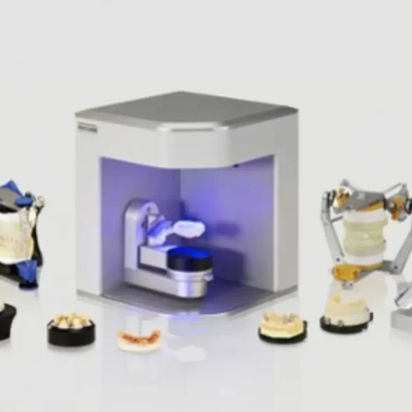
Identica T300 to meet all of your needs. With powerful scan engine and affordable pricing, the new Identica T300 is a perfect fit for both performance enthusiasts and entry level users. Its evolutionary design and high tech features will help your lab rediscover productivity. Bundled colLab 2017 scan software makes scan data processing easy and efficient.Quality & affordability- 2.0 MP cameras, Blue LED
- Accuracy : 7 μm (ISO)
- Full arch scan time 24 sec
- 2-Axis Manual
Identica T300 dramatically improves the most important aspects of scanner performance. The latest software technology provides you with the cleanest and sharpest data. T300 is specially designed to bring out the finest data quality using its 2.0 MP cameras. T300 will save your precious time and money by minimizing trial & error to adjust restoration fits.Identica T300 is an upgrade version of Identica Blue, provided at lowest pricing. Giving better value for the affordable scanner users. Identica T300 has enhanced data quality compared to Identica Blue.Introducing the Next Generation of 3D Dental Scanners: Our T-Series Medit represents the apex in 3D dental scanning. With unsurpassed speed, accuracy, and reliability, Medit helps you rediscover your productivity. The T-Series’ new powerful scan engine and affordable pricing are the perfect fit for performance enthusiasts and entry-level users, helping your lab rediscovering productivity thanks to its convenient design and high-tech features. Medit T300 is the latest Medit scanner with an enhanced SW scan. It uses Blue Light technology, which enables quick scanning and provides excellent precision (7μm).The program is user-friendly, because it takes you through simple and fast individual steps, from inserting the model into the scanner to exporting the file to the CAD design program. FEATURES:Medit Identica T300 Dental 3D Scanner- Scanned printing: 2 manual axes.
- Touch sensor: no.
- Full arcade scan speed: 24 ´´
- Resolution of the camera: 2 x 2 MP camera.
- Scan volume: 90 x 72 x 60 mm.
- Scanning principle: Optical change phase.
- Dimensions: 290 x 290 x 340 mm.
- Weight: 12Kg.
- Light source: Blue LED.
- Connectivity: Type USB 3.0 B.
- Power: 100- 240 vac, 50-60 HZ.
- Warranty: 2 years.
ADVANCED DATA PROCESSING AND HIGHER RESOLUTION DUAL CAMERAS The Identica T300 dramatically improves the fundamental aspects of scanner performance and the latest software technology provides the clearest, sharpest data. Identica T300 is customized to deliver the finest data quality using its dual 2.0 MP cameras. Identica T300 will save valuable time and money by minimizing trial & error when adjusting restoration fits. | Medit Identica Hybrid 3D Dental Scanner
Medit Identica Hybrid 3D dental scanner is fastest and most accurate scanner provides performance speeds of 16 seconds for full arch scans and allows technicians to perform auto double impression scansWith speed, accuracy, and reliability, Medit Identica Hybrid can helps doctor rediscover your productivity with 74% faster scan times, a flexible multi-die that cuts scan steps significantly, color scan texture for enhancing details, and more.Medit Identica Hybrid 3D dental scanner Benefits:- Very high scan speed;
- Flexible multi-die plate for the automatic scan of models and up to eight individual dies in one process;
- Three axes and three colour cameras with high resolution – measuring accuracy < 7 µm, automatic scanning of double-phase impressions;
- Integrated implant libraries with an optimised matching-process of the scan abutments for highest precision;
- Blue light LED scan technology reduces scan time by 74%;
- I.M.V (Intelligent Multi View) scan technology – areas that are difficult to capture like e.g. interproximal areas und undercut areas can be scanned safely and fast;
- Articulator scan with optionally available articulator plate, therefore no scan fixator required;
- Texture scan for the visualisation of markings on the model in the CAD software;
- Interface to dds dentalCAD software;
- Open system – data output in STL format.
Includes:- Exocad Dental CAD Software
- computer, which also has the implant module
- scan flags to send out for custom abutments.
- cable set; model holder; model plate; multi-die plate; flexible multi-die plate; impression holder; calibration plate; spacer plates and installation CD.
Specifications:- Light source: LED blue light
- Camera resolution: Grayscale 1.3 (MP) colour texture
- Axis control: 3-axis sync
- Scanning area: 80x60x60mm
- Dimension: 290x290x342mm
- Weight: 13kg
- Power: 150w
- Interface: USB 3.0 B type
Identica Hybrid ScannerImproving on earlier generations of Medit scanners, Identica Hybrid puts extreme precision and speed within any laboratory’s budget. Three cameras capture full color, 5 Megapixel images and multi-die scanning provides the capacity to capture a full arch, two partials or up to eight single-unit dies in a single scan, considerably speeding your workflow.- • Full-arch scans in 16 seconds
- • Three-axis full-color scanning
- • Scan up to 8 single units per scan cycle
Specifications- Light source: LED blue light
- Axis control: 3 Axis sync
- Scanning area: 80 x 60 x 60 mm
- Dimension: 290 x 290 x 342 mm
- Weight: 13 kg
- Power: 19.5v / 150w
- Interface: USB 3.0 B Types
|




Reviews
There are no reviews yet.