| Description | Ezer ERK-7800 Auto Refractor/KeratometerConfidently measure the human eye with a sophisticated cutting edge technology designed to enhance your ability to diagnose ocular errors faster, easier, and accurately. Ezers ERK-7800 is brilliantly designed technology measures the eye with ultra precision and speed to provide reliable data as the starting point of your refraction and contact lens fitting. It’s compact and modern design enables seamless operation and an enhanced patient comfort, making it the ideal choice in for any ophthalmic practice. Backed by an industry leading 2-years warranty! | The Auto “Refkeratometer” RC-800 is the latest in economical, user-friendly, highly accurate auto “refkeratometry”. The RC-800 provides simple, easy measurement by the operator at an economical cost, without compromising accuracy or reliability. | The Extra – 818 is the outcome of more than a decade of treatments and thousands of devices operated worldwide. It contains It contains unique features:
* Contemporary touch screen control
* Built-in Patch Test procedure for examining the optimal energy level
* A stunning number of 450,000 flashes in both lamps.
* The Large Handpiece has a Push Button in addition to the Foot Switch making
work easier and faster for the therapist. | Experience 6 analyses in 1 device: refraction, keratometry, topography, tonometry, pachymetry and dry eye analysis. | The Zeiss Humphrey Acuitus 5015 Auto Refractor Keratometer offers expanded refracting capabilities, allowing a practice to refract children and elderly patients alike. The Zeiss Acuitus 5015 also allows refraction one-day post operative of IOL and refractive surgery patients. The Zeiss Acuitus 5015 includes a flat color screen display which produces clear and concise images. | New Handpiece Design The Optima – 518 Classic is the first model to use the new Mark II which enables a multipurpose handpiece:
* Faster treatments – up to 0.5 Seconds Between pulses.
* Real-time handpiece temperature control.
* Contemporary and stylish design.
* Light weight design – 25% less weight to improve the therapists’ comfort. |
| Content | Ezer ERK-7800 Autorefractor KeratometerFeatures of the ERK-7800 Auto Refractor/Keratometer
 Auto Refractor/Keratometer
Enhanced Retro-illumination Imaging
Accurate Measurement of Iris & Pupils Diameters
Precise Temperature Sensor Compensation
Enhanced Measuring Speed
Comfortable Motorized Chin Rest
Automatic High Speed Cutting Printer
Simple One Touch Lock Knob
20 Touch & Tilt Color Monitor Display
Sophisticated TFT LCD Graphic User Interface
Easy Network Capability
Backed by an Industry Leading 2-years Warranty!
Auto Refractor/Keratometer
The ERK-7800 instantly reports the patient’s assessed eye power by combining the Auto Refractor/Keratometer functions into this one sharp instrument, making measurement a snap. This determines the refractive and keratometric properties of the human eye to assist eye care professionals in the process of prescribing corrective eyeglasses and contact lenses. Keratometry and Refractometry (RK Mode) Enhanced Retro-illumination Imaging Ezer ERK-7800 Autorefractor Keratometer
With the Enhanced Retro-illumination Imaging capability in the ERK-7800, you can treat and see abnormal crystalline lenses, cataracts, and scratches of corneas helping you to determine how healthy the patients eyes are with better clarity. This allows for observation of the eye condition of the cataract or contact lens surface. The system saves up to 2 images of each eye. Accurate Measurement of Iris & Pupils Diameters
The corneal diameter and base curve of contact lenses can be measured with this one instrument. Therefore, measurements of eye and prescriptions for glasses and contact lenses can be made more efficiently. Measuring Cornea Size (SIZE MODE) Wide Dioptric Measurement Range
Because the ERK-7800 covers a wide measurement range from -25D to +22D, even an examinee with strong myopia can be measured.
Enhanced Measuring Speed
Specially designed image chips have succeeded to make REF measuring speed faster reducing the overall examination time and increases throughput for complete examinations. Comfortable Motorized Chin Rest
Simply by pressing up/down buttons, user can set the height of measuring point comfortably. With the cutting edge auto sensor mechanism, you can track down, measure and focus the eye with out ever making a mistake. Automatic Printer Paper Cut
The printer is designed to execute, print, & cut data automatically at high speed for immediate results. Simple One Touch Lock Knob
With just the touch of a single button, the main body can easily be locked to its base. 20 Degrees Tilted LCD Monitor
The LCD Monitor tilts up to 20 degrees; a design made for comfort making. Excellent ergonomics makes it possible to operate the ERK-7800 while standing or sitting, for added convenience. Sophisticated TFT LCD Graphic User Interface
Sharp images with easy to understand icons make this 6.4-inch color TFT LCD interface user-friendly and enjoyable to work with. Auto Refractor/Keratometer
Enhanced Retro-illumination Imaging
Accurate Measurement of Iris & Pupils Diameters
Precise Temperature Sensor Compensation
Enhanced Measuring Speed
Comfortable Motorized Chin Rest
Automatic High Speed Cutting Printer
Simple One Touch Lock Knob
20 Touch & Tilt Color Monitor Display
Sophisticated TFT LCD Graphic User Interface
Easy Network Capability
Backed by an Industry Leading 2-years Warranty!
Auto Refractor/Keratometer
The ERK-7800 instantly reports the patient’s assessed eye power by combining the Auto Refractor/Keratometer functions into this one sharp instrument, making measurement a snap. This determines the refractive and keratometric properties of the human eye to assist eye care professionals in the process of prescribing corrective eyeglasses and contact lenses. Keratometry and Refractometry (RK Mode) Enhanced Retro-illumination Imaging Ezer ERK-7800 Autorefractor Keratometer
With the Enhanced Retro-illumination Imaging capability in the ERK-7800, you can treat and see abnormal crystalline lenses, cataracts, and scratches of corneas helping you to determine how healthy the patients eyes are with better clarity. This allows for observation of the eye condition of the cataract or contact lens surface. The system saves up to 2 images of each eye. Accurate Measurement of Iris & Pupils Diameters
The corneal diameter and base curve of contact lenses can be measured with this one instrument. Therefore, measurements of eye and prescriptions for glasses and contact lenses can be made more efficiently. Measuring Cornea Size (SIZE MODE) Wide Dioptric Measurement Range
Because the ERK-7800 covers a wide measurement range from -25D to +22D, even an examinee with strong myopia can be measured.
Enhanced Measuring Speed
Specially designed image chips have succeeded to make REF measuring speed faster reducing the overall examination time and increases throughput for complete examinations. Comfortable Motorized Chin Rest
Simply by pressing up/down buttons, user can set the height of measuring point comfortably. With the cutting edge auto sensor mechanism, you can track down, measure and focus the eye with out ever making a mistake. Automatic Printer Paper Cut
The printer is designed to execute, print, & cut data automatically at high speed for immediate results. Simple One Touch Lock Knob
With just the touch of a single button, the main body can easily be locked to its base. 20 Degrees Tilted LCD Monitor
The LCD Monitor tilts up to 20 degrees; a design made for comfort making. Excellent ergonomics makes it possible to operate the ERK-7800 while standing or sitting, for added convenience. Sophisticated TFT LCD Graphic User Interface
Sharp images with easy to understand icons make this 6.4-inch color TFT LCD interface user-friendly and enjoyable to work with. | Tomey RC-800 Auto Refractor Keratometer
Refractometry: Simple handling of the fine adjustment and measurement by yourself. Fogging is applied automatically for each measurement for more accurate results.Keratometry: Short consecutive measurements and time provide reliable data. Measurements at φ3.0mm (R = 8.0mm) give you enough information for contact lens fitting.Diameter Measurement of Cornea and Pupil: Easy measurement by moving the two cursors on the display to the boundaries of Cornea or Pupil.Language Selection: RC-800 has a Multi-language function. You can select your required language from “English”, “Japanese”, “German”, “Chinese”, “Spanish”, “Spanish Latin America”, “Italian”, “Portuguese”, “French” and “Russian”.Features:• Highly Accurate
• Simple to Use
• Diameter Measurement of Cornea and Pupil
• High Speed Printer
• Auto Shot
• Color Touch Screen
• USB connection
• Large Patient Database
• Easy Database Operation
• Multi-Language Operation
• Fourier Refractive Analysis
• Quick Data Reference
• Built-in LCD Alignment
• Keratoconus Screening & Other ApplicationsMeasurement Ranges:Refraction:• Sphere: -25.00D to +22.00D (VD=12.0mm)
• Cylinder: 0D to ±10.00D (VD=12.0mm)
• Axis: 0° to 180°
• Minimum Pupil Dia: Ø2.0mm
• Measurement time: 0.2 second/single eye (data taking time)Keratometry:• Range: 5.00mm to 11.00mm
• Corneal Refraction: 30.68D to 67.50D (n=1.3375)
• Corneal Astigmatism: 0D to 10D (n=1.3375)
• Axis: 0° to 180°
• Measurement area: Ø3mm (at 8.00mm corneal curvature)
• Measurement time: 0.1 second/single eye (data taking time)Other features:Tomey RC-800 Auto Refractor Keratometer• PD Measurement range 50 to 86mm
• Corneal diameter measurement
• Measurement range: 10.0 to 14.0mm
• Display unit: 0.1mm
• Pupil diameter measurement
• Measurement range: 1.0 to 10.0 mm
• Display unit: 0.1mm
• Built-in Printer Thermal printer
• Data output type RS232C / USB-D
• Display 5.7 inch color liquid crystal display
• Dimensions and weight 297(W)x 500(D) x 448(H)mm
• Approx. 17kg
• Power Requirements AC 100V to 240V, 50 / 60 Hz, 80VA to 100VAThe Auto “Refkeratometer” RC-800 is the latest in economical, user-friendly, highly accurate auto “refkeratometry”. The RC-800 provides simple, easy measurement by the operator at an economical cost, without compromising accuracy or reliability. | Active Optical Extra 818 IPL Skin Rejuvenation & Hair RemovalHandpieces:Integrated dual handpieces
Chilled Tip:Up to 4c
Pulse Interval:0.5-2 Seconds
Output Power:11-31 joulse/cm2, upto 4,000W
Cooling System:Revolutionary dual cooling
Control:Intuitive touch screen control
Lamp’s Life Span:250,000 Flashes (Universal) 200,000 Flashes (Facial)
Treatment Area:4.5 cm2 (15*50mm) Universal, 4.5 cm2 (15*30mm) Facial
Wavelength:420-100 nm
Weight:42Kg (including Stand)
Dimensions:44cm x 45cm x 100cmExtra-818 Active Optical Extra 818 IPL Skin Rejuvenation & Hair Removal
The Extra – 818 is the outcome of more than a decade of treatments and thousands of devices operated worldwide. It contains It contains unique features:
* Contemporary touch screen control
* Built-in Patch Test procedure for examining the optimal energy level
* A stunning number of 450,000 flashes in both lamps.
* The Large Handpiece has a Push Button in addition to the Foot Switch making
work easier and faster for the therapist.The Extra – 818, based on Active’s GEM-PL technology, sets sets a specific Pulse Pattern according to the selected treatment type, skin color, skin photosensitivity and treatment depth.
The treatment protocols and different pulse patterns take into account the tissue’s TRT (Thermal Relaxation Time) and the combined knowledge accumulated from thousands of treatments done during the years and clinical investigations done to confirm the findings.The combination of tested treatment protocols with high end engineering makes the Extra – 818 the perfect device for leading clinics.Revolutionary Cooling System
The Extra – 818 has a dual cooling system, controlled and monitored in order to ensure long lifetime to the lamp and a comfortable treatment to the patient.Competitive cooling systems “freeze” the skin so that during the treatment the skin temperature will rise to a bearable level. However, this “freezing” temperature might limit the results because the follicles will not reach the coagulation temperature. To ensure optimal results Active installed a special Processor that prevents overcooling the skin and maintains a uniform low temperature of the “glass” throughout the treatment. This revolutionary system enables the Extra – 818 to provide best results with optimal patient comfort.Research Program
For clinical investigations, the self setting program allows researchers to set the treatment’s parameters according to their needs. Allowing advanced research with the device.
The energy levels in the self setting program can reach 31Jcm2 with high intensity of 4,000W. | Rodenstock Alino Multifunction Refractometer
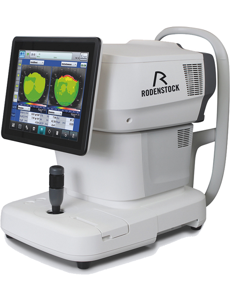
Tune-up your businessExperience 6 analyses in 1 device: Rodenstock Alino Multifunction Refractometer
Are you ready for something that is all-in-one, timesaving, space-saving and suits your business perfectly?Have a look at how powerful ALINO can be.6 in 1 solution01 RefractionHaving a good starting value for subjective refraction is essential. The QUICK REF MODE supports this even for uncooperative patients.02 KeratometryKAI (Kerato-asymmetry index) & KRI (Kerato-regularity index) give the first hint of possible irregularities on the cornea. This supports evaluation of the best vision correction or best-fitting contact lens.03 TonometryThe very gentle air puff created by a new generation of air-flow technology means that patients more readily accept the need for regular check-ups of the IOP measurement.04 PachymetryCorrecting the IOP by measuring the central corneal thickness is essential in interpreting the intraocular pressure.05 TopographyVarious topographic maps support you in carrying out vision screenings, contact lens fittings and in patient education.06 Dry EyeObserve the customers’ eye health and evaluate the long-term tolerability of contact lenses.Take advantage of the combination of tests:Additional servicesTonometry + PachymetryContact lens fitting andtolerance controlKeratometry + Topography + Dry EyeEye healthcareTopography + Tonometry + Pachymetry + Dry EyeSpecifications Rodenstock Alino Multifunction Refractometer
REFRACTIVE POWER MEASUREMENTSpherical refractive power (S) -30.00 D to + 25.00 D (at VD = 12.0 mm)Cylindrical power (C) 0.00 D to ± 12.50 D (at VD = 12.0 mm)Astigmatic axis (A) 0° to 180°KERATOMETRY MEASUREMENTCorneal curvature radius 5.00 mm to 13.00 mmCorneal astigmatic axis 0° to 180°INTRAOCULAR PRESSURE MEASUREMENTMeasurement range 1 mmHg to 60 mmHG (1 hPa to 80 hPa)PACHYMETRY MEASUREMENTMeasurement range 300 μm to 800 μmTOPOGRAPHY MEASUREMENTCorneal curvature radius 5.50 mm to 10.00 mmCorneal astigmatic axis 0° to 180°AUXILIARY FUNCTIONInterpupillary distance Measurement range: 20 mm to 85 mmCorneal diameter and pupil diameter Measurement range: 1.00 mm to 14.00 mmDry eye observation Blinking Analysis, Tear Meniscus height, Hyperemia, Meibomian glandsDATA MANAGEMENTInternal Database Integrated SD CardPrinter Integrated thermal printerData Output Type 3x USB 2.0, 1x LAN, 1x SD Card-slot, 1x WLANExport Format CSV, XML, JPG, PDFDIMENSIONS & ELECTRIC REQUIREMENTSDimensions WDH 312 × 491 × 450 mmWeight Approx. 23 kgVoltage 100 VAC to 240 VACFrequency 50/60 HzPower consumption 110 VA | Zeiss Humphrey Acuitus 5015 Autorefractor Keratometer
Autorefractor/Keratometer that
can take a combination of refraction, central and peripheral keratometry. Unit has been tested with a reference eye (not included) and as well with a test subject and
takes accurate OD/OS reading.
Great unit for your optometry practice.
Unit is patient ready. This has just been professionally calibrated by our optical engineer.
Also includes a printed out Acuitus 5015 User manual,power cord.The Zeiss Humphrey Acuitus 5015 Auto Refractor Keratometer offers expanded refracting capabilities, allowing a practice to refract children and elderly patients alike. The Zeiss Acuitus 5015 also allows refraction one-day post operative of IOL and refractive surgery patients. The Zeiss Acuitus 5015 includes a flat color screen display which produces clear and concise images. Additional features on the Zeiss Acuitus 5015 include auto acquisition, auto XYZ, making the measurements easily acquired. Additional benefits on the Zeiss Acuitus 5015 are automatic eye detection, thirty measurements per second, and RS 232 interface. The Zeiss Acuitus 5015 offers a multitude of features in a small compact footprint at an extremely affordable price point. | Active Optical Systems Optima 518 IPL LaserHandpiece (Standard):Multipurpose Wavelength Mark-II
Handpiece (Optional):Facial Treatments
Pulse Interval/Repetition Rate:0.5 – 2 Seconds
Maximum Output Treatment Pattern:22 Joule /cm 2
Output Power:11-31 Joule / cm 2 , up to 4000 W
Cooling System:Improved Air Cooling
Control:Advanced Software Interface
Lamp’s Life Span:200,000 Flashes (minimum)
Treatment Area/Spot Size:15 x 50 mm, (7.5 cm 2)
Wavelength:420 – 1,100 nm
Weight:88.18 lbs (40Kg) (including stand)
Dimensions:17.32 inches x 17.72 inches x 39.37 inches (44cm x 45cm x 100cm)
Mains:220-230 VAC, 50/60 Hz; 100-120 VAC, 50/60 Hz
Max Mains Power:600 VA
Treatments Variety:
Skin Rejuvenation
Pigmentation Treatment
Acne Treatment
Hair Removal
Hair Removal – Deep Follicles
Hair Removal – Sensitive Skin
Vascular (Facial) Treatment
Tattoo Removal
The Laser uses 12 preset programs. The programs are set in a consecutive, energy intensity increasing order, as specified by the tables of settings, covering all types of hair and skin combinations to be treated.
12 Programs
1 – Very Dark Skin, Fitzpatrick Skin Type IV
2 – Dark Skin, Fitzpatrick Skin Type IV
3 – Dark Skin, Fitzpatrick Skin Type III
4 – Dark Skin, Fitzpatrick Skin Type III
5 – Medium Skin, Fitzpatrick Skin Type II
6 – Medium Skin, Fitzpatrick Skin Type II
7 – Light Skin, Fitzpatrick Skin Type II
8 – Light Skin, Fitzpatrick Skin Type I
9 – Very Light Skin, Fitzpatrick Skin Type I
10 – Very Light Skin, Fitzpatrick Skin Type I
11 – Very Light Skin, Fitzpatrick Skin Type I
12 – High Power, Fitzpatrick Skin Type IOptima-518-Classic Active Optical Systems Optima 518 IPL LaserThe Optima – 518 Classic was designed to answer the needs of sophisticated practitioners who want to provide state of the art treatment to their patients on one hand, based on proven and tested treatment protocols and on the other hand to have the ability to reach new and improved protocols by conducting innovative clinical investigations.The Optima – 518 Classic, based on active’s GEM-PL technology, sets the pulse pattern according to the selected treatment type, skin color, skin photosensitivity and treatment depth. The treatment protocols and different pulse patterns take into account the tissue’s TRT (Thermal Relaxation Time) and the combined knowledge accumulated from thousands of treatments done during the years and clinical investigations done to confirm the findings.New Handpiece Design The Optima – 518 Classic is the first model to use the new Mark II which enables a multipurpose handpiece:
* Faster treatments – up to 0.5 Seconds Between pulses.
* Real-time handpiece temperature control.
* Contemporary and stylish design.
* Light weight design – 25% less weight to improve the therapists’ comfort.Research program for clinical investigations, the self setting program allows researchers to set the treatment’s parameters according to their needs. Allowing advanced research with the device. The energy levels in the self setting program can reach 31J cm 2 with high intensity of 4000 W.Approvals
* CE MDD Intl. Medical Standard
* FDA U.S. Safety Standard
* ISO-13485 Med. Production STD
* IEC 60-601-1 Medical Safety STD
* CFS Gov. Health Approval
* CSA Can. & U.S. Safety App
* TGA Australian health STD. |




Reviews
There are no reviews yet.